Deer Development
| Embryology - 19 Apr 2024 |
|---|
| Google Translate - select your language from the list shown below (this will open a new external page) |
|
العربية | català | 中文 | 中國傳統的 | français | Deutsche | עִברִית | हिंदी | bahasa Indonesia | italiano | 日本語 | 한국어 | မြန်မာ | Pilipino | Polskie | português | ਪੰਜਾਬੀ ਦੇ | Română | русский | Español | Swahili | Svensk | ไทย | Türkçe | اردو | ייִדיש | Tiếng Việt These external translations are automated and may not be accurate. (More? About Translations) |
Introduction
The two main groups of deer are the Cervinae, including the muntjac, the elk (wapiti), the fallow deer, and the chital; and the Capreolinae, including the reindeer (caribou), the roe deer, and the moose.
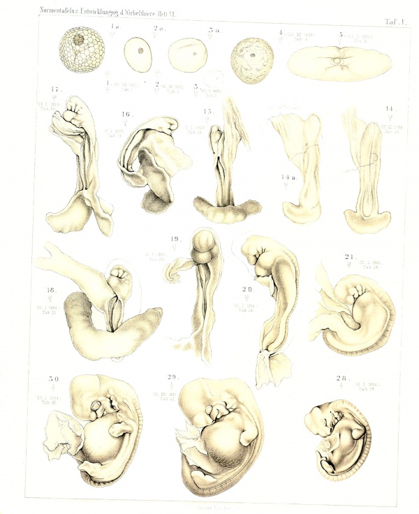
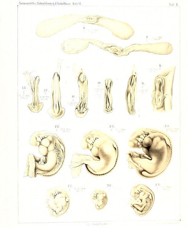
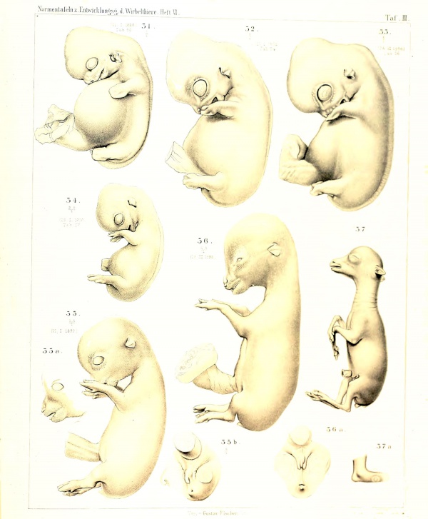
Deer are seasonally polyestrous animals that have multiple estrous cycles only during certain periods of the year, similar to horses, sheep, goats, and cats. A historic 1906 paper by Sakurai[1] characterised the prenatal development of the deer embryo (cervus capreolus). The average prenatal development period for deer (Mule) is 200 days.
Deer are typically a uniparental species, where the offspring (fawn) is only cared for by the mother (doe).
| Animal Development Time | ||||||||||||||||||||||||||||||||||||||||||||||||||||||||||||||||||||||||||||||||||||||||||||||||||||||||||||||||||||||||||||||||||||||||||||||
|---|---|---|---|---|---|---|---|---|---|---|---|---|---|---|---|---|---|---|---|---|---|---|---|---|---|---|---|---|---|---|---|---|---|---|---|---|---|---|---|---|---|---|---|---|---|---|---|---|---|---|---|---|---|---|---|---|---|---|---|---|---|---|---|---|---|---|---|---|---|---|---|---|---|---|---|---|---|---|---|---|---|---|---|---|---|---|---|---|---|---|---|---|---|---|---|---|---|---|---|---|---|---|---|---|---|---|---|---|---|---|---|---|---|---|---|---|---|---|---|---|---|---|---|---|---|---|---|---|---|---|---|---|---|---|---|---|---|---|---|---|---|---|
| ||||||||||||||||||||||||||||||||||||||||||||||||||||||||||||||||||||||||||||||||||||||||||||||||||||||||||||||||||||||||||||||||||||||||||||||
Animal Notes and Table Data Sources
|
- Links: estrous cycle | Category:Deer
| Animal Development: axolotl | bat | cat | chicken | cow | dog | dolphin | echidna | fly | frog | goat | grasshopper | guinea pig | hamster | horse | kangaroo | koala | lizard | medaka | mouse | opossum | pig | platypus | rabbit | rat | salamander | sea squirt | sea urchin | sheep | worm | zebrafish | life cycles | development timetable | development models | K12 |
Some Recent Findings
|
| More recent papers |
|---|
|
This table allows an automated computer search of the external PubMed database using the listed "Search term" text link.
More? References | Discussion Page | Journal Searches | 2019 References | 2020 References Search term: Deer Embryology | Deer Development |
| Older papers |
|---|
| These papers originally appeared in the Some Recent Findings table, but as that list grew in length have now been shuffled down to this collapsible table.
See also the Discussion Page for other references listed by year and References on this current page. |
Embryo Development
Plate 1
Plate 2
Plate 3
References
- ↑ Sakurai T. Normal Plates of the Development of the Deer Embryo (cervus capreolus). (1906) Vol. 6 in series by Keibel F. Normal plates of the development of vertebrates (Normentafeln zur Entwicklungsgeschichte der Wirbelthiere) Fisher, Jena., Germany.
- ↑ Franco A, Masot J & Redondo E. (2017). Comparative analysis of the merino sheep and Iberian red deer abomasum during prenatal development. Anim. Sci. J. , 88, 1575-1587. PMID: 28422357 DOI.
Reviews
Articles
Yanagawa Y, Matsuura Y, Suzuki M, Saga S, Okuyama H, Fukui D, Bandou G, Katagiri S, Takahashi Y & Tsubota T. (2009). Fetal age estimation of Hokkaido sika deer (Cervus nippon yesoensis) using ultrasonography during early pregnancy. J. Reprod. Dev. , 55, 143-8. PMID: 19106485 DOI.
Masot AJ, Franco AJ & Redondo E. (2007). Morphometric and immunohistochemical study of the abomasum of red deer during prenatal development. J. Anat. , 211, 376-86. PMID: 17645454 DOI.
Terms
- abomasum - (maw, rennet-bag, reed tripe) is the fourth and final stomach compartment in ruminants that secretes rennet.
- Hokkaido sika deer - (Cervus nippon yesoensis)
Cite this page: Hill, M.A. (2024, April 19) Embryology Deer Development. Retrieved from https://embryology.med.unsw.edu.au/embryology/index.php/Deer_Development
- © Dr Mark Hill 2024, UNSW Embryology ISBN: 978 0 7334 2609 4 - UNSW CRICOS Provider Code No. 00098G


