Corpus Luteum Development: Difference between revisions
mNo edit summary |
mNo edit summary |
||
| Line 14: | Line 14: | ||
|-bgcolor="F5FAFF" | |-bgcolor="F5FAFF" | ||
| | | | ||
* '''Absent or Excessive Corpus Luteum Number Is Associated With Altered Maternal Vascular Health in Early Pregnancy'''{{#pmid:30636549|PMID30636549}} "Identifying modifiable factors that contribute to preeclampsia risk associated with assisted reproduction can improve maternal health. Vascular dysfunction predates clinical presentation of preeclampsia. Therefore, we examined if a nonphysiological hormonal milieu, a modifiable state, affects maternal vascular health in early pregnancy. ...The number of angiogenic and nonangiogenic circulating endothelial progenitor cell numbers was lower in the absence of a CL in FETs ( P=0.01 and P=0.03). Vascular health in early pregnancy is altered in women with aberrant numbers of CL (0 or >3) and might represent insufficient cardiovascular adaptation contributing to an increased risk of preeclampsia." | |||
* '''MiR-29b affects the secretion of PROG and promotes the proliferation of bovine corpus luteum cells'''{{#pmid:29617446|PMID29617446}} "The regulatory role of miRNAs has been explored in ovarian cells, and their effects on gonadal development, apoptosis, ovulation, steroid production and corpus luteum (CL) development have been revealed. In this study, we analyzed the expression of miR-29b at different stages of bovine CL development and predicted the target genes of miR-29b. We confirmed that miR-29b reduces the expression of the oxytocin receptor (OXTR), affects progesterone (PROG) secretion and regulates the function of the CL. RT-PCR showed that the expression of miR-29b was significantly higher in functional CL phases than in the regressed CL phase. Immunohistochemistry showed that OXTR was expressed in both large and small CL cells and was mainly located in the cell membrane and cytoplasm of these cells. We analyzed the expression levels of OXTR and found that transfection with a miR-29b mimic decreased OXTR expression, but transfection with the inhibitor had a limited effect on the expression of the OXTR protein. At the same time, the secretion of PROG was significantly increased in the miR-29b mimic-transfected group. We also analyzed the effect of miR-29b on the apoptosis of CL cells. Finally, we found that miR-29b could promote the proliferation of bovine CL cells. In conclusion, we found that miR-29b reduces the expression of OXTR and can promote PROG secretion and the proliferation of CL cells via OXTR." | * '''MiR-29b affects the secretion of PROG and promotes the proliferation of bovine corpus luteum cells'''{{#pmid:29617446|PMID29617446}} "The regulatory role of miRNAs has been explored in ovarian cells, and their effects on gonadal development, apoptosis, ovulation, steroid production and corpus luteum (CL) development have been revealed. In this study, we analyzed the expression of miR-29b at different stages of bovine CL development and predicted the target genes of miR-29b. We confirmed that miR-29b reduces the expression of the oxytocin receptor (OXTR), affects progesterone (PROG) secretion and regulates the function of the CL. RT-PCR showed that the expression of miR-29b was significantly higher in functional CL phases than in the regressed CL phase. Immunohistochemistry showed that OXTR was expressed in both large and small CL cells and was mainly located in the cell membrane and cytoplasm of these cells. We analyzed the expression levels of OXTR and found that transfection with a miR-29b mimic decreased OXTR expression, but transfection with the inhibitor had a limited effect on the expression of the OXTR protein. At the same time, the secretion of PROG was significantly increased in the miR-29b mimic-transfected group. We also analyzed the effect of miR-29b on the apoptosis of CL cells. Finally, we found that miR-29b could promote the proliferation of bovine CL cells. In conclusion, we found that miR-29b reduces the expression of OXTR and can promote PROG secretion and the proliferation of CL cells via OXTR." | ||
Revision as of 13:35, 5 March 2019
| Embryology - 25 Apr 2024 |
|---|
| Google Translate - select your language from the list shown below (this will open a new external page) |
|
العربية | català | 中文 | 中國傳統的 | français | Deutsche | עִברִית | हिंदी | bahasa Indonesia | italiano | 日本語 | 한국어 | မြန်မာ | Pilipino | Polskie | português | ਪੰਜਾਬੀ ਦੇ | Română | русский | Español | Swahili | Svensk | ไทย | Türkçe | اردو | ייִדיש | Tiếng Việt These external translations are automated and may not be accurate. (More? About Translations) |
Introduction

The corpus luteum (Latin, corpus = body, luteum = yellow) develops from the remains of Graffian follicle after ovulation. Functions as an endocrine organ (produce progesterone and oestrogens) supporting pregnancy and preventing menstruation (loss of the endometrial lining). Formed during the luteal phase (secretory phase) of the menstrual cycle by proliferation of both follicular granulosa cells (granulosa lutein cells) and thecal cells (theca lutein cells), which together interact to produce progesterone and oestrogens. Ovarian luteal cells have an important role in progesterone (P4) production.
Peak luteal function during the menstrual cycle, determined by maximum luteal area, progesterone concentration and estradiol concentration, is observed about 6 days following ovulation.[1]
If fertilization and pregnancy does not occur, the corpus luteum degenerates to form the corpus albicans.
Some Recent Findings
|
| More recent papers |
|---|
 This table allows an automated computer search of the external PubMed database using the listed "Search term" text link.
More? References | Discussion Page | Journal Searches | 2019 References | 2020 References Search term: Corpus Luteum |
History
| Regnier de Graaf (1641 – 1673) | Ludwig Fraenkel (1870 - 1951) |

|

|
| Regnier de Graaf (1641 – 1673) was the first observer in the ovary of a cow as a yellow structure, the yellow colour was caused by accumulation of steroidal hormones. | Ludwig Fraenkel (1870 - 1951) first identified the endocrine function of the corpus luteum.[4] |
Histology

| |

|

|
| Corpus luteum histology | Corpus luteum lutein cells |

Human corpus luteum (large image)
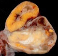
Human corpus luteum (small image)
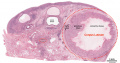
Corpus luteum histology
Corpus luteum histology
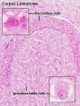
Corpus luteum lutein cells histology
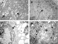
Human corpus luteum (light and electron micrograph)


Embryo Virtual Slide
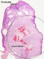
| Human Ovary and Corpus Luteum
|
| Ovary | Embryo Slides |

| Corpus Luteum Links: anatomy overview | image - histology overview | image - Layers granulosa and theca | image - Layers detail granulosa and theca | image - low power label | image - high power label | image - low power | image - high power | image - corpus albicans | theca and granulosa lutein cells | Granulosa cell | corpus luteum | granulosa lutein cells | theca lutein cells | corpus albicans | ovary | menstrual cycle |
| Historic Papers: 1969 corpus luteum ultrastructure 1 | 1969 corpus luteum ultrastructure 2 |
Corpus Albicans


Corpus albicans histology
(corpora albicantia) (Latin, corpus = body, albicans = whitish) The histological structure formed by luteolysis of the corpus luteum in the ovary. If implantation does not occur and the hormone hCG is not released the corpus luteum degenerates and the structure is white, not yellow, because of the absence of steroid hormone synthesis/accumulation.
| Corpus Luteum Links: anatomy overview | image - histology overview | image - Layers granulosa and theca | image - Layers detail granulosa and theca | image - low power label | image - high power label | image - low power | image - high power | image - corpus albicans | theca and granulosa lutein cells | Granulosa cell | corpus luteum | granulosa lutein cells | theca lutein cells | corpus albicans | ovary | menstrual cycle |
| Historic Papers: 1969 corpus luteum ultrastructure 1 | 1969 corpus luteum ultrastructure 2 |
Animal Models
References
- ↑ Baerwald AR, Adams GP & Pierson RA. (2005). Form and function of the corpus luteum during the human menstrual cycle. Ultrasound Obstet Gynecol , 25, 498-507. PMID: 15846762 DOI.
- ↑ von Versen-Höynck F, Narasimhan P, Selamet Tierney ES, Martinez N, Conrad KP, Baker VL & Winn VD. (2019). Absent or Excessive Corpus Luteum Number Is Associated With Altered Maternal Vascular Health in Early Pregnancy. Hypertension , 73, 680-690. PMID: 30636549 DOI.
- ↑ Xu MQ, Jiang H, Zhang LQ, Sun XL, Luo D, Fu Y, Gao Y, Yuan B & Zhang JB. (2018). MiR-29b affects the secretion of PROG and promotes the proliferation of bovine corpus luteum cells. PLoS ONE , 13, e0195562. PMID: 29617446 DOI.
- ↑ Simmer HH. (1971). The first experiments to demonstrate an endocrine function of the corpus luteum. On the occasion of the 100th birthday of Ludwig Fraenkel (1870-1951). Sudhoffs Arch , 55, 392-417. PMID: 4261581
Reviews
Sugino N, Matsuoka A, Taniguchi K & Tamura H. (2008). Angiogenesis in the human corpus luteum. Reprod. Med. Biol. , 7, 91-103. PMID: 29699289 DOI.
Stouffer RL, Bishop CV, Bogan RL, Xu F & Hennebold JD. (2013). Endocrine and local control of the primate corpus luteum. Reprod Biol , 13, 259-71. PMID: 24287034 DOI.
Bachelot A & Binart N. (2005). Corpus luteum development: lessons from genetic models in mice. Curr. Top. Dev. Biol. , 68, 49-84. PMID: 16124996 DOI.
Stouffer RL. (2003). Progesterone as a mediator of gonadotrophin action in the corpus luteum: beyond steroidogenesis. Hum. Reprod. Update , 9, 99-117. PMID: 12751773
Articles
Search Pubmed
Search Pubmed: corpus luteum Development
External Links
External Links Notice - The dynamic nature of the internet may mean that some of these listed links may no longer function. If the link no longer works search the web with the link text or name. Links to any external commercial sites are provided for information purposes only and should never be considered an endorsement. UNSW Embryology is provided as an educational resource with no clinical information or commercial affiliation.
Glossary Links
- Glossary: A | B | C | D | E | F | G | H | I | J | K | L | M | N | O | P | Q | R | S | T | U | V | W | X | Y | Z | Numbers | Symbols | Term Link
Genital Links: genital | Lecture - Medicine | Lecture - Science | Lecture Movie | Medicine - Practical | primordial germ cell | meiosis | endocrine gonad | Genital Movies | genital abnormalities | Assisted Reproductive Technology | puberty | Category:Genital
| ||||
|
| Menstrual Cycle | X Chromosome
Cite this page: Hill, M.A. (2024, April 25) Embryology Corpus Luteum Development. Retrieved from https://embryology.med.unsw.edu.au/embryology/index.php/Corpus_Luteum_Development
- © Dr Mark Hill 2024, UNSW Embryology ISBN: 978 0 7334 2609 4 - UNSW CRICOS Provider Code No. 00098G