Coelomic Cavity Development: Difference between revisions
| Line 9: | Line 9: | ||
:{{Template:Coelomic Cavity Links}} | [http://embryology.med.unsw.edu.au/Notes/coelom.htm original page] | :{{Template:Coelomic Cavity Links}} | [http://embryology.med.unsw.edu.au/Notes/coelom.htm original page] | ||
:{{Template:Systems}} | :{{Template:Systems}} | ||
==Some Recent Findings== | |||
Most triploblastic animals including vertebrates have a coelomic cavity that separates the outer and inner components of the body. The coelom is lined by two different tissue components, somatopleure and splanchnopleure, which are derived from the lateral plate region. Thus, the coelom is constructed as a result of a binary decision during early specification of the lateral plate. In this report we studied the molecular mechanisms of this binary decision. We first demonstrate that the splitting of the lateral plate into the two cell sheets progresses in an anteroposterior order and this progression is not coordinated with that of the somitic segmentation. By a series of embryological manipulations we found that young splanchnic mesoderm is still competent to be respecified as somatic mesoderm, and the ectoderm overlying the lateral plate is sufficient for this redirection. The lateral ectoderm is also required for maintenance of the somatic character of the mesoderm. Thus, the ectoderm plays at least two roles in the early subdivision of the lateral plate: specification and maintenance of the somatic mesoderm. We also show that the latter interactions are mediated by BMP molecules that are localized in the lateral ectoderm. Evolutionary aspects of the coelom formation are also considered. | |||
== Objectives == | == Objectives == | ||
Revision as of 16:03, 5 August 2010
Introduction

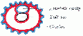
There are cavities, coeloms, that form and lie both outside (extraembryonic) and inside (intraembryonic) the embryo during development.
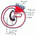
The intraembryonic coelom is the primitive cavity that lies within the developing embryo that will form the 3 major body cavities: pericardial, pleural, peritoneal. The coelom forms very early in embryogenesis and is much later paritioned inferiorly by the diaphragm and pleuroperitoneal membrane; and superiorly initially by the pleuropericardial fold between the heart and lungs. The intraembryonic coelom communicated through coelomic portals (at the level of midgut herniation) with the extraembryonic coelom.
All cavities are fluid filled and developing organs push against a wall of the cavity, generating a double coat (serosal/adventital) surrounding an organ (for example the lungs). The serous membrane is the epithelium (squamous) and its associated underlying loose connective tissue.
| Coelom Links: Introduction | Lecture - Week 3 Development | Lecture - Mesoderm Development | Placenta - Membranes | Category:Coelomic Cavity |
| Historic Embryology - Coelomic Cavity |
|---|
| 1891 peritoneal | 1897 human coelom | 1910 Coelom and Diaphragm | 1924 serous |
| System Links: Introduction | Cardiovascular | Coelomic Cavity | Endocrine | Gastrointestinal Tract | Genital | Head | Immune | Integumentary | Musculoskeletal | Neural | Neural Crest | Placenta | Renal | Respiratory | Sensory | Birth |
Some Recent Findings
Most triploblastic animals including vertebrates have a coelomic cavity that separates the outer and inner components of the body. The coelom is lined by two different tissue components, somatopleure and splanchnopleure, which are derived from the lateral plate region. Thus, the coelom is constructed as a result of a binary decision during early specification of the lateral plate. In this report we studied the molecular mechanisms of this binary decision. We first demonstrate that the splitting of the lateral plate into the two cell sheets progresses in an anteroposterior order and this progression is not coordinated with that of the somitic segmentation. By a series of embryological manipulations we found that young splanchnic mesoderm is still competent to be respecified as somatic mesoderm, and the ectoderm overlying the lateral plate is sufficient for this redirection. The lateral ectoderm is also required for maintenance of the somatic character of the mesoderm. Thus, the ectoderm plays at least two roles in the early subdivision of the lateral plate: specification and maintenance of the somatic mesoderm. We also show that the latter interactions are mediated by BMP molecules that are localized in the lateral ectoderm. Evolutionary aspects of the coelom formation are also considered.
Objectives
- Describe the development of the intra- and extra-embryonic coeloms.
- Describe the processes involved in the development of the three divisions of the intra-embryonic coelom; pericardium, pleural cavities and peritoneum.
- Describe the fate of the extra embryonic coelom.
- Describe the development of the diaphragm.
Development Overview
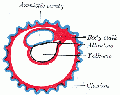
| Extraembryonic Coelom
|
Intraembryonic Coelom
|
Neural Tube
| |
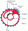
Intraembryonic Coelom

Percardial Cavity
Pleural Cavity
Peritoneal Cavity
Terms
- atresia- obstruction.
- eppiglottis- develops from hypobrachial eminence.
- fistula- abnormal comunication.
- hypopharyngeal eminence- fusion of 3rd pharyngeal arches, precursor of root of tongue.
- laryngotracheal groove- forms on anterior (ventral) wall of pharynx, gives rise to larynx, trachea, respiratory tree.
- larynx- lining from endoderm, cartilage from pharyngeal arch 4 and 6.
- lung buds- primordia of lungs.
- parietal pleura-outer lining of pleural cavity derived from epithelia of pericardioperitoneal canals from intraembryonic coelom.
- pleural cavity- walls derived from pericardioperitoneal canals -> intraembryonic coelom ->coelomic spaces -> lateral mesoderm -> mesoderm.
- pleuropericardial fold- restricts the communication between pleural cavity and pericardiac cavity, contains cardinal vein and phrenic nerve.
- pleuroperitoneal membrane- forms inferiorly at transverse septum to separate peritoneum from pleural cavity.
- septum transversum- mesoderm separating thoracic cavity and yolk sac, forms central tendon of diaphragm (and some of liver?).
- stenosis- narrowing
- surfactant- a detergent secreted by Type 2 alveolar cells between alveolar epithelium. Functions to lower surface tension, allowing lungs to remain inflated.
- visceral pleura- inner lining of pleural cavity derived from contact epithelia with lung bud of pericardioperitoneal canals from intraembryonic coelom.
Mesothelia
The epithelial covering of coelomic organs and also line their cavities.
Contribute to the vasculature of the heart and the intestinal tract. Undergo epithelial-mesenchymal transition (EMT), migration, and differentiation into endothelial cells, vascular smooth muscle cells, and pericytes.
References
Reviews
Articles
Search PubMed
Search Pubmed: Coelomic Cavity Development | pericardial cavity development | pleural cavity development | peritoneal cavity development
Additional Images
- Gray0031.gif


Glossary Links
- Glossary: A | B | C | D | E | F | G | H | I | J | K | L | M | N | O | P | Q | R | S | T | U | V | W | X | Y | Z | Numbers | Symbols | Term Link
Cite this page: Hill, M.A. (2024, April 19) Embryology Coelomic Cavity Development. Retrieved from https://embryology.med.unsw.edu.au/embryology/index.php/Coelomic_Cavity_Development
- © Dr Mark Hill 2024, UNSW Embryology ISBN: 978 0 7334 2609 4 - UNSW CRICOS Provider Code No. 00098G