Cloaca Development: Difference between revisions
mNo edit summary |
mNo edit summary |
||
| Line 61: | Line 61: | ||
:'''Links:''' [[Amniotic Cavity Development Movie|Early Endoderm Movie]] | [[Week 3 Development Movie|Week 3 Folding Movie]] | :'''Links:''' [[Amniotic Cavity Development Movie|Early Endoderm Movie]] | [[Week 3 Development Movie|Week 3 Folding Movie]] | ||
==Development== | ==Embryonic Development== | ||
===Stage 10=== | ===Stage 10=== | ||
{| | {| | ||
| Line 140: | Line 140: | ||
===Historic=== | ===Historic=== | ||
{{#pmid:14444236}} | {{#pmid:14444236}} | ||
{{Ref-Felix1912}} | |||
===Search Pubmed=== | ===Search Pubmed=== | ||
| Line 158: | Line 160: | ||
Fig.8. Section through body of Harvard Embryo {{HE714}} near the {{cloaca}}. | Fig.8. Section through body of Harvard Embryo {{HE714}} near the {{cloaca}}. | ||
{{Ref-Felix1912}} | |||
<gallery> | <gallery> | ||
| Line 165: | Line 169: | ||
File:Keibel_Mall_2_602.jpg|Fig. 602. Cloaca human embryo 7 mm GL (embryo Chr. I), separation of the rectum from the ventral remains of the cloaca is shown | File:Keibel_Mall_2_602.jpg|Fig. 602. Cloaca human embryo 7 mm GL (embryo Chr. I), separation of the rectum from the ventral remains of the cloaca is shown | ||
File:Keibel_Mall_2_603.jpg|Fig. 603 a and b. Human embryo 7 mm GL (Embryo Chr. I) division of cloaca into rectum and ventral remains of the cloaca is almost complete, the saddle between the two being immediately over the cloacal membrane. | File:Keibel_Mall_2_603.jpg|Fig. 603 a and b. Human embryo 7 mm GL (Embryo Chr. I) division of cloaca into rectum and ventral remains of the cloaca is almost complete, the saddle between the two being immediately over the cloacal membrane. | ||
File:Keibel_Mall_2_604.jpg|Fig. 604 a and b. Human embryo 11 mm GL (Embryo P. I.) The rectum is completely separated from the ventral remains of the cloaca, and the cloacal membrane has been divided into the canal and urogenital membranes. | |||
File:Keibel_Mall_2_605.jpg|Fig. 605. Human embryo 5.3 mm GL (Embryo {{KE1420}}) Primary excretory duct and opening with reference to cloaca and cloacal membrane, anlage of ureteric bud. | |||
File:Keibel_Mall_2_606.jpg|Fig. 606. Human embryo 4.25 mm vertex-breech length, 28 somites (Embryo H. M. 1) Primary excretory duct and position of orifice relatively to cloaca and cloacal membrane. | |||
File:Keibel_Mall_2_607.jpg|Fig. 607. Human embryo 18 mm Between the umbilicus and the coccygeal tubercle cranio-caudally and the two lower extremities laterally, the cloacal tubercle has appeared. | |||
[[File:Keibel_Mall_2_652.jpg|Fig. 652. The division of the ventral remains of the cloaca is completed. | |||
</gallery> | </gallery> | ||
| Line 182: | Line 191: | ||
==Terms== | ==Terms== | ||
* '''cloacal duct of Reichel''' - | * '''cloacal duct of Reichel''' - Reichel P. The development of the bladder and urinary tubes (''Die Entwicklung der Harnblase und der Harnrohre''), (1893) Verb. phys. med. Gesellsch. Wiirzburg. | ||
* '''cloacal tubercle''' - (genital tubercle) | |||
{{GIT terms}} | {{GIT terms}} | ||
Revision as of 13:34, 14 November 2018
| Embryology - 24 Apr 2024 |
|---|
| Google Translate - select your language from the list shown below (this will open a new external page) |
|
العربية | català | 中文 | 中國傳統的 | français | Deutsche | עִברִית | हिंदी | bahasa Indonesia | italiano | 日本語 | 한국어 | မြန်မာ | Pilipino | Polskie | português | ਪੰਜਾਬੀ ਦੇ | Română | русский | Español | Swahili | Svensk | ไทย | Türkçe | اردو | ייִדיש | Tiếng Việt These external translations are automated and may not be accurate. (More? About Translations) |
Introduction

The initial cloaca is the common early endoderm lined space of the hindgut that will later become partitioned by a septum into a dorsal gastrointestinal component (rectum) and ventral renal/genital component (urogenital sinus). Note that the cloaca in mammals is an early embryonic transient structure and only persists in birds and reptiles. Located at the superior end of the cloaca is the allantois, that extends into the connecting stalk and later the placental cord. Located at the inferior end of the cloaca is the cloacal membrane, that also forms part of the embryo surface.
The gastrointestinal tract ends at this cloacal membrane, equivalent to the beginning of the tract at the buccopharyngeal membrane at the upper end. The cloacal membrane is formed during gastrulation by ectoderm and endoderm without a middle (intervening) layer of mesoderm, that later degenerates after cloacal septation.
The hindgut component will contribute to the gastrointestinal tract intestine of the distal transverse colon, descending colon, sigmoid colon, rectum.
The urogenital sinus component will contribute the renal urinary bladder and participate in genital development.
Some Recent Findings
|
| More recent papers |
|---|
 This table allows an automated computer search of the external PubMed database using the listed "Search term" text link.
More? References | Discussion Page | Journal Searches | 2019 References | 2020 References Search term: Cloaca <pubmed limit=5>Cloaca</pubmed>
<pubmed limit=5>Cloacal Membrane</pubmed> |
| Older papers |
|---|
| These papers originally appeared in the Some Recent Findings table, but as that list grew in length have now been shuffled down to this collapsible table.
See also the Discussion Page for other references listed by year and References on this current page. |
Movies
| Early Endoderm | Week 3 Folding |
|---|---|
| <html5media height="400" width="350">File:Amnion 001.mp4</html5media> | <html5media height="500" width="350">File:Week3_folding.mp4</html5media> |
- Links: Early Endoderm Movie | Week 3 Folding Movie
Embryonic Development
Stage 10

|

|
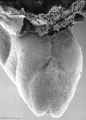
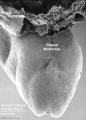
Caudal end of embryo showing primitive streak region, cloacal membrane, and connecting stalk. |
- Links: Carnegie stage 10 | Stage 10 Movie
Stage 11

|
Historic image<ref name=Low1908>Low A. Description of a human embryo of 13-14 mesodermic somites. (1908) J Anat Physiol. 42(3): 237-51. PMID 17232769 | PMC1289161 of an embryo model (sagittal section, viewed from the left) showing hindgut and cloaca. |
- Links: Carnegie stage 11
Stage 12
- ===Stage 13===
- ===Stage 22===
- ==Abnormalities==
- ===Persistent Cloaca Perineum===
- ==References==
- ===Books===
- ===Reviews===
- ===Articles===
- ===Historic===
- ===Search Pubmed===
- ==Additional Images==
- ===Historic===
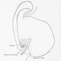
Fig. 599. Embryo Pfannenstiel-Kromer, 1.38 mm long, 5-6 somites, shows relation of cloaca and cloacal membrane to allantoic stalk.
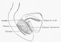
Fig. 600. Embryo Pfannenstiel III 2.6 mm GL 13-14 somites, allantoic stalk is given off at a right angle from the cloaca, immediately above cloacal membrane. Ventral wall of the cloaca is formed only by the cloacal membrane.
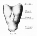
Fig. 601. Cloacal membrane human embryo 3 mm GL (Embryo E. B.) forms a rhomboidal groove slightly depressed below the surface of the embryo, between the umbilicus and the coccygeal tubercle.
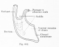
Fig. 602. Cloaca human embryo 7 mm GL (embryo Chr. I), separation of the rectum from the ventral remains of the cloaca is shown
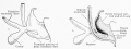
Fig. 603 a and b. Human embryo 7 mm GL (Embryo Chr. I) division of cloaca into rectum and ventral remains of the cloaca is almost complete, the saddle between the two being immediately over the cloacal membrane.
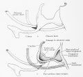
Fig. 604 a and b. Human embryo 11 mm GL (Embryo P. I.) The rectum is completely separated from the ventral remains of the cloaca, and the cloacal membrane has been divided into the canal and urogenital membranes.
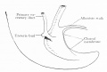
Fig. 605. Human embryo 5.3 mm GL (Embryo 1420) Primary excretory duct and opening with reference to cloaca and cloacal membrane, anlage of ureteric bud.
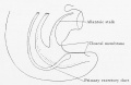
Fig. 606. Human embryo 4.25 mm vertex-breech length, 28 somites (Embryo H. M. 1) Primary excretory duct and position of orifice relatively to cloaca and cloacal membrane.
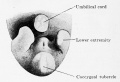
Fig. 607. Human embryo 18 mm Between the umbilicus and the coccygeal tubercle cranio-caudally and the two lower extremities laterally, the cloacal tubercle has appeared.


Florian J. The early development of man, with special reference to the development of the mesoderm and cloacal membrane. (1933) J. Anat., 67(2): 263-76. PMID 17104422
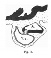
Fig.1
Fig.2
Fig.3
Fig.4
Fig.5
Fig.6
Fig.7
Fig.8

Terms
- cloacal duct of Reichel - Reichel P. The development of the bladder and urinary tubes (Die Entwicklung der Harnblase und der Harnrohre), (1893) Verb. phys. med. Gesellsch. Wiirzburg.
- cloacal tubercle - (genital tubercle)
| Gastrointestinal Tract Terms | ||
|---|---|---|
| ||
|
Glossary Links
- Glossary: A | B | C | D | E | F | G | H | I | J | K | L | M | N | O | P | Q | R | S | T | U | V | W | X | Y | Z | Numbers | Symbols | Term Link
Cite this page: Hill, M.A. (2024, April 24) Embryology Cloaca Development. Retrieved from https://embryology.med.unsw.edu.au/embryology/index.php/Cloaca_Development
- © Dr Mark Hill 2024, UNSW Embryology ISBN: 978 0 7334 2609 4 - UNSW CRICOS Provider Code No. 00098G