Category:Ultrasound: Difference between revisions
From Embryology
(Created page with 'This page lists UNSW Embryology content related to ultrasound imaging during development.') |
No edit summary |
||
| Line 1: | Line 1: | ||
This page lists UNSW Embryology content related to ultrasound imaging during development. | This page lists UNSW Embryology content related to ultrasound imaging during development. | ||
:'''Links:''' [[Ultrasound]] | [[Prenatal Diagnosis]] | |||
[[Category:Prenatal Diagnosis]] | |||
Revision as of 23:09, 9 August 2012
This page lists UNSW Embryology content related to ultrasound imaging during development.
- Links: Ultrasound | Prenatal Diagnosis
Pages in category 'Ultrasound'
The following 38 pages are in this category, out of 38 total.
U
- Ultrasound
- Template:Ultrasound
- Ultrasound - Abnormal
- Ultrasound - Cleft Lip Movie 1
- Ultrasound - Cleft Lip Movie 2
- Ultrasound - Dandy-Walker Movie 1
- Ultrasound - Ectopic Movie 1
- Ultrasound - Ectopic Movie 2
- Ultrasound - Fetal Heart Rate
- Ultrasound - Fetus Movie 1
- Ultrasound - Fetus Movie 2
- Ultrasound - Nuchal Translucency
- Ultrasound - Placenta Previa Movie 1
- Ultrasound - Rabbit Embryo Movie
- Ultrasound - Spina Bifida Movie 1
- Ultrasound - Transposition Great Arteries Movie 1
- Ultrasound - Vasa Previa Movie 1
- Ultrasound - Velamentous Cord Movie 1
- Template:Ultrasound Movies
- Template:Ultrasound Movies - abnormal
- Template:Ultrasound Movies - normal
- Template:Ultrasound Placenta Previa
- Template:Ultrasound Spina-Bifida
- Template:Ultrasound terms
- Template:Ultrasound Transposition Great Arteries
- Template:Ultrasound Vasa Previa
- Template:Ultrasound Velamentous Cord
Media in category 'Ultrasound'
The following 69 files are in this category, out of 69 total.
 Anencephaly ultrasound.jpg 900 × 658; 108 KB
 Aorta coarctation echocardiogram.jpg 601 × 283; 28 KB
 Cervical ectopic ultrasound.jpg 800 × 541; 62 KB
 Cleft lip 01.jpg 585 × 438; 34 KB
 Cleft lip 03.jpg 585 × 439; 38 KB
 Complete atrioventricular canal.jpg 320 × 240; 22 KB
 Complete hydatidiform mole 01.jpg 748 × 560; 50 KB
 Dichorionic twins ultrasound 01.gif 401 × 282; 2.51 MB
 Echidna egg ultrasound.jpg 484 × 390; 83 KB
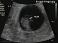 Ectopic 01 heart.jpg 655 × 491; 34 KB
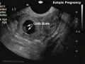 Ectopic 01 limbs.jpg 655 × 491; 37 KB
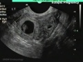 Ectopic 01 zoom.jpg 655 × 491; 42 KB
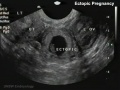 Ectopic 01.jpg 655 × 491; 40 KB
 Fetal blood flow liver and brain.jpg 677 × 790; 69 KB
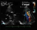
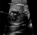
 Fetal cardiac ultrasound 01.jpg 900 × 600; 88 KB
 Fetal ductus venosus pressure wave 01.jpg 706 × 755; 52 KB
 Fetal ductus venosus ultrasound 01.jpg 783 × 1,000; 68 KB
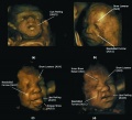 Fetal facial expression 01.jpg 1,200 × 1,094; 122 KB
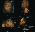 Fetal facial expression 02.jpg 1,914 × 1,762; 205 KB
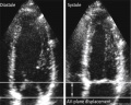 Fetal heart atrioventricular plane displacement 01.jpg 915 × 733; 55 KB
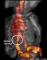
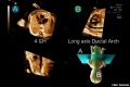 Fetal ultrasound ductal arch 01.jpg 800 × 533; 27 KB
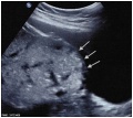
 Gastroschisis 01.jpg 493 × 439; 23 KB
 Hydatidiform mole 02.jpg 585 × 1,100; 237 KB
 Hypospadia 3D ultrasound 01.jpg 1,150 × 497; 87 KB
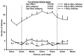
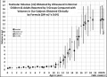 Male puberty testicular volume graph.jpg 1,140 × 826; 126 KB
 Mouse ovarian follicle size.jpg 600 × 407; 36 KB
 Patent ductus arteriosus echocardiogram.jpg 1,200 × 960; 133 KB
 Placenta accreta ultrasound bladder wall interface.jpg 600 × 537; 48 KB
 Placenta accreta ultrasound retroplacental clear space loss.jpg 600 × 510; 43 KB
 Placenta previa - anterior.jpg 730 × 547; 107 KB
 Placenta previa ultrasound 01.jpg 739 × 554; 68 KB
 Placental chorioangioma ultrasound 01.jpg 1,183 × 837; 101 KB
 Placental chorioangioma ultrasound 02.jpg 1,183 × 837; 92 KB
 Placental cord ultrasound 01.jpg 585 × 1,309; 192 KB
 Placental cord ultrasound 02.jpg 731 × 479; 101 KB
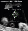 Placental cord ultrasound 03.jpg 598 × 646; 61 KB
 Placental cord ultrasound 04.jpg 729 × 572; 65 KB
 Postnatal persistant ductus venosus ultrasound 01.jpg 1,370 × 600; 96 KB
 Postnatal persistant ductus venosus ultrasound 03.jpg 694 × 600; 54 KB
 Robert Rushmer.jpg 390 × 469; 47 KB
 Septate uterus ultrasound 01.jpg 1,280 × 525; 95 KB
 Tetralogy of Fallot 02.jpg 800 × 796; 63 KB
 Trisomy 10 mosaicism cardiovascular abnormalities.jpg 1,418 × 489; 147 KB
 Trisomy 21 - nuchal translucency graph 01.jpg 638 × 1,000; 69 KB
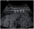
 Ultrasound - fetal abdominal circumference.jpg 672 × 512; 23 KB
 Ultrasound - Hypoplastic left heart syndrome 01.jpg 800 × 600; 53 KB
 Ultrasound - Hypoplastic left heart syndrome 02.jpg 890 × 626; 54 KB
 Ultrasound - Hypoplastic left heart syndrome 03.jpg 874 × 612; 51 KB
 Ultrasound - Hypoplastic left heart syndrome 04.jpg 919 × 618; 68 KB
 Ultrasound 12 week icon.jpg 500 × 374; 46 KB
 Ultrasound 12wk 4D.mov ; 776 KB
- Ultrasound 12wk 4Dv2.mov ; 1.46 MB
 Ultrasound nuchal translucency.jpg 668 × 598; 58 KB
 Ultrasound placenta previa 01.jpg 1,024 × 692; 61 KB
 Ultrasound twinning 01.jpg 1,024 × 692; 72 KB
 Ultrasound twinning 02.jpg 1,024 × 692; 71 KB
 Ultrasound uterine and ovarian vascularity.jpg 531 × 780; 132 KB
 US Circumvallate placenta 01.jpg 1,024 × 692; 61 KB
 US Circumvallate placenta 02.jpg 1,024 × 692; 72 KB
 Velamentous cord insertion 01.jpg 1,024 × 692; 78 KB
 Velamentous cord insertion 02.jpg 1,024 × 692; 104 KB
 Ventricular septal defect 01.jpg 1,024 × 692; 84 KB
 Z3DCleft Lip Picture.jpg 640 × 480; 32 KB
 ZPulmonary Atresia.jpg 653 × 618; 85 KB
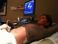 ZUltrasound Exam.JPG 1,600 × 1,200; 846 KB
 ZUltrasound Image of Fetal Aorta.jpg 640 × 480; 47 KB
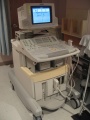 ZUltrasound machine.jpg 480 × 640; 93 KB
 ZUltrasound System.jpg 262 × 400; 49 KB


