Category:Skull
From Embryology
The pages and media shown below relate to development of the skull.
Pages in category 'Skull'
The following 119 pages are in this category, out of 119 total.
A
B
F
J
M
P
- Palate Development
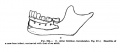
- Paper - A model of the left half of the human mandible at the 17 mm CRL stage
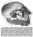
- Paper - Description of a reconstruction of the head of a thirty-millimetre embryo (1910)
- Paper - Development of the malleus of the human ear - Illustrated in atlas series
- Paper - Development of the otic capsule 2
- Paper - Further observations on the ossification of the human lower jaw
- Paper - Notes on the development of the human sphenoid (1910)
- Paper - On the development and morphology of the human sphenoid bone
- Paper - On the premature obliteration of sutures in the human skull (1915)
- Paper - Pharyngeal end of Rathke's pouch (1911)
- Paper - Preliminary note on the skull of a human fetus of 43 mm greatest length
- Paper - Some observations on the roof of the primordial human cranium (1923)
- Paper - Structure and development of the pig skull
- Paper - The cartilaginous skull of a human embryo twenty-one millimeters in length (1920)
- Paper - The chondrocranium of a 20 mm human embryo
- Paper - The development of the cochlear fenestra, fossula and secondary tympanic membrane
- Paper - The development of the human maxilla, vomer, and paraseptal cartilages (1911)
- Paper - The development of the otic capsule in the region of the vestibular aqueduct
- Paper - The developmental and adult anatomy of the air-cells in the petrous part of the temporal bone
- Paper - The fontanella metopica and its remnants in an adult skull (1918)
- Paper - The genesis and development of the nasolacrimal passages in man
- Paper - The lateral wall of the cavum nasi in man, with especial reference to the various developmental stages
- Paper - The Long Fox lecture - The development of the human skull (1910)
- Paper - The Monotreme Skull - A Contribution to Mammalian Morphogenesis
- Paper - The ossification of the human frontal bone with special reference to its presumed pre- and post-frontal elements
- Paper - The primordial cranium of miniopterus schreibersi at the 17 millimetre total length stage (1919)
- Paper - The skull of a human fetus of 40 mm 1
- Paper - The skull of a human fetus of 40 mm 2
- Template:Pharyngeal arch
R
- Template:Ref-AnsonBast1958e
- Template:Ref-AnsonBastRichany1956
- Template:Ref-Bolk1915
- Template:Ref-Covell1927
- Template:Ref-Fawcett1910lecture
- Template:Ref-Fawcett1910sphenoid
- Template:Ref-Fawcett1911
- Template:Ref-Fawcett1917
- Template:Ref-Fawcett1918
- Template:Ref-Fawcett1918a
- Template:Ref-Fawcett1919
- Template:Ref-Fawcett1923
- Template:Ref-Gilse1927
- Template:Ref-GladstoneWakeley1923
- Template:Ref-Hayes1922
- Template:Ref-Inman1937
- Template:Ref-InmanSaunders1937
- Template:Ref-Kernan1916
- Template:Ref-Lewis1920
- Template:Ref-Low1909
- Template:Ref-Macklin1914a
- Template:Ref-Macklin1914b
- Template:Ref-Macklin1921
- Template:Ref-Macklin1921a
- Template:Ref-Murray1943
- Template:Ref-Parker1874
- Template:Ref-RichanyBastAnson1956
- Template:Ref-Schaeffer1910a
- Template:Ref-Schaeffer1910b
- Template:Ref-Schaeffer1911
- Template:Ref-Schaeffer1912
- Template:Ref-Schultz1918
- Template:Ref-StelterBastAnson1960
- Template:Ref-Sutton1885
- Template:Ref-Terry1909
- Template:Ref-Tomes1853
- Template:Ref-Walusch1906
- Template:Ref-Watson1915
S
Media in category 'Skull'
The following 200 files are in this category, out of 247 total.
(previous page) (next page)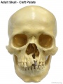 Adult skull cleft palate 01.jpg 750 × 1,000; 89 KB
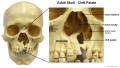 Adult skull cleft palate 02.jpg 1,280 × 720; 139 KB
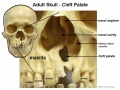 Adult skull cleft palate 03.jpg 904 × 678; 104 KB
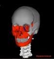 Adult Skull Movie 1 icon.jpg 442 × 480; 33 KB
 Adult Skull Movie 2 icon.jpg 479 × 480; 39 KB
 Adult Skull Movie 3 icon.jpg 445 × 480; 36 KB
 Adult Skull Movie 4 icon.jpg 446 × 480; 34 KB
 Axial skeleton.jpg 1,000 × 1,434; 111 KB
 Bailey132+133.jpg 940 × 570; 101 KB
 Bailey132.jpg 466 × 413; 43 KB
 Bailey133.jpg 806 × 655; 85 KB
 Bailey135.jpg 940 × 965; 216 KB
 Bailey136.jpg 835 × 566; 114 KB
 Bailey137.jpg 672 × 539; 73 KB
 Bailey138.jpg 831 × 400; 62 KB
 Bailey139.jpg 961 × 671; 96 KB
 Bailey140.jpg 793 × 505; 58 KB
 Brown024.jpg 558 × 800; 52 KB
 Craniofrontonasal syndrome.jpg 1,280 × 543; 130 KB
 Craniosynostosis .jpg 375 × 430; 85 KB
 Fawcett 1910 fig01.jpg 986 × 1,085; 256 KB
 Fawcett 1910 fig02.jpg 824 × 690; 56 KB
 Fawcett 1910 fig03.jpg 970 × 996; 202 KB
 Fawcett 1910 fig04.jpg 789 × 807; 101 KB
 Fawcett 1910 fig05.jpg 1,031 × 727; 103 KB
 Fawcett 1910 fig06.jpg 1,064 × 1,022; 285 KB
 Fawcett 1910 fig07.jpg 968 × 958; 228 KB
 Fawcett 1910 fig08.jpg 1,087 × 1,050; 261 KB
 Fawcett 1910 fig09.jpg 932 × 901; 207 KB
 Fawcett 1910 fig10.jpg 782 × 747; 118 KB
 Fawcett 1910 fig11.jpg 768 × 719; 124 KB
 Fawcett 1910 fig12.jpg 1,068 × 970; 282 KB
 Fawcett 1910 fig13.jpg 1,056 × 922; 289 KB
 Fawcett 1910 fig14.jpg 1,058 × 989; 271 KB
 Fawcett 1910 fig15.jpg 962 × 973; 200 KB
 Fawcett 1910 fig16.jpg 1,238 × 909; 333 KB
 Fawcett1910 fig01.jpg 891 × 639; 148 KB
 Fawcett1910 fig02.jpg 1,035 × 949; 265 KB
 Fawcett1910 fig03.jpg 905 × 978; 160 KB
 Fawcett1910 fig04.jpg 969 × 1,110; 115 KB
 Fawcett1923 fig01.jpg 749 × 585; 62 KB
 Fawcett1923 fig02.jpg 1,280 × 649; 63 KB
 Fetal head lateral.jpg 632 × 447; 34 KB
 Fetal head medial.jpg 632 × 447; 34 KB
 Fetal head section 01.jpg 1,200 × 821; 186 KB
 Fetal head section.jpg 1,200 × 821; 167 KB
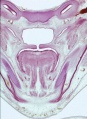
 Fetal week 10 hard palate 01.jpg 800 × 532; 77 KB
 Fetal week 10 hard palate 02.jpg 398 × 633; 66 KB
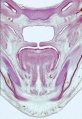
 Fetal week 10 hard palate 03.jpg 600 × 450; 122 KB
 Fetal week 10 hard palate 04.jpg 1,198 × 795; 196 KB
 Fetal week 10 hard palate 06.jpg 534 × 778; 88 KB
 Fetal week 10 hard palate 07.jpg 534 × 778; 97 KB
 Fetal week 10 palate 01.gif 534 × 778; 1.14 MB
 Fetal week 10 palate 01.mp4 ; 427 KB
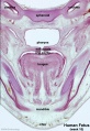
 Fetal week 10 palate icon.jpg 534 × 778; 100 KB
 Fetal week 10 soft palate 01.jpg 571 × 784; 95 KB
 Fetal week 10 soft palate 02.jpg 534 × 778; 87 KB
 Fetal week 10 soft palate 03.jpg 534 × 778; 95 KB
 Fetal week 14 head bone lateral 01.jpg 1,000 × 773; 107 KB
 Fetal week 9 hard palate fusion 01.jpg 661 × 400; 51 KB
 Fetal week 9 head lateral 01.jpg 700 × 600; 78 KB
 Frazer1911 fig01.jpg 1,280 × 766; 187 KB
 Frazer1911 fig02.jpg 1,280 × 872; 112 KB
 Frazer1911 fig03.jpg 1,280 × 600; 153 KB
 Gray0130.jpg 535 × 600; 95 KB
 Gray0178.jpg 617 × 368; 44 KB
 Gray0179.jpg 617 × 368; 47 KB
 Gray0180.jpg 617 × 368; 48 KB
 Gray0181.jpg 617 × 368; 52 KB
 Gray0193.jpg 719 × 1,057; 293 KB
 Gray0769.jpg 600 × 454; 101 KB
 Gray0913.jpg 671 × 600; 98 KB
 Gray0914.jpg 708 × 500; 102 KB
 Gray0915.jpg 720 × 600; 94 KB
 Human fetal temporal bone and mandible 01.jpg 1,200 × 805; 170 KB
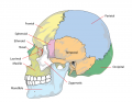 Human skull lateral simplified.png 740 × 576; 138 KB
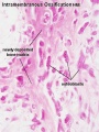 Intramembranous ossification centre.jpg 450 × 600; 69 KB
 Keibel Mall 2 265.jpg 1,439 × 1,200; 408 KB
 Keibel Mall 223.jpg 727 × 1,100; 89 KB
 Keibel Mall 224.jpg 706 × 1,112; 105 KB
 Keibel Mall 224A.jpg 671 × 377; 38 KB
 Keibel Mall 224B.jpg 685 × 563; 56 KB
 Keibel Mall 224C.jpg 706 × 306; 20 KB
 Keibel Mall 308.jpg 677 × 578; 43 KB
 Keibel Mall 309.jpg 687 × 479; 52 KB
 Keibel Mall 310.jpg 686 × 760; 161 KB
 Keibel Mall 311.jpg 700 × 906; 181 KB
 Keibel Mall 312.jpg 704 × 677; 79 KB
 Keibel Mall 313.jpg 702 × 652; 84 KB
 Keibel Mall 314.jpg 701 × 625; 89 KB
 Keibel Mall 315.jpg 736 × 433; 84 KB
 Keibel Mall 316.jpg 716 × 327; 57 KB
 Keibel Mall 317.jpg 688 × 376; 53 KB
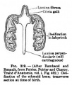
 Keibel Mall 318.jpg 687 × 423; 79 KB
 Keibel Mall 319.jpg 300 × 352; 27 KB
 Keibel Mall 320.jpg 693 × 313; 33 KB
 Keibel Mall 321.jpg 746 × 651; 122 KB
 Keibel Mall 322.jpg 735 × 717; 133 KB
 Keibel Mall 323.jpg 700 × 211; 27 KB
 Keibel Mall 324.jpg 722 × 377; 47 KB
 Keith1902 fig003.jpg 1,000 × 700; 113 KB
 Keith1902 fig005.jpg 650 × 557; 36 KB
 Keith1902 fig007.jpg 946 × 700; 80 KB
 Keith1902 fig011.jpg 954 × 500; 66 KB
 Keith1902 fig015b.jpg 1,000 × 719; 97 KB
 Keith1902 fig130.jpg 827 × 700; 139 KB
 Keith1902 fig131.jpg 800 × 632; 89 KB
 Keith1902 fig132.jpg 800 × 621; 119 KB
 Keith1902 fig133.jpg 437 × 700; 56 KB
 Keith1902 fig134.jpg 590 × 697; 66 KB
 Keith1902 fig135.jpg 634 × 700; 58 KB
 Keith1902 fig136.jpg 710 × 600; 91 KB
 Keith1902 fig137.jpg 800 × 482; 81 KB
 Keith1902 fig138.jpg 523 × 600; 27 KB
 Keith1902 fig139.jpg 598 × 600; 28 KB
 Keith1902 fig140.jpg 886 × 700; 85 KB
 Keith1921 fig130.jpg 710 × 402; 65 KB
 Keith1921 fig131.jpg 755 × 621; 102 KB
 Leopoldo Maggi.jpg 1,501 × 1,997; 1.75 MB
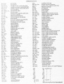 Lewis1920 abbreviations.jpg 609 × 800; 84 KB
 Lewis1920 fig01.jpg 800 × 896; 63 KB
 Lewis1920 fig02.jpg 575 × 800; 32 KB
 Lewis1920 fig03.jpg 686 × 981; 71 KB
 Lewis1920 fig04.jpg 705 × 800; 90 KB
 Lewis1920 fig05.jpg 900 × 834; 68 KB
 Lewis1920 fig06.jpg 800 × 789; 58 KB
 Lewis1920 fig07.jpg 1,000 × 894; 92 KB
 Lewis1920 fig08.jpg 1,000 × 765; 81 KB
 Lewis1920 fig09.jpg 1,000 × 765; 79 KB
 Lewis1920 fig10.jpg 800 × 576; 59 KB
 Lewis1920 fig11.jpg 800 × 686; 44 KB
 Lewis1920 fig12.jpg 800 × 483; 47 KB
 Lewis1920 fig13.jpg 800 × 520; 40 KB
 Lewis1920 fig14.jpg 1,200 × 761; 130 KB
 Lewis1920 fig15.jpg 1,200 × 722; 142 KB
 Lewis1920 fig16.jpg 1,000 × 743; 101 KB
 Lewis1920 Plate 1.jpg 917 × 1,200; 123 KB
 Lewis1920 Plate 2.jpg 939 × 1,200; 95 KB
 Lewis1920 Plate 3.jpg 881 × 1,200; 103 KB
 Lewis1920 Plate 4.jpg 903 × 1,200; 131 KB
 Lewis1920 Plate 5.jpg 969 × 1,200; 145 KB
 Lewis1920 table 1.jpg 1,000 × 299; 32 KB
 Lewis1920 table 2.jpg 1,000 × 581; 83 KB
 Low1909 fig01.jpg 900 × 371; 120 KB
 Low1909 fig02.jpg 1,059 × 889; 184 KB
 Low1909 fig03.jpg 904 × 896; 216 KB
 Low1909 fig04.jpg 1,043 × 927; 141 KB
 Low1909 fig05.jpg 893 × 891; 229 KB
 Low1909 fig06.jpg 1,371 × 1,213; 310 KB
 Low1909 fig07.jpg 897 × 893; 202 KB
 Low1909 plate01.jpg 3,574 × 2,331; 749 KB
 Low1909 plate01fig01.jpg 1,280 × 696; 115 KB
 Low1909 plate01fig02.jpg 1,280 × 518; 81 KB
 Low1909 plate01fig03.jpg 1,280 × 572; 95 KB
 Low1909 plate01fig04.jpg 1,280 × 600; 92 KB
 Low1909 plate01fig05.jpg 1,280 × 535; 73 KB
 Low1909 plate01fig06.jpg 1,280 × 546; 90 KB
 Macklin-plate01.jpg 2,331 × 3,061; 1.13 MB
 Macklin-plate02.jpg 2,331 × 3,061; 1.08 MB
 Macklin-plate03.jpg 2,331 × 3,061; 1,010 KB
 Macklin-plate04.jpg 2,331 × 3,061; 956 KB
 Macklin-plate05.jpg 2,331 × 3,061; 925 KB
 Macklin-plate1a.jpg 1,142 × 1,500; 374 KB
 Macklin-plate2a.jpg 1,142 × 1,500; 361 KB
 Macklin-plate3a.jpg 1,142 × 1,500; 321 KB
 Macklin-plate4a.jpg 1,142 × 1,500; 335 KB
 Macklin-plate5a.jpg 1,142 × 1,500; 324 KB
 Macklin1914 fig06.jpg 1,809 × 1,270; 251 KB
 Macklin1914 fig07.jpg 1,808 × 1,268; 289 KB
 Macklin1914 fig08.jpg 1,790 × 1,004; 170 KB
 Macklin1914 fig09.jpg 1,685 × 1,045; 180 KB
 Macklin1914 fig10.jpg 2,125 × 1,321; 318 KB
 Macklin1914 fig11.jpg 1,739 × 983; 152 KB
 Macklin1914 fig12.jpg 1,700 × 939; 176 KB
 Macklin1914 fig14.jpg 2,141 × 1,397; 434 KB
 Macklin1914 fig15.jpg 1,857 × 1,281; 342 KB
 Macklin1914 fig16.jpg 2,226 × 1,571; 506 KB
 Macklin1914 fig17.jpg 2,228 × 1,674; 530 KB
 Macklin1914 fig18.jpg 1,651 × 1,113; 284 KB
 Macklin1914 plate01.jpg 2,184 × 2,602; 673 KB
 Macklin1914 plate02.jpg 2,196 × 2,608; 746 KB
 Macklin1914 plate03.jpg 2,095 × 2,550; 597 KB
 Macklin1914 plate04.jpg 2,435 × 2,640; 810 KB
 Macklin1914 plate05.jpg 2,176 × 2,397; 522 KB
 Macklin1914 plate06.jpg 1,928 × 2,837; 567 KB
 Macklin1914 plate07.jpg 1,813 × 2,647; 384 KB
 Macklin1914 plate08.jpg 2,125 × 2,116; 355 KB
 Macklin1914 plate09.jpg 1,909 × 2,781; 487 KB
 Macklin1914 plate10.jpg 1,994 × 2,267; 465 KB
 Macklin1914 plate11.jpg 2,141 × 2,337; 569 KB
 Macklin1914 plate12.jpg 1,857 × 2,234; 433 KB
 Macklin1914 plate13.jpg 2,226 × 2,399; 575 KB
 Macklin1914 plate14.jpg 2,228 × 2,862; 773 KB
 Macklin1914 plate15.jpg 1,866 × 2,805; 767 KB
 Mall1906 fig03.jpg 797 × 800; 47 KB
 Mall1906 fig04.jpg 1,114 × 1,044; 137 KB
 Mall1906 table02.jpg 2,000 × 858; 404 KB
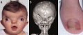
 Microcephaly and short stature 01.jpg 1,280 × 414; 92 KB
 Rugh 143.jpg 898 × 800; 175 KB
 Rugh 144.jpg 890 × 600; 107 KB


