Category:Sensory
From Embryology
This page lists UNSW Embryology content related to sensory development. See also special sensory systems.
Subcategories
This category has the following 12 subcategories, out of 12 total.
Pages in category 'Sensory'
The following 185 pages are in this category, out of 185 total.
2
A
B
- Template:Balance
- BGD Lecture - Face and Ear Development
- BGDB Face and Ear - Early Embryo
- BGDB Practical - Face and Ear Development
- Book - Anatomical and physiological studies on the growth of the inner ear of the albino rat (1923)
- Book - Contributions to Embryology Carnegie Institution No.20
- Book - Contributions to Embryology Carnegie Institution No.20 part 1
- Book - Contributions to Embryology Carnegie Institution No.20 part 2
- Book - Contributions to Embryology Carnegie Institution No.20 part 3
- Book - Contributions to Embryology Carnegie Institution No.20 part 4
- Book - Contributions to Embryology Carnegie Institution No.20 part 5
- Book - Contributions to Embryology Carnegie Institution No.20 part 6
- Book - Contributions to Embryology Carnegie Institution No.20 part 7
- Book - Contributions to Embryology Carnegie Institution No.21
- Book - Contributions to Embryology Carnegie Institution No.69
- Book - Manual of Human Embryology 16
- Book - Manual of Human Embryology 16-1
- Book - Manual of Human Embryology 16-2
- Book - Manual of Human Embryology 16-3
- Book - Manual of Human Embryology 16-4
C
E
H
I
O
P
- Paper - 1880 The Platypus Cochlea
- Paper - 1917 The Typical Form of the Cochlea and Its Variations
- Paper - A model to illustrate the probable action of the tectorial membrane (1915)
- Paper - A note on the length of the basilar membrane in man and in various mammals (1940)
- Paper - Abnormal ossification of Meckel's cartilage
- Paper - Adult form of the human stapes in the light of its development
- Paper - Blood supply of the otic capsule of a 150 mm (C.R.) human fetus
- Paper - Comparative morphology of the ear 3
- Paper - Contribution to the structure and development of the vertebrate head
- Paper - Contribution to the structure and development of the vertebrate head 1
- Paper - Contribution to the structure and development of the vertebrate head 2
- Paper - Contribution to the structure and development of the vertebrate head 3
- Paper - Development of the aquaductus cochleae and the periotic (perilymphatic) duct
- Paper - Development of the aquaeductus cochleae and its contained periotic duct and cochlear vein in human embryos
- Paper - Development of the incus of the human ear - illustrated in atlas series
- Paper - Development of the malleus of the human ear - Illustrated in atlas series
- Paper - Development of the Otic Capsule 1
- Paper - Development of the otic capsule 2
- Paper - Development of the Otic Capsule 3
- Paper - Development of the Otic Capsule 4
- Paper - Development of the otic capsule of the human ear - illustrated in atlas series
- Paper - Development of the stapes of the human ear - illustrated in atlas series
- Paper - Experimental observations on the development of the amphibian ear vesicle (1909)
- Paper - Histogenesis of the otic capsule (1917)
- Paper - Major features in the developmental history of the human stapes (1940)
- Paper - Migration of the ear vesicle in the tadpole during normal development (1921)
- Paper - On the development of the external ear passages
- Paper - On the development of the membrana tectoria with reference to its structure and attachments
- Paper - On the development of the membranous labyrinth and the acoustic and facial nerves in the human embryo
- Paper - On the development of the retina and optic nerve, and of the membranous labyrinth and auditory nerve
- Paper - On the proportions, development and attachment of the tectorial membrane (1915)
- Paper - Ossification of the otic capsule in human fetuses
- Paper - Perichondrial ossification and the fate of the perichondrium with special reference to that of the otic capsule
- Paper - Postnatal growth and adult structure of the otic (endolymphatic) sac
- Paper - Some experiments on the developing ear vesicle of the tadpole with relation to equilibration
- Paper - Some factors in the development of the amphibian ear vesicle and further experiments on equilibration
- Paper - Some features of the auditory apparatus of a 16 mm human embryo
- Paper - Some uniform characteristics of the primate auricle (1922)
- Paper - Stapes, fissula ante fenestram and associated structures in man 1
- Paper - Stapes, fissula ante fenestram and associated structures in man 2
- Paper - Stapes, fissula ante fenestram and associated structures in man 3
- Paper - Stapes, fissula ante fenestram and associated structures in man 4
- Paper - Stapes, fissula ante fenestram and associated structures in man 5
- Paper - The comparison of auricular height determinations (1925)
- Paper - The cytological processes involved in the formation of the scalae of the internal ear
- Paper - The development and structure of the otic (endolymphatic) sac
- Paper - The development of the auditory nerve in vertebrates (1910)
- Paper - The development of the auditory ossicles and associated structures in man
- Paper - The development of the auditory ossicles, the otic capsule and the extracapsular tissues
- Paper - The development of the cochlear fenestra, fossula and secondary tympanic membrane
- Paper - The development of the ear-bones in the mouse
- Paper - The development of the external ear (1934)
- Paper - The development of the first branchial arch in man and the fate of Meckel's cartilage
- Paper - The development of the otic capsule in the region of surgical fenestration 1
- Paper - The development of the otic capsule in the region of surgical fenestration 2
- Paper - The development of the otic capsule in the region of the vestibular aqueduct
- Paper - The development of the pillar cells, tunnel space, and Nuel's spaces in the organ of Corti (1919)
- Paper - The Development of the Scala Tympani, Scala Vestibuli and Perioticular Cistern in the Human Embryo
- Paper - The development of the second branchial arch (Reichert's cartilage), facial canal and associated structures in man
- Paper - The developmental and adult anatomy of the air-cells in the petrous part of the temporal bone
- Paper - The developmental course of the human auditory vesicle
- Paper - The distal projection of the endolymphatic sac in human embryos
- Paper - The early development of the membranous labyrinth in mammalian embryos
- Paper - The early development of the otic vesicle in staged human embryos
- Paper - The early embryology of the auditory ossicles in man
- Paper - The early formations of the middle ear and eustachian tube - a criticism
- Paper - The early relation of the auditory vesicle to the ectoderm in human embryos
- Paper - The Factors Involved in the Excavation of the Cavities in the Cartilaginous Capsule of the Ear in the Human Embryo
- Paper - The fissula ante fenestram of the human otic capsule; aberrant form and contents
- Paper - The form and structure of the endolymphatic and associated ducts in the child
- Paper - The genesis and structure of the membrana tectoria and the crista spiralis of the cochlea (1918)
- Paper - The Origin of the Otic and Optic Primordia in Man
- Paper - The vascular drainage of the endolymphatic sac and its topographical relation to the transverse sinus in the human
- Paper - Vertebrate cephalogenesis 1 (1890)
- Paper - Vertebrate cephalogenesis 2 (1892)
- Template:Placode
- Template:Placodes
R
S
- Template:Senses Links
- Template:Sensorimotor cortex
- Template:Sensorimotor Cortex Timeline table
- Template:Sensory
- Sensory - Balance Development
- Sensory - Carnegie Stage 22
- Sensory - Hearing Abnormalities
- Sensory - Hearing and Balance Development
- Sensory - Magnetoreception Development
- Sensory - Smell Development
- Sensory - Taste Development
- Sensory - Touch Development
- Sensory - Vision Development
- Sensory System Development
- Site Map
- Talk:Site Map
- Template:Smell
- Template:Stapes
Media in category 'Sensory'
The following 77 files are in this category, out of 77 total.
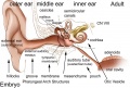 Adult hearing embryonic origins.jpg 1,000 × 675; 80 KB
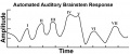 Automated Auditory Brainstem Response.jpg 1,239 × 521; 37 KB
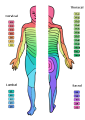 Dermatomes.png 424 × 600; 82 KB
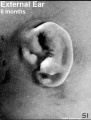 External Ear 5 months-icon.jpg 352 × 464; 63 KB
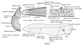
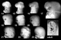 External ear stages-14-23-adult a.jpg 800 × 524; 30 KB
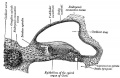 Gray0903.jpg 600 × 389; 42 KB
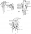
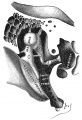 Gray0919.jpg 500 × 731; 93 KB
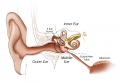 Hearing cartoon.jpg 800 × 553; 43 KB
 Human embryonic tongue 01.jpg 1,200 × 818; 443 KB
 Human embryonic tongue 02.jpg 650 × 850; 220 KB
 Human embryonic tongue 03.jpg 650 × 470; 159 KB
 Human embryonic tongue 04.jpg 650 × 470; 131 KB
 Human embryonic tongue 05.jpg 650 × 470; 146 KB
 Human embryonic tongue 06.jpg 650 × 470; 99 KB
 Human embryonic tongue 07.jpg 650 × 470; 187 KB
 Human embryonic tongue 08.jpg 650 × 470; 185 KB
 Human embryonic tongue 09.jpg 650 × 470; 180 KB
 Human embryonic tongue 10.jpg 650 × 470; 118 KB
 Human embryonic tongue 11.jpg 650 × 470; 155 KB
 Human embryonic-fetal tongue 01.jpg 1,000 × 1,129; 490 KB
 Human fetal tongue 01.jpg 1,500 × 672; 350 KB
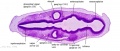
 Keibel Mall 2 126.jpg 1,000 × 598; 66 KB
 Keibel Mall 2 158.jpg 563 × 700; 47 KB
 Keibel Mall 2 159.jpg 679 × 699; 47 KB
 Keibel Mall 2 160.jpg 501 × 700; 35 KB
 Keibel Mall 2 161.jpg 621 × 700; 46 KB
 Keibel Mall 2 162.jpg 579 × 800; 69 KB
 Keibel Mall 2 163.jpg 675 × 500; 51 KB
 Keibel Mall 2 164.jpg 610 × 500; 44 KB
 Keibel Mall 2 165.jpg 1,037 × 900; 166 KB
 Keibel Mall 2 166.jpg 969 × 1,000; 144 KB
 Keibel Mall 2 167.jpg 1,000 × 496; 86 KB
 Keibel Mall 2 168.jpg 583 × 1,000; 65 KB
 Keibel Mall 2 169.jpg 634 × 800; 97 KB
 Keibel Mall 2 170.jpg 1,000 × 793; 65 KB
 Keibel Mall 2 171.jpg 1,000 × 696; 84 KB
 Keibel Mall 2 172.jpg 822 × 800; 82 KB
 Keibel Mall 2 173.jpg 818 × 800; 142 KB
 Keibel Mall 2 174.jpg 607 × 600; 56 KB
 Keibel Mall 2 175.jpg 496 × 597; 40 KB
 Keibel Mall 2 176.jpg 992 × 800; 134 KB
 Keibel Mall 2 177.jpg 965 × 389; 48 KB
 Keith1902 fig036a.jpg 678 × 639; 93 KB
 Keith1902 fig036b.jpg 678 × 639; 70 KB
 Max Brödel-cochlea drawing1934.jpg 531 × 395; 69 KB
 Meissner corpuscle 01.jpg 793 × 662; 86 KB
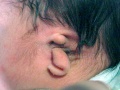 Microtia.jpg 600 × 450; 27 KB
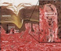
 Mouse circumvallate papilla 01.jpg 712 × 955; 114 KB
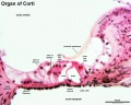
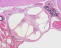 Mouse organ of corti 01.jpg 1,280 × 1,024; 339 KB
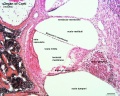 Mouse organ of corti 02.jpg 1,280 × 1,024; 320 KB
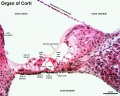 Mouse organ of corti 03.jpg 1,280 × 1,024; 207 KB
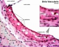 Mouse organ of corti 04.jpg 1,280 × 1,024; 202 KB
 Mouse organ of corti 05.jpg 1,280 × 1,024; 171 KB
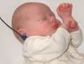 Newborn hearing test.jpg 200 × 153; 6 KB
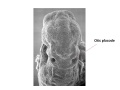 Otic placode label 1.jpg 960 × 720; 53 KB
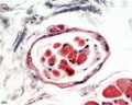
 Pacinian corpuscle histology 01.jpg 800 × 640; 227 KB
 Pacinian corpuscle histology 02.jpg 1,280 × 1,024; 207 KB
 Pacinian corpuscle histology 03.jpg 1,280 × 1,024; 133 KB
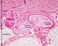
 Patten045.jpg 800 × 968; 156 KB
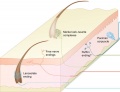
 Preplacodal development model.jpg 800 × 416; 41 KB
 Rugh 129.jpg 1,200 × 649; 138 KB
 Rugh 130.jpg 901 × 1,000; 130 KB
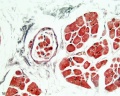 Skeletal muscle histology 009.jpg 1,280 × 1,024; 274 KB
 Skeletal muscle histology 010.jpg 1,280 × 1,024; 207 KB
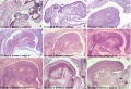
 Stage 13 image 001.jpg 1,000 × 357; 44 KB
 Stage 13 image 003.jpg 1,000 × 436; 71 KB
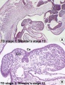
 Stage 13 image 051.jpg 1,000 × 382; 55 KB
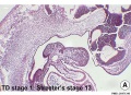
 Stage 13 image 052.jpg 1,000 × 476; 81 KB
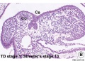
 Stage 13 image 053.jpg 1,000 × 423; 76 KB
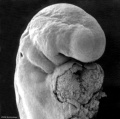
 Stage12 sem6.jpg 1,620 × 1,612; 190 KB
 Stage12 sem6a.jpg 1,000 × 995; 100 KB
 Stage12 sem6b.jpg 800 × 796; 74 KB
 Stage12 sem6c.jpg 600 × 597; 49 KB
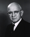 Theodore H. Bast.jpg 800 × 978; 108 KB
 Touch receptors in mammalian skin cartoon 01.jpg 600 × 459; 38 KB
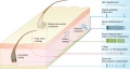 Touch receptors in mammalian skin cartoon.jpg 800 × 429; 57 KB
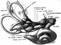
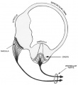 Vestibular labyrinth cartoon.jpg 600 × 657; 44 KB


