Category:Renal
From Embryology
This Embryology category shows media and pages related to renal system development. This includes the kidney, ureters, urinary bladder and urethra.
Subcategories
This category has the following 4 subcategories, out of 4 total.
Pages in category 'Renal'
The following 200 pages are in this category, out of 205 total.
(previous page) (next page)2
A
B
- Template:Bladder
- Template:Bladder exstrophy
- Book - A Laboratory Manual and Text-book of Embryology 8
- Book - Contributions to Embryology Carnegie Institution No.131
- Book - Manual of Human Embryology 19
- Book - Manual of Human Embryology 19-1
- Book - Manual of Human Embryology 19-2
- Book - Manual of Human Embryology 19-3
- Book - Manual of Human Embryology 19-4
- Book - Quain's Embryology 10
- Book - Text-Book of Embryology 15
C
E
H
I
- Template:ICD Renal (Q60-Q64)
- Template:ICD-11 Renal Agenesis or Dysgenesis anomalies collapse table
- Template:ICD-11 Renal Agenesis or Dysgenesis anomalies table
- Template:ICD-11 Renal anomalies header table
- Template:ICD-11 Ureter and Urethra anomalies collapse table
- Template:ICD-11 Ureter and Urethra anomalies table
- Template:Intermediate mesoderm
M
P
- Paper - A case of congenital absence of the left kidney and ureter (1915)
- Paper - A case of congenital cystic kidney, associated with several other abnormalities (1925)
- Paper - A case of duplication of the ureters (1915)
- Paper - A note on the post-natal growth of the kidney, thyroid gland and liver (1924)
- Paper - A three weeks human embryo with especial reference to the brain and nephric system (1905)
- Paper - An experimental study of the development of the amphibian cloaca (1940)
- Paper - Anomalies of the genito-urinary tract
- Paper - Congenital absence of the kidney
- Paper - Congenital cystic dilatation of the renal collecting tubules (1951)
- Paper - Congenital malformations of the ureters (1912)
- Paper - Congenital renal anomalies: with special reference to horseshoe kidney (1939)
- Paper - Congenital strictures and spiral twists of the ureters (1917)
- Paper - Congenital urogenital anomalies in rats including unilateral renal agenesia (1936)
- Paper - Congenital urogenital anomalies in rats including unilateral renal agenesia: further data in support of their inheritance (1937)
- Paper - Cystic disease of the kidneys (1935)
- Paper - Cysts of the genital ducts Müllerian and Wolffian (1946)
- Paper - Cysts of the Wolffian body (1924)
- Paper - Description of a horseshoe kidney
- Paper - Detailed form of the Wolffian body in human embryos of the first eight weeks
- Paper - Development and growth of the metanephros or permanent kidney in chick embryos eight to ten days incubation (1922)
- Paper - Development of the trigone of the bladder and the termination of the mesonephric ducts (1946)
- Paper - Development of the Wolffian body in Sus Scrofa Domesticus
- Paper - Double ureters in human and pig embryos
- Paper - Dystocia due to distension of the urinary bladder of the fœtus (1909)
- Paper - Horseshoe Kidney
- Paper - Morphology of the human urinogenital tract (1901)
- Paper - Notes on the Wolffian body of higher mammals (1902)
- Paper - Obstructions about the mesentery in infants (1936)
- Paper - On the development and shape of uriniferous tubules of certain of the higher mammals (1905)
- Paper - Polycystic disease of the kidney (1927)
- Paper - Polycystic disease of the kidney (1934)
- Paper - Spindle-shaped dilatations and tortuosity of the ureters in the fetus (1902)
- Paper - The changes in the mesonephric tubules of human embryos ten to twelve weeks old
- Paper - The clinical diagnosis of congenital anomaly in the kidney and ureter (1912)
- Paper - The development of the cloaca in human embryos
- Paper - The early development of the human nephros
- Paper - The evolutionary significance of the embryology of the amphibian nephric system (1940)
- Paper - The genesis of exstrophy of the bladder and epispadias
- Paper - The interrelations of the mesonephros, kidney and placenta in different classes of animals
- Paper - The interrelations of the mesonephros, kidney, and placenta in different classes of animals (1916)
- Paper - The mechanism of kidney development in human embryos as revealed by an early stage in the agenesis of the ureteric buds
- Paper - The normal changes in the position of the embryonic kidney
- Paper - The origin and growth of renal calculi (1937)
- Paper - The origin of the renal artery in mammals and its anomalies
- Paper - The Terminal Part of the Wolffian Duct
- Paper - The veins of the Wolffian body in the pig
- Template:Polycystic kidney disease
- Template:Pronephros
R
- Template:Ref-Abbott1930
- Template:Ref-Altschule1930
- Template:Ref-Angle1918
- Template:Ref-Bell1935
- Template:Ref-Boyden1931
- Template:Ref-Boyden1932
- Template:Ref-Braasch1912
- Template:Ref-Brambell1927b
- Template:Ref-Bremer1915
- Template:Ref-Bremer1916
- Template:Ref-Bremer1918
- Template:Ref-Eisendrath1912
- Template:Ref-Eisendrath1917
- Template:Ref-Eisendrath1925
- Template:Ref-Gage1905
- Template:Ref-Gladstone1915a
- Template:Ref-Gladstone1924b
- Template:Ref-Gruenwald1937
- Template:Ref-Gruenwald1939
- Template:Ref-Gruenwald1943
- Template:Ref-Hain1936
- Template:Ref-Hain1937
- Template:Ref-Hamann1902
- Template:Ref-Hill1905
- Template:Ref-HinmanGibsonKutzmann1924
- Template:Ref-Howland1916
- Template:Ref-Huber1905
- Template:Ref-JacobYusufJacob2012
- Template:Ref-LewisPapez1915
- Template:Ref-MacCallum1902
- Template:Ref-Mall1901a
- Template:Ref-Minot1898
- Template:Ref-Monie1946
- Template:Ref-MortonJones1936
- Template:Ref-Nagel1889
- Template:Ref-O'Connor1940a
- Template:Ref-O'Connor1940b
- Template:Ref-Oconnor1938
- Template:Ref-OConnor1939
- Template:Ref-Oppenheimer1934
- Template:Ref-PapinEisendrath1927
- Template:Ref-Patten1952b
- Template:Ref-Piersol1927
- Template:Ref-Pohlman1905a
- Template:Ref-Pohlman1905b
- Template:Ref-Pohlman1919
- Template:Ref-Randall1937
- Template:Ref-Rienhoff1922
- Template:Ref-RuscheBacon1939
- Template:Ref-Shikinami1926
- Template:Ref-Siddiqi1937
- Template:Ref-SmithStrasberg1946
- Template:Ref-Spicer1909
- Template:Ref-Stoerk1904
- Template:Ref-Torrey1954
- Template:Ref-Vermooten1951
- Template:Ref-Wakeley1915
- Template:Ref-West1925
- REI - Reproductive Medicine Seminar 2018
- Template:Renal
- Template:Renal abnormalities
- Renal Blood Supply Movie
- Template:Renal drug clearance table
- Template:Renal Histology
- Template:Renal histology
- Template:Renal Links
- Renal System - Abnormalities
- Renal System - Carnegie Stage 13
- Renal System - Carnegie Stage 22
- Renal System - Fetal
- Renal System - Molecular
- Renal System Development
- Renal System Histology
- Template:Renal terms
- Template:Renal Vascular Anomalies
- Royal Hospital for Women - Reproductive Medicine Seminar 2018
U
Media in category 'Renal'
The following 74 files are in this category, out of 283 total.
(previous page) (next page) Rat kidney HIM 03.jpg 561 × 1,000; 169 KB
 Rat kidney HIM 04.jpg 599 × 602; 142 KB
 Renal - early glomerulus.jpg 1,155 × 432; 52 KB
 Renal - podocyte development 01.jpg 1,200 × 908; 155 KB
 Renal - S-shaped body stage.jpg 1,155 × 432; 99 KB
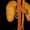
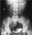 Renal agenesis 01.jpg 600 × 662; 44 KB
 Renal agenesis.jpg 600 × 778; 35 KB
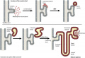 Renal development cartoon01.jpg 900 × 612; 116 KB
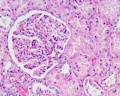
 Renal histology 01.jpg 1,280 × 1,024; 684 KB
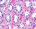
 Renal histology 02.jpg 1,280 × 1,024; 376 KB
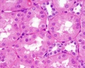
 Renal histology 03.jpg 1,280 × 1,024; 280 KB
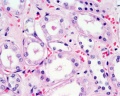
 Renal histology 04.jpg 1,280 × 1,024; 266 KB
 Renal histology 05.jpg 1,280 × 1,024; 275 KB
 Renal histology 06.jpg 1,280 × 1,024; 579 KB
 Renal histology 07.jpg 1,280 × 1,024; 396 KB
 Renal histology 08.jpg 1,280 × 1,024; 293 KB
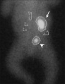
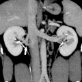
 Renal outflow obstruction.jpg 600 × 407; 33 KB
 Rugh 022.jpg 755 × 1,200; 216 KB
 Rugh 147.jpg 1,200 × 663; 137 KB
 Rugh 148.jpg 994 × 800; 240 KB
 Rugh 153.jpg 800 × 498; 67 KB
 Stage 11 historic-Atwell1930-3.jpg 1,000 × 679; 87 KB
 Stage 11 historic-Atwell1930-3a.jpg 800 × 543; 57 KB
 Stage 11 historic-Atwell1930-3b.jpg 600 × 407; 32 KB
 Stage 11 historic-Atwell1930-3c.jpg 400 × 271; 15 KB
 Stage 11 historic-Heuser1930-1.jpg 521 × 1,000; 94 KB
 Stage 11 historic-Heuser1930-1a.jpg 417 × 800; 57 KB
 Stage 11 historic-Heuser1930-1b.jpg 313 × 600; 30 KB
 Stage 11 historic-Heuser1930-1c.jpg 209 × 400; 15 KB
 Stage 13 kidney sections 2.jpg 600 × 400; 53 KB
 Stage 22 image 188.jpg 1,000 × 665; 112 KB
 Stage 22 image 189.jpg 1,000 × 672; 204 KB
 Stage 22 image 190.jpg 1,000 × 657; 211 KB
 Stage 22 image 191.jpg 1,000 × 653; 100 KB
 Stage 22 image 192.jpg 1,000 × 658; 180 KB
 Stage 22 image 193.jpg 1,000 × 667; 182 KB
 Stage 22 image 194.jpg 1,000 × 671; 210 KB
 Stage 22 image 196.jpg 1,000 × 671; 102 KB
 Stage 22 image 197.jpg 1,000 × 668; 135 KB
 Stage 22 image 198.jpg 1,000 × 664; 192 KB
 Stage 22 image 201.jpg 1,200 × 754; 324 KB
 Stage 22 image 202.jpg 1,455 × 920; 617 KB
 Stage 22 image 210.jpg 1,101 × 794; 447 KB
 Stage 22 image 210a.jpg 1,000 × 644; 310 KB
 Stage 22 image 210b.jpg 800 × 515; 207 KB
 Stage 22 image 210c.jpg 400 × 257; 52 KB
 Stage 22 image 213.jpg 738 × 554; 198 KB
 Stage 22 image 214.jpg 738 × 554; 203 KB
 Stage 22 image 301.jpg 1,200 × 754; 329 KB
 Stage 22 image 302.jpg 1,455 × 920; 625 KB
 Stage22 mesonephros.jpg 600 × 397; 83 KB
 Streeter1957 plate04.jpg 1,500 × 1,986; 736 KB
 Supernumerary renal vein 01.jpg 800 × 798; 72 KB
 Supernumerary renal vein 02.jpg 800 × 795; 89 KB
 Supernumerary renal vein 03.jpg 800 × 794; 80 KB
 Supernumerary renal vein 04.jpg 800 × 850; 76 KB
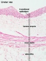 Ureter histology 001.jpg 375 × 500; 50 KB
 Ureter histology 002.jpg 375 × 500; 34 KB
 Ureter.jpg 1,050 × 684; 226 KB
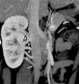
 Ureteral duplication 01.jpg 600 × 812; 61 KB
 Urinary Bladder Histology.jpg 581 × 399; 42 KB
 Urogenital septum 001.mov ; 180 KB
 Waterston14.jpg 438 × 680; 66 KB
 Wen1928-Fig16.jpg 702 × 1,500; 195 KB
 Wen1928-Fig17.jpg 877 × 1,000; 186 KB
 Wen1928-Fig18.jpg 1,000 × 962; 331 KB
 Wen1928-Fig29.jpg 1,200 × 884; 133 KB
 West06.jpg 308 × 802; 39 KB
 West07.jpg 984 × 153; 15 KB
 William Bowman.jpg 600 × 665; 49 KB
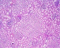
 Wilms tumor.jpg 776 × 512; 310 KB
 Windle1940 fig43.jpg 1,000 × 1,003; 119 KB
 Xenopus golph2 expression.jpg 616 × 800; 147 KB
 Zebrafish nephrogenesis signaling01.jpg 508 × 324; 29 KB


