Category:Pituitary
From Embryology
This Embryology category shows content related to the endocrine pituitary (hypophysis) development.
Pages in category 'Pituitary'
The following 50 pages are in this category, out of 50 total.
P
- Paper - A quantitative study of the hypophysis of the human anencephalic fetus (1927)
- Paper - Differentiation of pituicytes in the human foetus
- Paper - Experimental evidence regarding the role of the anterior pituitary in the development and regulation of the genital system
- Paper - Growth of the human prenatal hypophysis and the hypophyseal fossa (1927)
- Paper - Pharyngeal end of Rathke's pouch (1911)
- Paper - Sexual differences of the hypophyses and their determination by the gonads
- Paper - Some factors influencing the early development of the mammalian hypophysis (1935)
- Paper - The development of the hypophysis cerebri in man (1926)
- Paper - The development of the hypophysis cerebri of the rabbit
- Paper - The development of the mammalian pituitary and its morphological significance (1908)
- Paper - The histological appearances of the mammalian pituitary body (1908)
- Paper - The nerve supply to the pituitary body (1913)
- Template:Pituitary
- Template:Pituitary Vignette
- Template:Placode
- Template:Placodes
- Template:Posterior pituitary
R
- Template:Rathke's pouch
- Template:Ref-Andersen1971
- Template:Ref-Andriezen1894
- Template:Ref-Atwell1918
- Template:Ref-Atwell1926
- Template:Ref-AtwellSitler1918
- Template:Ref-Covell1927
- Template:Ref-Covell1927b
- Template:Ref-Espinasse1933
- Template:Ref-Gilbert1935
- Template:Ref-Herring1908a
- Template:Ref-Herring1908b
- Template:Ref-Hill1934
- Template:Ref-Pfeiffer1936
- Template:Ref-Shanklin1940
- Template:Ref-SmithEngle1927
Media in category 'Pituitary'
The following 113 files are in this category, out of 113 total.
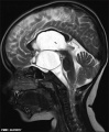
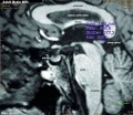 Adult human brain MRI01.jpg 700 × 607; 81 KB
 Atwell1918 fig01.jpg 600 × 432; 61 KB
 Atwell1918 fig02.jpg 800 × 582; 83 KB
 Atwell1918 fig03.jpg 800 × 604; 117 KB
 Atwell1918 fig04.jpg 409 × 550; 37 KB
 Atwell1918 fig05.jpg 704 × 550; 56 KB
 Atwell1918 fig06.jpg 600 × 641; 84 KB
 Atwell1918 fig07.jpg 425 × 378; 44 KB
 Atwell1918 fig08.jpg 638 × 553; 46 KB
 Atwell1918 fig09.jpg 559 × 635; 84 KB
 Atwell1918 fig10.jpg 627 × 703; 90 KB
 Atwell1918 fig11.jpg 665 × 904; 131 KB
 Atwell1918 fig12.jpg 453 × 603; 36 KB
 Atwell1918 fig13.jpg 625 × 800; 122 KB
 Atwell1918 fig14.jpg 800 × 788; 117 KB
 Atwell1918 fig15.jpg 701 × 900; 171 KB
 Atwell1918 fig18.jpg 1,000 × 1,193; 168 KB
 Atwell1918 fig19.jpg 316 × 515; 21 KB
 Atwell1918 fig20.jpg 702 × 515; 48 KB
 Atwell1918 fig21.jpg 800 × 477; 108 KB
 Atwell1918 fig22.jpg 1,000 × 887; 100 KB
 Atwell1918 fig23.jpg 1,000 × 953; 123 KB
 Atwell1918 fig24.jpg 506 × 399; 43 KB
 Atwell1918 fig25.jpg 532 × 496; 48 KB
 Atwell1918 fig26.jpg 566 × 498; 57 KB
 Atwell1918 fig27.jpg 900 × 802; 113 KB
 Atwell1918 fig28.jpg 1,000 × 886; 348 KB
 Atwell1918 fig29.jpg 791 × 780; 120 KB
 Atwell1918 fig30.jpg 601 × 451; 45 KB
 Atwell1918 fig31.jpg 1,000 × 700; 267 KB
 Atwell1918 fig32.jpg 510 × 303; 28 KB
 Atwell1918 fig33.jpg 387 × 336; 29 KB
 Atwell1918 fig34.jpg 353 × 406; 32 KB
 Atwell1918 fig35.jpg 1,200 × 849; 386 KB
 Atwell1918 fig38.jpg 1,000 × 377; 129 KB
 Atwell1918 fig39.jpg 600 × 709; 139 KB
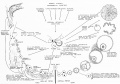
 Drosophila and mouse placode similarity.jpg 499 × 1,086; 337 KB
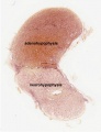
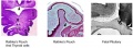 Embryonic and fetal pituitary.jpg 450 × 166; 14 KB
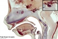
 Fetal head section 01.jpg 1,200 × 821; 186 KB
 Fetal head section 02.jpg 1,200 × 821; 171 KB
 Fetal head section 03.jpg 1,200 × 821; 174 KB
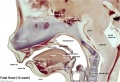
 Fetal head section.jpg 1,200 × 821; 167 KB
 Frazer1911 fig01.jpg 1,280 × 766; 187 KB
 Frazer1911 fig02.jpg 1,280 × 872; 112 KB
 Frazer1911 fig03.jpg 1,280 × 600; 153 KB
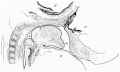
 Gray1180.jpg 1,200 × 712; 185 KB
 Gray1181.jpg 800 × 487; 77 KB
 Gray1182.jpg 800 × 982; 119 KB
 Herring1908b fig06.jpg 1,280 × 1,234; 283 KB
 Herring1908b fig07.jpg 1,280 × 974; 290 KB
 Historic-pituitary.jpg 639 × 367; 54 KB
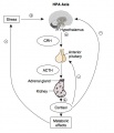 HPA axis.jpg 600 × 700; 46 KB
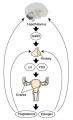
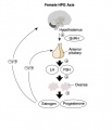 HPG female axis.jpg 600 × 700; 41 KB
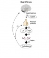 HPG male axis.jpg 600 × 700; 36 KB
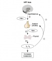 HPT axis.jpg 600 × 700; 35 KB
 Human week 10 fetus 10.jpg 1,200 × 900; 291 KB
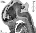 Human- fetal week 10 head D.jpg 600 × 544; 111 KB
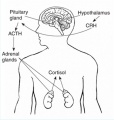
 Hypothalamus pituitary adrenal cartoon.jpg 653 × 600; 82 KB
 Hypothalamus pituitary adrenal pathway cartoon.jpg 490 × 516; 31 KB
 Hypothalamus pituitary cartoon.jpg 653 × 600; 81 KB
 Keith1902 fig015a.jpg 971 × 600; 74 KB
 Keith1902 fig117.jpg 561 × 800; 46 KB
 Keith1921 fig099.jpg 446 × 416; 39 KB
 Keith1921 fig101.jpg 1,144 × 652; 128 KB
 Keith1921 fig102.jpg 1,257 × 845; 172 KB
 Keith1921 fig103.jpg 758 × 495; 91 KB
 Kollmann355.jpg 861 × 460; 75 KB
 Kollmann356.jpg 867 × 584; 102 KB
 Martin Rathke.jpg 359 × 465; 20 KB
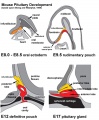 Mouse-pituitary development.jpg 660 × 800; 85 KB
 Mouse-pituitary Sox4 expression.jpg 596 × 448; 55 KB
 Nelsen1953 fig022.jpg 1,200 × 839; 207 KB
 Pituitary development animation.gif 600 × 400; 272 KB
 Pituitary histology 001.jpg 450 × 600; 72 KB
 Pituitary histology 002.jpg 450 × 600; 81 KB
 Pituitary histology 003.jpg 450 × 600; 94 KB
 Pituitary histology 004.jpg 1,280 × 1,024; 342 KB
 Pituitary histology 005.jpg 1,280 × 1,024; 326 KB
 Pituitary histology 006.jpg 1,280 × 1,024; 450 KB
 Pituitary histology 007.jpg 1,280 × 1,024; 325 KB
 Pituitary histology 008.jpg 1,280 × 1,024; 340 KB
 Pituitary histology 009.jpg 466 × 610; 64 KB
 Pituitary histology 010.jpg 1,005 × 961; 249 KB
 Pituitary histology 011.jpg 900 × 1,388; 309 KB
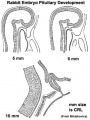 Pituitary rabbit development.jpg 374 × 500; 33 KB
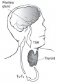 Pituitary thyroid pathway.jpg 454 × 632; 32 KB
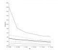 Postnatal thyrotropin levels graph.jpg 852 × 729; 35 KB
 Rathke cleft cyst 01.jpg 493 × 600; 46 KB
 Rugh 116.jpg 1,015 × 1,000; 270 KB
 Shanklin1940 fig01.jpg 1,000 × 657; 136 KB
 Shanklin1940 fig02.jpg 1,000 × 682; 131 KB
 Shanklin1940 fig04.jpg 884 × 1,018; 112 KB
 Shanklin1940 fig06.jpg 1,280 × 837; 92 KB
 Shanklin1940 fig07.jpg 1,280 × 1,080; 125 KB
 Shanklin1940 plate01.jpg 1,522 × 2,138; 362 KB
 Shanklin1940 plate02.jpg 1,411 × 2,131; 210 KB
 Stage 13 image 057.jpg 1,000 × 511; 99 KB
 Stage 13 image 058.jpg 1,000 × 481; 94 KB
 Stage 13 image 059.jpg 1,000 × 513; 92 KB
 Stage 22 image 158.jpg 1,000 × 646; 273 KB
 Stage 22 image 220.jpg 1,200 × 699; 334 KB
 Stage 22 image 221.jpg 1,200 × 699; 336 KB
 Stage17 bf11.jpg 1,375 × 2,048; 166 KB
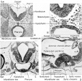
 Streeter1957 fig08-19.jpg 800 × 1,102; 109 KB
 Streeter1957 fig08-20.jpg 800 × 1,102; 104 KB
 Streeter1957 fig08-21.jpg 800 × 1,102; 114 KB
 Streeter1957 fig08-22.jpg 800 × 1,102; 189 KB
 Streeter1957 fig08-23.jpg 800 × 1,102; 206 KB
 Streeter1957 fig08.jpg 1,280 × 1,712; 507 KB
 Streeter1957 plate02.jpg 1,500 × 2,001; 653 KB
 XXhpgaxis.gif 300 × 495; 22 KB
 XXhpgaxis.jpg 300 × 495; 24 KB


