Category:Pharyngeal Arch
From Embryology
This Embryology category covers content related to pharyngeal arch (branchial arch) development. This is generally covered in Head Development notes.
Main page - Pharyngeal Arches
| Neural Crest Links: neural crest | Lecture - Early Neural | Lecture - Neural Crest Development | Lecture Movie | Schwann cell | adrenal | melanocyte | peripheral nervous system | enteric nervous system | cornea | cranial nerve neural crest | head | skull | cardiac neural crest | Nicole Le Douarin | Neural Crest Movies | neural crest abnormalities | Category:Neural Crest | |||
|
Pages in category 'Pharyngeal Arch'
The following 54 pages are in this category, out of 54 total.
C
P
- Paper - Evolutionary factors in the production of pharyngeal diverticula
- Paper - On the relation of the head chorda to the pharyngeal epithelium in the pig embryo
- Paper - The aortic arch derivatives in human adult (1951)
- Paper - The development of the first branchial arch in man and the fate of Meckel's cartilage
- Paper - The Disappearance of the Precervical Sinus
- Paper - The fifth aortic arch of mammalian embryos; the nature of the last pharyngeal evagination
- Paper - The pharyngeal pouches and their derivatives in the mammalia
- Paper - The potency of the pharyngeal entoderm (1932)
- Paper - The second visceral arch and groove in the tubo-tympanic region
- Paper - Three demonstrations on congenital melformations of palate, face, and neck
- Paper - Transformation of the aortic-arch system during the development of the human embryo (1922)
- Template:Pharyngeal arch
- Template:Pharyngeal Arch collapse table
- Template:Pharyngeal Arch table
- Pharyngeal arches
R
- Template:Ref-Allis1923
- Template:Ref-Anderson1922
- Template:Ref-Barry1951
- Template:Ref-Coulter1909
- Template:Ref-Frazer1914
- Template:Ref-Frazer1926
- Template:Ref-Keith1909
- Template:Ref-Kingsbury1914b
- Template:Ref-Negus1925
- Template:Ref-Rand1917
- Template:Ref-Reagan1912
- Template:Ref-Shaner1921
- Template:Ref-Woollard1932
- Template:Reichert’s cartilage
Media in category 'Pharyngeal Arch'
The following 51 files are in this category, out of 51 total.
 Congdon1922-31.jpg 1,063 × 1,000; 93 KB
 Congdon1922-32.jpg 1,133 × 1,000; 132 KB
 Congdon1922-34.jpg 920 × 1,000; 122 KB
 Congdon1922-35.jpg 920 × 1,000; 97 KB
 Congdon1922-36.jpg 920 × 1,000; 113 KB
 Foster136.jpg 556 × 420; 33 KB
 Gray0947.jpg 600 × 398; 56 KB
 Gray0978.jpg 483 × 600; 67 KB
 Gray0979.jpg 500 × 446; 56 KB
 Gray0980.jpg 542 × 450; 57 KB
 Gray0981.jpg 538 × 340; 50 KB
 Head and heart muscle cartoon.jpg 874 × 800; 129 KB
 Heart outflow tract stage 14 03.jpg 989 × 996; 134 KB
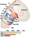 Hindbrain neural crest migration.jpg 450 × 545; 48 KB
 Human stage16 face 01.jpg 500 × 504; 20 KB
 Human stage17 face 01.jpg 500 × 504; 21 KB
 Human stage18 face 01.jpg 500 × 504; 23 KB
 Keibel Mall 2 314.jpg 760 × 1,000; 103 KB
 Keibel Mall 2 315.jpg 604 × 800; 61 KB
 Keibel Mall 2 316.jpg 899 × 1,000; 148 KB
 Keibel Mall 2 317.jpg 805 × 1,000; 95 KB
 Keibel Mall 2 318.jpg 1,077 × 1,000; 111 KB
 Keibel Mall 2 319.jpg 811 × 800; 82 KB
 Keibel Mall 2 320.jpg 973 × 800; 110 KB
 Keibel Mall 2 321.jpg 904 × 800; 76 KB
 Keibel Mall 2 322.jpg 729 × 800; 47 KB
 Keibel Mall 2 323.jpg 718 × 800; 54 KB
 Keibel Mall 2 324.jpg 1,280 × 930; 125 KB
 Keibel Mall 2 325.jpg 1,280 × 882; 225 KB
 Keibel Mall 2 326.jpg 1,280 × 730; 207 KB
 Keibel Mall 2 327.jpg 1,100 × 676; 114 KB
 Keibel Mall 2 328.jpg 1,100 × 534; 96 KB
 Keibel Mall 2 329.jpg 930 × 582; 74 KB
 Keibel Mall 2 330.jpg 1,280 × 1,009; 342 KB
 Meckels cartilage - middle ear from the jaw.jpg 1,161 × 1,280; 259 KB
 Pharyngeal arch cartilages.jpg 400 × 324; 26 KB
 Stage 13 image 005.jpg 1,000 × 451; 81 KB
 Stage 13 image 006.jpg 1,000 × 439; 83 KB
 Stage 13 image 007.jpg 1,000 × 514; 93 KB
 Stage 13 image 056.jpg 1,000 × 516; 102 KB
 Stage 13 image 057.jpg 1,000 × 511; 99 KB
 Stage 13 image 058.jpg 1,000 × 481; 94 KB
 Stage 13 image 059.jpg 1,000 × 513; 92 KB
 Stage 13 image 060.jpg 1,000 × 486; 96 KB
 Stage 13 image 061.jpg 1,000 × 600; 101 KB
 Stage13 face ventral view01.jpg 1,290 × 2,048; 152 KB
 Stage13 oral cavity floor01.jpg 1,315 × 2,048; 234 KB
 Stage13 oral cavity floor02.jpg 1,315 × 2,048; 371 KB
 Stage13 pharyngeal arch excerpts.gif 600 × 300; 86 KB
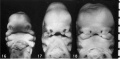 Stage16-18 face.jpg 800 × 393; 34 KB
 West02.jpg 619 × 549; 34 KB


