Category:Pfannenstiel III: Difference between revisions
(Created page with " == Carnegie Stage 11 == This {{Embryology}} category shows pages and media related to the historic human embryo Pfannenstiel III. Corresponding to Carnegie stage 11 deve...") |
|||
| Line 2: | Line 2: | ||
== Carnegie Stage 11 == | == Carnegie Stage 11 == | ||
This {{Embryology}} category shows pages and media related to the historic human embryo Pfannenstiel III. Corresponding to [[Carnegie stage 11]] development in [[week 4]], 23 - 26 days, {{GA}} week 6-7. The embryos have a crown rump length (CRL) of 2.5 - 4.5 mm and somite number 13 - 20 pairs. | This {{Embryology}} category shows pages and media related to the historic human embryo Pfannenstiel III, named after Professor Pfannenstiel of Griefswald. | ||
Corresponding to [[Carnegie stage 11]] development in [[week 4]], 23 - 26 days, {{GA}} week 6-7. The embryos have a crown rump length (CRL) of 2.5 - 4.5 mm and somite number 13 - 20 pairs. | |||
| Line 10: | Line 12: | ||
* two pharyngeal arches, clearly marked. | * two pharyngeal arches, clearly marked. | ||
* neural lips are closed rostrally as far as the collicular region. | * neural lips are closed rostrally as far as the collicular region. | ||
Revision as of 03:42, 29 May 2017
Carnegie Stage 11
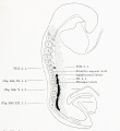
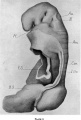
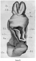
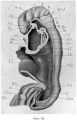
This Embryology category shows pages and media related to the historic human embryo Pfannenstiel III, named after Professor Pfannenstiel of Griefswald.
Corresponding to Carnegie stage 11 development in week 4, 23 - 26 days, GA week 6-7. The embryos have a crown rump length (CRL) of 2.5 - 4.5 mm and somite number 13 - 20 pairs.
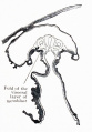
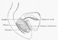
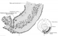
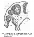
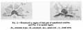
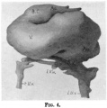
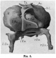
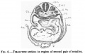
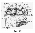
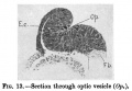
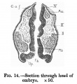
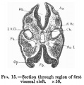
Pfannenstiel III - (14 somites) Pfannenstiel III (Professor Pfannenstiel of Griefswald) described by Keibel, F., and Elze, C. 1908. Normentafeln zur Entwicklungsgeschichte der Wirbeltiere.(Normal Plates for Evolution of Vertebrates) 8. Heft Normentafeln zur Entwicklungsgeschichte des Menschen. (Vol. 8. Normal Plates of the Development of the Human Embryo) Fisher, Jena., Germany. Shown as Embryo No.6 (Plate 1 fig. Vr. and Plate 2 fig. Vv.).
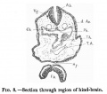
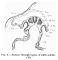
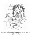
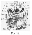
- described also by Low, A.[1]
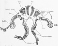
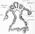
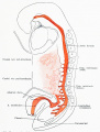
- Plastic models available of - external form, central nervous system, digestive system, heart and large vessels.
- two pharyngeal arches, clearly marked.
- neural lips are closed rostrally as far as the collicular region.
There is also a specific Carnegie stage 11 resource page.
| Week: | 1 | 2 | 3 | 4 | 5 | 6 | 7 | 8 |
| Carnegie stage: | 1 2 3 4 | 5 6 | 7 8 9 | 10 11 12 13 | 14 15 | 16 17 | 18 19 | 20 21 22 23 |
- Carnegie Stages: 1 | 2 | 3 | 4 | 5 | 6 | 7 | 8 | 9 | 10 | 11 | 12 | 13 | 14 | 15 | 16 | 17 | 18 | 19 | 20 | 21 | 22 | 23 | About Stages | Timeline
| Carnegie Collection - Stage 11 | ||||||||||
|---|---|---|---|---|---|---|---|---|---|---|
| Serial No. | Pairs of somites | Size (mm) | Grade | Fixative | Embedding Medium | Plane | Thinness (µm) | Stain | Year | Notes |
| 12 | 14 | E, 2.1 Ch, 13 | Poor | P | Transverse | 10 | Al. carm. | 1893 | ||
| 164 | 18 | E, 3.5 Ch, 14 | Good | Formalin | P | Transverse | 20 | Al. carm. | 1913 | |
| 318 | 13/14 | E, 2.5 Ch, 16 | Good | P | Transverse | 25 | Al. carm. | 1905 | ||
| 470 | 17 | E, 4.3 Ch, 16 | Good | Formalin | P | Transverse | 10 | Al. carm. . | 1910 | |
| 779 | 14 | E, 2.75 | Good | C | Transverse | 15 | Al. coch. | 1913 | Dysraphism. Noted by Dekaban (1964)[2] | |
| 1182b | E, 3 Ch, 15x12x5 | Good | Formalin | ? | Transverse | 20 | Al. carm. | 1915 | ||
| 2053 | 20 | E, 3.1 Ch, 12 | Exc. | Formalin | P | Transverse | 10 | Al. coch. | 1918 | Most advanced in group. Ag added to slide 2 Monographs by Davis (1923)[3] and Congdon (1922)[4] |
| 4315 | 17 | E, 4.7 Ch, 23x10.4X11 | Excellent | ? | C-P | Transverse | 10 | I.H. & E. | 1923 | Univ. Chicago No. 951. Wen (1928)[5] |
| 4529 | 14 | E, 2.4 Ch, 21 | Excellent | Formalin | P | Transverse | 10 | Al. coch, or. G. | 1924 | Heuser (1930)[6] |
| 4783 | 13 | E, 2.3 | Fair | ? | ? | Transverse | 5 | I.H. | 1924 | Wallin (1913)[7] |
| 4877 | 13 | E, 2 Ch, 15 | Good | Formalin | P | Transverse | 15 | Al. coch. | 1925 | |
| 5072 | 17 | E, 3 | Good | Formalin | P | Transverse | 10 | (Stain - Haematoxylin Eosin) | 1925 | Tubal Type specimen. Atwell (1930)[8] |
| 6050 | 19/21 | E.,3 Ch, 10 | Good | Formalin | C-P | Coronal | 10 | Al. coch. | 1930 | Advanced |
| 6344 | 13 | E, 2.5 Ch, 17 | Excellent | Formalin | C-P | Transverse | 6 | Al. coch. | 1931 | Least advanced in group |
| 6784 | 17 | E, 5 Ch, 16 | Excellent | Formalin | C-P | Transverse | 6 | I.H, or. G. | 1933 | |
| 7358 | 16 | E, ? Ch, 15 | Poor | Alc, formol | p | Oblique | 25 | (Stain - Haematoxylin Eosin) | 1936 | |
| 7611 | 16 | E., 2.4 Ch., 12 | Excellent | Bouin | C-P | Transverse | 8 | (Stain - Haematoxylin Eosin) | 1938 | |
| 7665 | 19 | E., 4.36 | Excellent | ? | C-P | Transverse | 6 | 1939 | Univ. Chicago No. H 1516 | |
| 7702 | 17 | E, 3.7 Ch., 14 | Good | Formalin | C-P | Transverse | 10 | Al. coch. | 1940 | Returned to B M Patten |
| 7851 | 13 | E., 4.3 Ch, 18 | Excellent | Formalin | C-P | Transverse | 8 | (Stain - Haematoxylin Eosin) | 1940 | Slightly injured |
| 8005 | 16/17 | E, 3 | Excellent | Bouin | C-P | Transverse | 8 | (Stain - Haematoxylin Eosin) | 1942 | Tubal |
| 8116 | 17 | E, 14 Ch.. 17 | Good | Formalin | p | Sagittal | 8 | Azan | 1953 | |
| 8962 | 15 | E, 1.55 | Good | ? | * | Sagittal | ? | ? | 1952 | Tubal Univ. Chicago No. H 810 |
Abbreviations
| ||||||||||
References
| ||||||||||
References
Pages in category 'Pfannenstiel III'
The following 7 pages are in this category, out of 7 total.
Media in category 'Pfannenstiel III'
The following 24 files are in this category, out of 24 total.
 Keibel Mall 2 526.jpg 1,280 × 1,391; 186 KB
 Keibel Mall 2 528.jpg 1,280 × 1,014; 214 KB
 Keibel Mall 2 529.jpg 1,280 × 1,223; 293 KB
 Keibel Mall 2 541.jpg 1,280 × 1,683; 313 KB
 Keibel Mall 2 545.jpg 761 × 1,093; 104 KB
 Keibel Mall 2 600.jpg 1,000 × 692; 77 KB
 Keibel Mall 2 609.jpg 1,200 × 753; 130 KB
 Low 01.jpg 502 × 535; 60 KB
 Low 02.jpg 900 × 328; 58 KB
 Low 04.jpg 535 × 541; 40 KB
 Low 05.jpg 529 × 552; 45 KB
 Low 06.jpg 751 × 484; 59 KB
 Low 07.jpg 711 × 619; 56 KB
 Low 08.jpg 566 × 499; 48 KB
 Low 09.jpg 589 × 609; 50 KB
 Low 10.jpg 519 × 586; 61 KB
 Low 11.jpg 414 × 451; 49 KB
 Low 12.jpg 450 × 419; 43 KB
 Low 13.jpg 637 × 443; 52 KB
 Low 14.jpg 508 × 554; 60 KB
 Low 15.jpg 565 × 570; 73 KB
 Low plate 01.jpg 661 × 981; 137 KB
 Low plate 02.jpg 581 × 980; 118 KB
 Low plate 03.jpg 707 × 1,099; 166 KB


