Category:Pancreas
From Embryology
This Embryology category shows content related to pancreas development (both exocrine and endocrine).
The Embryology template term thyroid links directly to the Thyroid Development page.
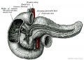
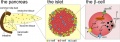
- Links: Endocrine Pancreas | Exocrine Pancreas
Pages in category 'Pancreas'
The following 45 pages are in this category, out of 45 total.
G
H
P
- Template:Pancreas
- Template:Pancreas Histology Images
- Template:Pancreas islet species comparison table
- Template:Pancreatic islet
- Paper - Cytogenesis of the human fetal pancreas (1962)
- Paper - Cytological studies of Langerhans's islets, with special reference to the problem of their relation to the pancreatic acinus tissue (1920)
- Paper - Models of the pancreas in embryos of the pig, rabbit, cat, and man (1908)
- Paper - Some Observations on the Development of the Ventral Pancreas in Man
- Paper - The bi-lobed form of the ventral pancreas in mammals
- Paper - The development of the islands of Langerhans in the human embryo (1903)
- Paper - The early morphogenesis and histogenesis of the liver in Sus scrofa domesticus, including notes on the morphogenesis of the ventral pancreas
R
- Template:Ref-Baldwin1910a
- Template:Ref-Bell1922
- Template:Ref-Bloom1931
- Template:Ref-Conklin1962
- Template:Ref-Corner1914
- Template:Ref-Delmas1939
- Template:Ref-Dewitt1906
- Template:Ref-Hard1944
- Template:Ref-Hilton1903
- Template:Ref-Lane1907
- Template:Ref-Lewis1905a
- Template:Ref-Lewis1911
- Template:Ref-Odgers1930
- Template:Ref-Pearce1903
- Template:Ref-Politzer1952
- Template:Ref-RussuVaida1959
- Template:Ref-Saguchi1920
- Template:Ref-Thyng1908
Media in category 'Pancreas'
The following 60 files are in this category, out of 60 total.
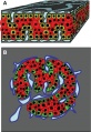
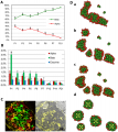
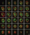 3D Human pancreatic islet.jpg 1,088 × 1,280; 295 KB
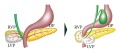 Annular pancreas.jpg 600 × 265; 16 KB
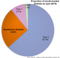 Australia - insulin-treated diabetes by type 2015.jpg 800 × 774; 59 KB
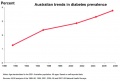 Australian trends diabetes prevalence 19990-2008.jpg 767 × 518; 42 KB
 Bailey273.jpg 560 × 522; 58 KB
 Bailey274.jpg 558 × 442; 32 KB
 Bailey275.jpg 485 × 483; 48 KB
 Bailey278 279.jpg 669 × 812; 85 KB
 Bailey280.jpg 866 × 581; 133 KB
 Blood test result for glucose and iron.jpg 879 × 345; 54 KB
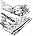 Diabetes record blood sugar.jpg 300 × 332; 27 KB
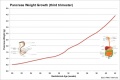 Fetal pancreas weight growth graph.jpg 1,000 × 669; 49 KB
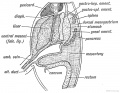
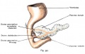
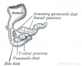
 Human fetal pancreas anatomy cartoon.jpg 455 × 376; 93 KB
 Human stem cell pancreas implants 01.jpg 690 × 730; 109 KB
 Human- pancreatic adult islet-glucagon.jpg 600 × 460; 67 KB
 Human- pancreatic adult islet-insulin.jpg 600 × 460; 68 KB
 Human- pancreatic adult islet.jpg 600 × 460; 74 KB
 Keith1902 fig216.jpg 1,000 × 771; 170 KB
 Keith1902 fig218.jpg 800 × 475; 70 KB
 Kollmann392.jpg 919 × 561; 62 KB
 Kollmann397.jpg 862 × 554; 64 KB
 Model of human pancreatic islet.jpg 482 × 702; 237 KB
 Molecular endocrine pancreas cells 01.jpg 800 × 804; 71 KB
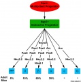
 Mouse pancreas cell lineage.jpg 1,855 × 2,039; 291 KB
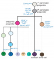
 Mouse pancreas development.jpg 600 × 939; 261 KB
 Mouse-pancreas duct formation.jpg 1,000 × 709; 154 KB
 Mouse-pancreas islets-sweet taste receptor.jpg 1,050 × 261; 38 KB
 Odgers1930 fig01.jpg 979 × 883; 106 KB
 Odgers1930 fig02.jpg 985 × 745; 119 KB
 Odgers1930 fig03.jpg 926 × 755; 122 KB
 Odgers1930 plate01.jpg 1,621 × 2,260; 819 KB
 Pancreas acinar cell em01.jpg 1,280 × 928; 496 KB
 Pancreas adult.jpg 600 × 427; 50 KB
 Pancreas cartoon.jpg 1,000 × 347; 51 KB
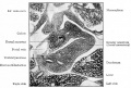
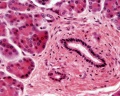
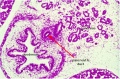
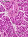 Pancreas histology 001.jpg 375 × 500; 90 KB
 Pancreas histology 002.jpg 375 × 500; 50 KB
 Pancreas histology 003.jpg 375 × 500; 90 KB
 Pancreas histology 004.jpg 400 × 267; 50 KB
 Pancreas histology 005.jpg 375 × 500; 64 KB
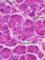
 Pancreas histology 101.jpg 1,280 × 1,024; 505 KB
 Pancreas histology 102.jpg 1,280 × 1,024; 309 KB
 Pancreas histology 103.jpg 1,280 × 1,024; 313 KB
 Pancreas histology 104.jpg 1,280 × 1,024; 371 KB
 Pancreas histology 105.jpg 1,280 × 1,024; 277 KB
 Pancreas histology 106.jpg 1,280 × 1,024; 314 KB
 Pancreas islet - structure human and rat.jpg 945 × 930; 259 KB
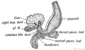
 Pancreas rotation.jpg 652 × 320; 23 KB
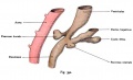
 Pancreatic duct developing.jpg 400 × 322; 15 KB
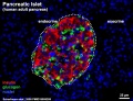
 Pancreatic islet.png 600 × 462; 425 KB
 Pearce1903 fig01.jpg 900 × 809; 335 KB
 Pearce1903 fig02.jpg 730 × 1,198; 377 KB
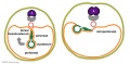
 Rat- pancreatic islet development.jpg 1,200 × 524; 230 KB
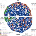
 Ratio of alpha & beta cells at different phases of fetal development.png 537 × 600; 609 KB
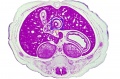 Stage 22 image 183.jpg 1,000 × 662; 147 KB
 Stage 22 image 185.jpg 1,000 × 656; 109 KB
 Stage 22 image 186.jpg 1,000 × 658; 209 KB
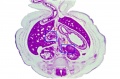
 Stage22 pancreas a.jpg 800 × 640; 99 KB
 Stage22 pancreas b.jpg 600 × 480; 63 KB
 Stage22 pancreas c.jpg 400 × 320; 38 KB
 Stage22 pancreas.jpg 1,000 × 800; 264 KB


