Category:Mouse E16.5: Difference between revisions
From Embryology
No edit summary |
No edit summary |
||
| Line 4: | Line 4: | ||
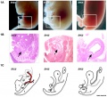
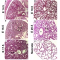
* Mouse (C57BL/6J) - '''mouse eccrine glands''' anlagen first apparent at 16.5 days postconception (DPC) and found only on the footpads (when mature resemble human eccrine glands) PMID 22135020 | * Mouse (C57BL/6J) - '''mouse eccrine glands''' anlagen first apparent at 16.5 days postconception (DPC) and found only on the footpads (when mature resemble human eccrine glands) PMID 22135020 | ||
'''Links:''' [[Mouse Development]] | [[Mouse Stages]] | [[Mouse_Timeline_Detailed|Mouse Timeline]] | |||
[[Category:Animal Development]] [[Category:Mouse]] | |||
Revision as of 11:49, 14 July 2012
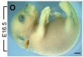
The pages and media listed below relate to E16.5 mouse development.
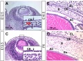
- Mouse (129/Balb/c) - crural Pacinian corpuscles develop between embryonic day (E) 16.5 and postnatal day (P) 0 (with the delivery occurring on day E19) PMID 15376326
- Mouse (C57BL/6J) - mouse eccrine glands anlagen first apparent at 16.5 days postconception (DPC) and found only on the footpads (when mature resemble human eccrine glands) PMID 22135020
Links: Mouse Development | Mouse Stages | Mouse Timeline
Pages in category 'Mouse E16.5'
The following 2 pages are in this category, out of 2 total.
Media in category 'Mouse E16.5'
The following 21 files are in this category, out of 21 total.
 Mouse bladder development E12.5-E16.5.jpg 1,105 × 1,000; 202 KB
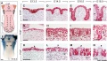
 Mouse cornea development 01.jpg 1,200 × 880; 325 KB
 Mouse cornea E16.5.jpg 701 × 562; 113 KB
 Mouse CT E16.5.jpg 221 × 344; 9 KB
 Mouse gonad Gcnf expression 01.jpg 1,947 × 843; 304 KB
 Mouse gonad Gcnf expression E16.5.jpg 325 × 786; 40 KB
 Mouse lung development 01.jpg 1,000 × 1,254; 791 KB
 Mouse lung development 01a.jpg 800 × 1,003; 495 KB
 Mouse lung development 02.jpg 922 × 922; 239 KB
 Mouse lung development 03.jpg 540 × 1,200; 349 KB
 Mouse melanoblast distribution 01.jpg 697 × 1,000; 192 KB
 Mouse melanoblast distribution 06.jpg 761 × 524; 74 KB
 Mouse model of ovarian cord formation 01.jpg 800 × 491; 85 KB
 Mouse model of ovarian cord formation.jpg 800 × 491; 85 KB
 Mouse placenta 01.jpg 429 × 463; 63 KB
 Mouse posterior neuropore Axd mutant.jpg 475 × 1,074; 95 KB
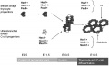 Mouse thyroid Hes1 model.jpg 600 × 364; 33 KB
 Mouse tongue Pax9 expression 01.jpg 1,200 × 687; 259 KB
 Mouse tongue Pax9 expression 02.jpg 1,200 × 833; 299 KB
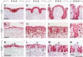
 Mouse ventral body wall development 01.jpg 1,200 × 635; 130 KB
 Mouse-adrenal gland E16.5.jpg 600 × 463; 93 KB


