Category:Mouse E10.5: Difference between revisions
m (→Events) |
m (→Events) |
||
| (13 intermediate revisions by the same user not shown) | |||
| Line 1: | Line 1: | ||
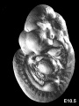
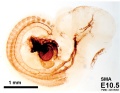
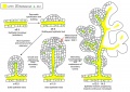
[[File:Day 10.5 Deep Lens Indentation.JPG|thumb|Mouse E10.5]] | |||
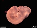
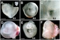
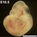
This {{Embryology}} category shows pages and media related to mouse embryonic day 10.5 ('''E10.5''') of development. This staging by "days" relate to in the female presence of a vaginal plug indicating that the mating occurred, see [[Mouse_Timeline_Detailed#Timed_Pregnancy|timed pregnancy]]. | This {{Embryology}} category shows pages and media related to mouse embryonic day 10.5 ('''E10.5''') of development. This staging by "days" relate to in the female presence of a vaginal plug indicating that the mating occurred, see [[Mouse_Timeline_Detailed#Timed_Pregnancy|timed pregnancy]]. | ||
{{Mouse E days}} | {{Mouse E days}} | ||
| Line 6: | Line 8: | ||
==Events== | ==Events== | ||
* | * {{Neural crest}} - {{Enteric nervous system}} cholinergic myenteric neurons appear within even within the migration wavefront of neural precursors.{{#pmid:24712519|PMID24712519}} | ||
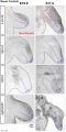
* | * {{Musculoskeletal}} - dermomyotome visible adjacent to the ectoderm. The mesoderm of the primary body wall was non-compact and coalesced in the ventral midline.{{#pmid:22976993|PMID22976993}} | ||
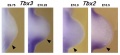
* [[ | ** [[Musculoskeletal System - Limb Development|Limb Development]] - '''Sall4''' integrates Gli3 and the Plzf-Hox system in hindlimb patterning.{{#pmid:25848055|PMID25848055}} | ||
* [[ | * {{Respiratory}} - bronchial branching commences from left and right bronchi.{{#pmid:25114215|PMID25114215}} | ||
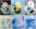
* {{Genital}} | |||
** First appearance beside the cloacal membrane of a pair of genital swellings. | |||
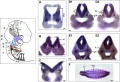
** '''Sry''' expression starts in somatic cells of XY genital ridges.{{#pmid:22439850|PMID22439850}} | |||
* [[Thymus Development]]{{#pmid:15057786|PMID15057786}} See also [[:File:Mouse thymus development 02.jpg|image]] | |||
** '''{{Hox}}a3''' (homeobox A3) - [[:Category:Mouse E9.5|E9.5]] to [[:Category:Mouse E10.5|E10.5]], third cleft surface ectoderm. | |||
** '''{{Pax}}1''' (paired box gene 1) [[:Category:Mouse E9.5|E9.5]] to [[:Category:Mouse E10.5|E10.5]], all pharyngeal pouch endoderm. | |||
** '''{{Pax}}9''' (paired box gene 9) [[:Category:Mouse E9.5|E9.5]] to [[:Category:Mouse E10.5|E10.5]], all pharyngeal pouch endoderm. | |||
** '''Eya1''' (eyes absent 1 homologue ) - Early [[:Category:Mouse E9.5|E9.5 to [[:Category:Mouse E10.5|E10.5]], all pharyngeal pouch endoderm, cleft ectoderm and neural crest cell mesenchyme organogenesis. | |||
* {{Cardiovascular}} - {{Heart}} first sign of atrial septation. | |||
* {{Renal}} vasculature - endothelial cells grow coordinately with the kidney bud.{{#pmid:29627966|PMID29627966}} | |||
===References=== | ===References=== | ||
Latest revision as of 06:56, 12 December 2018

This Embryology category shows pages and media related to mouse embryonic day 10.5 (E10.5) of development. This staging by "days" relate to in the female presence of a vaginal plug indicating that the mating occurred, see timed pregnancy.
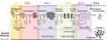
- Mouse Stages: E1 | E2.5 | E3.0 | E3.5 | E4.5 | E5.0 | E5.5 | E6.0 | E7.0 | E7.5 | E8.0 | E8.5 | E9.0 | E9.5 | E10 | E10.5 | E11 | E11.5 | E12 | E12.5 | E13 | E13.5 | E14 | E14.5 | E15 | E15.5 | E16 | E16.5 | E17 | E17.5 | E18 | E18.5 | E19 | E20 | Timeline | About timed pregnancy
| Carnegie | Stage | |||||||||||||||||||||||
| Human | Days | 1 | 2-3 | 4-5 | 5-6 | 7-12 | 13-15 | 15-17 | 17-19 | 20 | 22 | 24 | 28 | 30 | 33 | 36 | 40 | 42 | 44 | 48 | 52 | 54 | 55 | 58 |
| Mouse | Days | 1 | 2 | 3 | E4.5 | E5.0 | E6.0 | E7.0 | E8.0 | E9.0 | E9.5 | E10 | E10.5 | E11 | E11.5 | E12 | E12.5 | E13 | E13.5 | E14 | E14.5 | E15 | E15.5 | E16 |
| Rat | Days | 1 | 3.5 | 4-5 | 5 | 6 | 7.5 | 8.5 | 9 | 10.5 | 11 | 11.5 | 12 | 12.5 | 13 | 13.5 | 14 | 14.5 | 15 | 15.5 | 16 | 16.5 | 17 | 17.5 |
| Note these Carnegie stages are only approximate day timings for average of embryos. Links: Carnegie Stage Comparison | ||||||||||||||||||||||||
| ||||||||||||||||||||||||
| Timeline Links: human timeline | mouse timeline | mouse detailed timeline | chicken timeline | rat timeline | Medaka | Category:Timeline |
Events
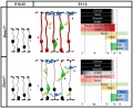
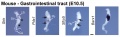
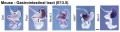
- neural crest - enteric nervous system cholinergic myenteric neurons appear within even within the migration wavefront of neural precursors.[1]
- musculoskeletal - dermomyotome visible adjacent to the ectoderm. The mesoderm of the primary body wall was non-compact and coalesced in the ventral midline.[2]
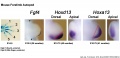
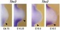
- Limb Development - Sall4 integrates Gli3 and the Plzf-Hox system in hindlimb patterning.[3]
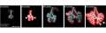
- respiratory - bronchial branching commences from left and right bronchi.[4]
- genital
- First appearance beside the cloacal membrane of a pair of genital swellings.
- Sry expression starts in somatic cells of XY genital ridges.[5]
- Thymus Development[6] See also image
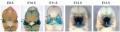
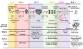
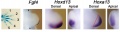
- Hoxa3 (homeobox A3) - E9.5 to E10.5, third cleft surface ectoderm.
- Pax1 (paired box gene 1) E9.5 to E10.5, all pharyngeal pouch endoderm.
- Pax9 (paired box gene 9) E9.5 to E10.5, all pharyngeal pouch endoderm.
- Eya1 (eyes absent 1 homologue ) - Early [[:Category:Mouse E9.5|E9.5 to E10.5, all pharyngeal pouch endoderm, cleft ectoderm and neural crest cell mesenchyme organogenesis.
- cardiovascular - heart first sign of atrial septation.
- renal vasculature - endothelial cells grow coordinately with the kidney bud.[7]
References
- ↑ Erickson CS, Lee SJ, Barlow-Anacker AJ, Druckenbrod NR, Epstein ML & Gosain A. (2014). Appearance of cholinergic myenteric neurons during enteric nervous system development: comparison of different ChAT fluorescent mouse reporter lines. Neurogastroenterol. Motil. , 26, 874-84. PMID: 24712519 DOI.
- ↑ Nichol PF, Corliss RF, Yamada S, Shiota K & Saijoh Y. (2012). Muscle patterning in mouse and human abdominal wall development and omphalocele specimens of humans. Anat Rec (Hoboken) , 295, 2129-40. PMID: 22976993 DOI.
- ↑ Akiyama R, Kawakami H, Wong J, Oishi I, Nishinakamura R & Kawakami Y. (2015). Sall4-Gli3 system in early limb progenitors is essential for the development of limb skeletal elements. Proc. Natl. Acad. Sci. U.S.A. , 112, 5075-80. PMID: 25848055 DOI.
- ↑ Kadzik RS, Cohen ED, Morley MP, Stewart KM, Lu MM & Morrisey EE. (2014). Wnt ligand/Frizzled 2 receptor signaling regulates tube shape and branch-point formation in the lung through control of epithelial cell shape. Proc. Natl. Acad. Sci. U.S.A. , 111, 12444-9. PMID: 25114215 DOI.
- ↑ de Lau WB, Snel B & Clevers HC. (2012). The R-spondin protein family. Genome Biol. , 13, 242. PMID: 22439850 DOI.
- ↑ Blackburn CC & Manley NR. (2004). Developing a new paradigm for thymus organogenesis. Nat. Rev. Immunol. , 4, 278-89. PMID: 15057786 DOI.
- ↑ Daniel E, Azizoglu DB, Ryan AR, Walji TA, Chaney CP, Sutton GI, Carroll TJ, Marciano DK & Cleaver O. (2018). Spatiotemporal heterogeneity and patterning of developing renal blood vessels. Angiogenesis , 21, 617-634. PMID: 29627966 DOI.
Search Pubmed: Mouse E10.5
Pages in category 'Mouse E10.5'
The following 7 pages are in this category, out of 7 total.
Media in category 'Mouse E10.5'
The following 55 files are in this category, out of 55 total.
 Anderson2016-fig03.jpg 800 × 800; 130 KB
 Anderson2016-fig04.jpg 800 × 800; 99 KB
 Anderson2016-fig06.jpg 800 × 800; 123 KB
 Anderson2016-fig08a.jpg 800 × 800; 112 KB
 Anderson2016-fig08b.jpg 800 × 800; 107 KB
 Anderson2016-fig13a.jpg 800 × 800; 115 KB
 Day 10.5 Deep Lens Indentation.JPG 514 × 483; 28 KB
 Hindlimb Tbx2 model.jpg 1,000 × 607; 84 KB
 Limb patterning factors 08.jpg 1,200 × 576; 79 KB
 Limb patterning factors 09.jpg 1,200 × 601; 82 KB
 Mouse - stomach 01.png 599 × 600; 1.45 MB
 Mouse Bmp4 expression face 01.jpg 1,200 × 322; 58 KB
 Mouse Bmp4 expression limb and face 01.jpg 1,200 × 513; 91 KB
 Mouse CT E10.5 head 01.jpg 1,000 × 636; 154 KB
 Mouse CT E10.5 head.jpg 1,200 × 449; 83 KB
 Mouse CT E10.5.jpg 602 × 800; 67 KB
 Mouse CT E9.5-E12 head.jpg 1,000 × 568; 56 KB
 Mouse E10.5 gene expression.jpg 1,747 × 1,650; 404 KB
 Mouse E10.5 hindlimb gene expression.jpg 1,000 × 336; 49 KB
 Mouse E10.5 Hoxa3.jpg 1,200 × 854; 225 KB
 Mouse E10.5 Nav2 expression.jpg 1,200 × 818; 186 KB
 Mouse E10.5 pecam1 detail.jpg 600 × 450; 76 KB
 Mouse E10.5 pecam1.jpg 600 × 450; 59 KB
 Mouse E10.5 smooth muscle actin detail.jpg 600 × 465; 65 KB
 Mouse E10.5 smooth muscle actin.jpg 600 × 465; 56 KB
 Mouse E8.5-E10.5 Hoxa3.jpg 1,210 × 1,000; 356 KB
 Mouse embryo E10.5.jpg 716 × 540; 30 KB
 Mouse external genital development.jpg 800 × 701; 75 KB
 Mouse face Bmp4.mp4 ; 480 KB
 Mouse forelimb E10.5 to E11.5.jpg 1,001 × 2,000; 331 KB
 Mouse head-neural crest 01.jpg 900 × 339; 48 KB
 Mouse limb bone development timeline.jpg 1,256 × 469; 107 KB
 Mouse limb skeleton cartoon.jpg 1,000 × 487; 64 KB
 Mouse limb tissue development.jpg 1,280 × 767; 161 KB
 Mouse Lmx1b gene expression.jpg 1,624 × 550; 104 KB
 Mouse pancreas development.jpg 600 × 939; 261 KB
 Mouse respiratory 36 to 60 somites.jpg 1,200 × 383; 58 KB
 Mouse telencephalon radial glia model.jpg 1,000 × 816; 80 KB
 Mouse thymus development 01.jpg 600 × 584; 69 KB
 Mouse thymus development 02.jpg 600 × 445; 50 KB
 Mouse Wnt signaling 01.jpg 600 × 450; 156 KB
 Mouse yolk sac 01.jpg 1,002 × 669; 115 KB
 Mouse- embryo E10.5.jpg 324 × 324; 14 KB
 Mouse- facial branchiomotor neuron migration.jpg 600 × 841; 104 KB
 Mouse- forelimb-bud-Tbx3-Tbx2.jpg 955 × 436; 45 KB
 Mouse- X-linked gene expression in primordial germ cells.jpg 800 × 632; 90 KB
 Mouse-E10.5 ganglia Sox10.jpg 540 × 685; 51 KB
 Mouse-E10.5-Sox10.jpg 400 × 463; 24 KB
 Mouse-E9.5 E10.5 E11.5 E12.5.png 599 × 407; 170 KB
 Mouse-forelimb bud Fgf4-Hoxd13-Hoxa13.jpg 881 × 237; 30 KB
 Mouse-Gastrointestinal-tract-E10.5-01.jpg 1,000 × 306; 43 KB
 Mouse-Gastrointestinal-tract-E13.5-01.jpg 1,000 × 266; 48 KB
 Mouse-neural crest Sox10 E10.5.jpg 1,000 × 420; 55 KB
 Mouse-pancreas duct formation.jpg 1,000 × 709; 154 KB
 Primordial germ cell 003.mov ; 695 KB


