Category:Limb
From Embryology
This page lists Embryology content related to limb development and limb abnormalities. The limb is also an excellent model for how tissue pattern is established.
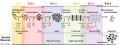
| Musculoskeletal Links: Introduction | mesoderm | somitogenesis | limb | cartilage | bone | bone timeline | bone marrow | shoulder | pelvis | axial skeleton | skull | joint | skeletal muscle | muscle timeline | tendon | diaphragm | Lecture - Musculoskeletal | Lecture Movie | musculoskeletal abnormalities | limb abnormalities | developmental hip dysplasia | cartilage histology | bone histology | Skeletal Muscle Histology | Category:Musculoskeletal | ||
|
Pages in category 'Limb'
The following 148 pages are in this category, out of 148 total.
B
- Template:Bardeen1906 figures
- BGDA Practical 7 - Week 6
- Book - An Atlas of Topographical Anatomy 19
- Book - An Atlas of Topographical Anatomy 20
- Book - An Atlas of Topographical Anatomy 21
- Book - An Atlas of Topographical Anatomy 22
- Book - An Atlas of Topographical Anatomy 23
- Book - An Atlas of Topographical Anatomy 24
- Book - An Atlas of Topographical Anatomy 26
- Book - An Atlas of Topographical Anatomy 27
- Book - An Atlas of Topographical Anatomy 28
- Book - Human Embryology and Morphology 20
- Book - Manual of Human Embryology 11D
- Book - Manual of Human Embryology 12
C
D
L
- Lecture - Limb Development
- Template:Limb
- Template:Limb abnormalities
- Template:Limb axis
- Template:Limb Links
- Template:Limb Rotation table
- Template:Limb skeleton mesenchyme-cartilage-bone table
- Template:Lower limb ossification collapse table
- Template:Lower limb ossification collapsible table
- Template:Lower limb ossification table
- Template:Lower limb ossification timeline
M
P
- Paper - A contribution to the embryology of the fore-limb
- Paper - A study of the development of the mammalian pelvis
- Paper - An unusual form of brachyphalangy and syndactyly with double proximal phalanx in the middle fingers (1932)
- Paper - Anatomy of a seven months foetus exhibiting bilateral absence of the ulna accompanied by monodactyly
- Paper - Chondrification in the hands and feet of staged human embryos
- Paper - Development and variation of the nerves and the musculature of the inferior extremity and of the neighboring regions of the trunk in man
- Paper - Development of the innervation pattern in the upper limb of staged human embryos (1990)
- Paper - Extra digits and digital reductions
- Paper - Familial abnormalities of the middle phalanges of each hand (1923)
- Paper - On an instance of two subclavian arteries of the early arm bud of man and its fundamental significance
- Paper - Rare congenital malformation of hands and feet (1924)
- Paper - Studies of the development of the human skeleton (1905)
- Paper - The arrangement of the bursae in the superior extremities of the full-time foetus
- Paper - The development and evolution of the "papillary" ridges and patterns on the volar surface of the hand (1906)
- Paper - The development of joints
- Paper - The development of the arm in man (1902)
- Paper - The development of the arteries of the human lower extremity
- Paper - The development of the limbs, body-wall and back
- Paper - The development of the limbs, body-wall and back (1901)
- Paper - The development of the patella
- Paper - The development of the principal arterial stems in the forelimb of the pig (1922)
- Paper - The development of the veins in the limbs of rabbit embryos
- Paper - The early development of the knee joint in staged human embryos
- Paper - The embryology of the human hip joint (1943)
- Paper - The epiphysis of the head of the femur (1915)
- Paper - The growth of the human foot
- Paper - The proximo-distal sequence of origin of the parts of the chick wing and the role of the ectoderm
- Paper - The sexual differences of the fetal pelvis
- Paper - The sexual differences of the fetal pelvis (1899)
- Template:Pelvis
- Template:Pelvis Timeline table
R
- Template:Ref-Bardeen1907
- Template:Ref-BardeenLewis1901
- Template:Ref-Brailsford1943
- Template:Ref-Carey1922
- Template:Ref-Cockayne1932
- Template:Ref-Cummins1929
- Template:Ref-Davenport1932
- Template:Ref-deLima1928
- Template:Ref-Evans1908
- Template:Ref-Evatt1906
- Template:Ref-GardnerO'Rahilly1968
- Template:Ref-Geddes1912b
- Template:Ref-GrayGardener1950
- Template:Ref-Haines1947
- Template:Ref-Haines1953
- Template:Ref-Hann1923
- Template:Ref-Harrison1915
- Template:Ref-Johnson1899
- Template:Ref-Keith1910
- Template:Ref-Lewis1902
- Template:Ref-Lewis1905b
- Template:Ref-Lewis1910a
- Template:Ref-Manson1924
- Template:Ref-O'Rahilly1957
- Template:Ref-PMID55906
- Template:Ref-Prentiss1906
- Template:Ref-Rutherford1914
- Template:Ref-Saunders1947a
- Template:Ref-Saunders1948
- Template:Ref-ShinoharaTanaka1990
- Template:Ref-Stacey1938
- Template:Ref-Walmsley1915
- Template:Ref-Walmsley1940
- Template:Ref-Watt1917
- Template:Ref-Whittaker1910
- Template:Ref-Wigoder1928
- Template:Ref-Woollard1922
S
U
V
Media in category 'Limb'
The following 200 files are in this category, out of 274 total.
(previous page) (next page)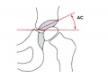 Acetabular angle.jpg 600 × 433; 22 KB
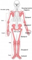 Appendicular skeleton developmental regions.jpg 1,000 × 1,849; 143 KB
 Appendicular skeleton small.jpg 220 × 407; 12 KB
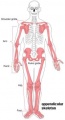
 Appendicular skeleton.jpg 1,000 × 1,849; 125 KB
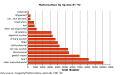 Australian abnormalities graph allsystem.png 509 × 320; 7 KB
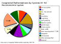 Australian abnormalities pie skmus.png 481 × 344; 9 KB
 Axolotl developing limb Bmp2 and Sox9.jpg 1,159 × 638; 105 KB
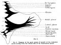
 Bailey143.jpg 911 × 673; 114 KB
 Bailey144.jpg 491 × 398; 39 KB
 Bailey145.jpg 777 × 654; 80 KB
 Bailey149.jpg 576 × 520; 38 KB
 Bailey150.jpg 406 × 596; 42 KB
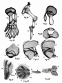
 Bailey189.jpg 810 × 632; 63 KB
 Bailey190.jpg 801 × 584; 69 KB
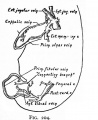 Bailey204.jpg 534 × 653; 62 KB
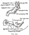 Bailey205.jpg 534 × 653; 68 KB
 Bardeen1905 plate13.jpg 1,000 × 1,337; 85 KB
 Bardeen1906-fig02.jpg 1,598 × 1,183; 228 KB
 Bardeen1906-fig03.jpg 1,598 × 1,166; 231 KB
 Bardeen1906-plate01.jpg 1,565 × 2,322; 238 KB
 Bardeen1906-plate02.jpg 1,719 × 2,302; 512 KB
 Bardeen1906-plate06.jpg 1,568 × 2,299; 379 KB
 Bardeen1906-plate31.jpg 1,571 × 2,330; 257 KB
 Bardeen1906-plate32.jpg 1,588 × 2,341; 292 KB
 Bardeen1906-plate41.jpg 1,555 × 2,323; 261 KB
 Bardeen1906-plate42.jpg 1,570 × 2,331; 240 KB
 Bardeen1906-plate51.jpg 1,570 × 2,330; 392 KB
 Bardeen1906-plate52.jpg 1,596 × 2,350; 404 KB
 Bat - adult and fetal limbs.jpg 600 × 612; 80 KB
 Bat and mouse limb comparison.jpg 1,000 × 591; 77 KB
 Bat limb 01.jpg 1,688 × 699; 149 KB
 Bat limb 02.jpg 1,200 × 430; 59 KB
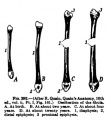
 Bone-femur.jpg 798 × 1,000; 150 KB
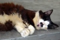 Cat 6 toes.jpg 420 × 280; 13 KB
 Chicken limb BMP modulator expression 01.jpg 1,200 × 1,224; 211 KB
 Chicken limb gene expression 01.jpg 1,200 × 758; 85 KB
 Chicken limb gene expression 02.jpg 1,000 × 478; 42 KB
 Chicken limb gene expression 03.jpg 1,200 × 711; 108 KB
 Chicken- limb bud chondrogenesis.jpg 600 × 1,032; 163 KB
 Chicken- wing cartilage.jpg 1,200 × 1,022; 218 KB
 Chicken-limb sox9 wnt6.jpg 1,173 × 373; 68 KB
 Clefthand-apical-defect.jpg 500 × 568; 19 KB
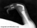 Congenital limb reduction xray.jpg 793 × 600; 18 KB
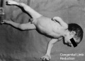 Congenital limb reduction.jpg 400 × 289; 11 KB
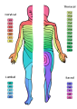 Dermatomes.png 424 × 600; 82 KB
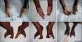 Ectrodactyly 01.jpg 1,200 × 619; 113 KB
 Ectrodactyly.jpg 500 × 244; 9 KB
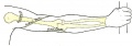
 Embryonic upper limb - brachial and superficial brachial artery.jpg 1,280 × 314; 122 KB
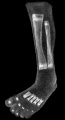
 Fetal limb abnormalities X-ray-02.jpg 685 × 931; 62 KB
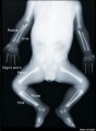 Fetal limb abnormalities X-ray-03.jpg 685 × 931; 57 KB
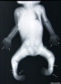
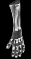
 Fetal limb X-ray-01.jpg 685 × 931; 57 KB
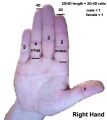 Finger length ratio - 2D4D.jpg 428 × 480; 26 KB
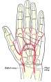
 Gray0595.jpg 600 × 491; 94 KB
 Gray0606.jpg 686 × 800; 93 KB
 Gray0608.jpg 400 × 623; 74 KB
 Gray0609.jpg 527 × 500; 69 KB
 Gray0610.jpg 303 × 1,000; 81 KB
 Gray0807.gif 587 × 500; 44 KB
 Gray0807.jpg 704 × 600; 97 KB
 Gray0822.jpg 599 × 600; 69 KB
 Gray1235.jpg 800 × 389; 41 KB
 Gray1236.jpg 800 × 281; 29 KB
 Gray1237.jpg 371 × 600; 57 KB
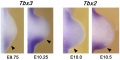
 Hindlimb Tbx2 model.jpg 1,000 × 607; 84 KB
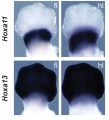
 Hoxa gene expression in limb bud 01.jpg 1,376 × 533; 85 KB
 Hoxa gene expression in limb bud 02.jpg 546 × 600; 40 KB
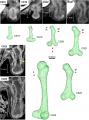 Human embryo femur CS18 to CS23.png 1,200 × 1,624; 1.42 MB
 Human embryonic shoulder girdle 01.jpg 1,000 × 726; 81 KB
 Human embryonic shoulder girdle 02.jpg 1,025 × 713; 109 KB
 Human embryonic shoulder girdle 04.jpg 1,000 × 755; 71 KB
 Human upper limb mri-01.jpg 1,409 × 936; 108 KB
 Keibel Mall 077.jpg 424 × 700; 33 KB
 Keibel Mall 078.jpg 278 × 236; 18 KB
 Keibel Mall 2 451.jpg 1,000 × 714; 73 KB
 Keibel Mall 215.jpg 727 × 379; 53 KB
 Keibel Mall 216.jpg 366 × 356; 30 KB
 Keibel Mall 274-278.jpg 717 × 1,072; 123 KB
 Keibel Mall 279-284.jpg 709 × 707; 84 KB
 Keibel Mall 285-288.jpg 703 × 674; 72 KB
 Keibel Mall 289.jpg 682 × 645; 85 KB
 Keibel Mall 290.jpg 800 × 452; 59 KB
 Keibel Mall 291.jpg 800 × 421; 54 KB
 Keibel Mall 292.jpg 400 × 439; 30 KB
 Keibel Mall 293.jpg 687 × 857; 137 KB
 Keibel Mall 301.jpg 734 × 945; 94 KB
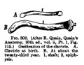 Keibel Mall 302.jpg 300 × 233; 18 KB
 Keibel Mall 307.jpg 800 × 803; 112 KB
 Keibel Mall 346.jpg 685 × 670; 71 KB
 Keibel Mall 347.jpg 700 × 706; 68 KB
 Keibel Mall 348.jpg 635 × 642; 72 KB
 Keibel Mall 349.jpg 661 × 692; 69 KB
 Keibel Mall 352.jpg 611 × 558; 52 KB
 Keibel Mall 353.jpg 649 × 489; 44 KB
 Keibel Mall 354.jpg 800 × 459; 55 KB
 Keibel Mall 355.jpg 767 × 800; 66 KB
 Keibel Mall 356.jpg 775 × 516; 50 KB
 Keibel Mall 357.jpg 870 × 535; 60 KB
 Keibel Mall 358.jpg 700 × 693; 49 KB
 Keibel Mall 359.jpg 654 × 460; 46 KB
 Keibel Mall 360.jpg 765 × 490; 55 KB
 Keibel Mall 361.jpg 673 × 468; 37 KB
 Keibel Mall 362.jpg 801 × 516; 57 KB
 Keibel Mall 363.jpg 605 × 407; 34 KB
 Keibel Mall 364.jpg 594 × 378; 35 KB
 Keibel Mall 365.jpg 747 × 550; 59 KB
 Keibel Mall 366.jpg 780 × 535; 60 KB
 Keith1902 fig233.jpg 933 × 800; 122 KB
 Keith1902 fig234.jpg 1,200 × 387; 84 KB
 Keith1902 fig235.jpg 1,200 × 329; 74 KB
 Keith1902 fig236.jpg 1,200 × 652; 109 KB
 Keith1902 fig237.jpg 896 × 800; 111 KB
 Keith1902 fig238.jpg 1,000 × 752; 166 KB
 Keith1902 fig239.jpg 595 × 800; 58 KB
 Keith1902 fig240.jpg 1,000 × 491; 55 KB
 Keith1902 fig241.jpg 458 × 800; 50 KB
 Keith1902 fig242.jpg 1,000 × 458; 75 KB
 Keith1902 fig243.jpg 1,000 × 631; 98 KB
 Keith1902 fig244.jpg 1,000 × 503; 100 KB
 Keith1902 fig245.jpg 1,000 × 614; 88 KB
 Keith1902 fig246.jpg 943 × 600; 98 KB
 Keith1902 fig247.jpg 1,000 × 488; 68 KB
 Keith1902 fig248.jpg 800 × 480; 41 KB
 Keith1902 fig249.jpg 1,000 × 713; 108 KB
 Keith1902 fig250.jpg 1,000 × 457; 81 KB
 Keith1902 fig251.jpg 600 × 446; 31 KB
 Keith1902 fig252.jpg 600 × 510; 39 KB
 Left foot polydactyly 01.jpg 481 × 600; 27 KB
 Left foot polydactyly 02.jpg 307 × 396; 10 KB
 Left hand reduction defect 01.jpg 400 × 293; 14 KB
 Left hand reduction defect 02.jpg 396 × 509; 12 KB
 Limb 3D map of cell proliferation rates.jpg 1,000 × 794; 242 KB
 Limb bud geometry and patterning.jpg 583 × 765; 76 KB
 Limb bud growth model 01.jpg 600 × 261; 30 KB
 Limb bud growth model 02.jpg 600 × 637; 80 KB
 Limb changing 3D geometry.jpg 1,000 × 1,062; 196 KB
 Limb comparison cartoon 01.jpg 1,780 × 2,203; 379 KB
 Limb comparison cartoon 02.jpg 1,200 × 778; 119 KB
 Limb hairy2 expression model.jpg 1,280 × 523; 76 KB
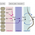 Limb induction-initiation signal 01.jpg 664 × 664; 204 KB
 Limb induction-initiation signal 02.jpg 1,280 × 1,009; 124 KB
 Limb mesenchymal cell shape.jpg 775 × 1,000; 180 KB
 Limb patterning factors 01.jpg 800 × 794; 76 KB
 Limb patterning factors 02.jpg 800 × 794; 73 KB
 Limb patterning factors 03.jpg 800 × 794; 36 KB
 Limb patterning factors 04.jpg 800 × 794; 67 KB
 Limb patterning factors 05.jpg 800 × 794; 37 KB
 Limb patterning factors 06.jpg 800 × 794; 41 KB
 Limb patterning factors 07.jpg 800 × 794; 44 KB
 Limb patterning factors 08.jpg 1,200 × 576; 79 KB
 Limb patterning factors 09.jpg 1,200 × 601; 82 KB
 Limb patterning factors 11.jpg 1,200 × 655; 78 KB
 Lizard embryo 08.jpg 1,200 × 900; 177 KB
 Malformation1.jpg 354 × 570; 40 KB
 Mall Meyer1921 fig90.jpg 479 × 791; 64 KB
 Mall Meyer1921 fig91.jpg 352 × 191; 14 KB
 Mall Meyer1921 fig92.jpg 359 × 734; 46 KB
 Mall Meyer1921 fig93.jpg 413 × 734; 42 KB
 Mall1906 fig05.jpg 561 × 1,055; 45 KB
 Mall1906 fig06.jpg 522 × 966; 41 KB
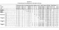 Mall1906 table06.jpg 2,117 × 1,074; 469 KB
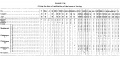 Mall1906 table07.jpg 2,118 × 1,034; 432 KB
 McMurrich1930 fig88.jpg 1,280 × 1,509; 218 KB
 McMurrich1930 fig89.jpg 1,280 × 681; 98 KB
 Model digit origin.png 599 × 505; 128 KB
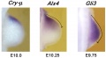
 Model GATA6 hindlimb.jpg 737 × 600; 60 KB
 Morgan 1925 fig65.jpg 1,175 × 766; 189 KB
 Morgan 1925 fig66.jpg 649 × 969; 108 KB
 Morgan 1925 fig68.jpg 1,000 × 744; 188 KB
 Mouse Bmp4 expression limb and face 01.jpg 1,200 × 513; 91 KB
 Mouse Cited1.jpg 446 × 550; 37 KB
 Mouse Dmrt2.jpg 446 × 550; 35 KB
 Mouse E10.5 gene expression.jpg 1,747 × 1,650; 404 KB
 Mouse E10.5 hindlimb gene expression.jpg 1,000 × 336; 49 KB
 Mouse E11.5 gene expression.jpg 1,760 × 1,650; 396 KB
 Mouse E12.5 gene expression.jpg 1,823 × 1,100; 272 KB
 Mouse E13.5 gene expression.jpg 1,764 × 1,100; 266 KB
 Mouse E9.5 gene expression.jpg 1,757 × 1,100; 269 KB
 Mouse Echdc1.jpg 446 × 550; 29 KB
 Mouse Egr1.jpg 446 × 550; 34 KB
 Mouse embryo cortex radial expansion.jpg 1,200 × 624; 249 KB
 Mouse Etv2.jpg 446 × 550; 33 KB
 Mouse Fbxo41.jpg 446 × 550; 24 KB
 Mouse Figf.jpg 446 × 550; 25 KB
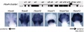
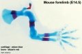
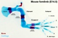
 Mouse forelimb cartilage and bone E14.5 E18.5.jpg 1,000 × 1,300; 156 KB
 Mouse forelimb cartilage and bone E18.5.jpg 774 × 600; 70 KB
 Mouse forelimb E10.5 to E11.5.jpg 1,001 × 2,000; 331 KB
 Mouse forelimb gene expression.jpg 2,205 × 1,650; 456 KB
 Mouse forelimb.jpg 600 × 812; 71 KB
 Mouse hindlimb cartilage and bone E18.5.jpg 774 × 600; 75 KB
 Mouse hindlimb gene expression.jpg 2,237 × 550; 147 KB
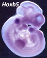 Mouse Hoxb5.jpg 446 × 550; 36 KB
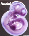 Mouse Hoxb6.jpg 446 × 550; 35 KB
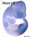 Mouse Hoxc10.jpg 446 × 550; 23 KB
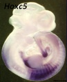 Mouse Hoxc5.jpg 446 × 550; 30 KB
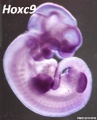 Mouse Hoxc9.jpg 446 × 550; 34 KB
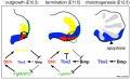
 Mouse interdigit apoptosis 01.jpg 800 × 800; 81 KB
 Mouse interdigit apoptosis 02.jpg 764 × 764; 61 KB
 Mouse limb bone development timeline.jpg 1,256 × 469; 107 KB
 Mouse limb cartilage and bone E14.5.jpg 800 × 526; 42 KB
 Mouse limb cartilage and bone E14.5L.jpg 1,000 × 658; 73 KB


