Category:Implantation: Difference between revisions
From Embryology
No edit summary |
mNo edit summary |
||
| Line 1: | Line 1: | ||
This page lists | This page lists Embryology pages and media related to the process of implantation. In human development, this process commences in week 2 ({{GA}} week 4). | ||
:'''Links:''' [[Implantation]] | |||
[[Category:Week 2]] | [[Category:Week 2]] | ||
[[Category:Uterus]] | [[Category:Uterus]] | ||
Latest revision as of 10:35, 18 March 2014
This page lists Embryology pages and media related to the process of implantation. In human development, this process commences in week 2 (GA week 4).
- Links: Implantation
Pages in category 'Implantation'
The following 38 pages are in this category, out of 38 total.
B
I
P
- Paper - A previllous human embryo (1946)
- Paper - Development of the human placenta in the first three months of gestation (1960)
- Paper - Implantation of the human ovum in the uterus
- Paper - Implantation of the ovum in mammals in the light of recent research (1937)
- Paper - The embedding of the embryo guinea-pig in the uterine wall and its nutrition at that stage of development
- Pregnancy Test
- Template:Pregnancy test
R
Media in category 'Implantation'
The following 43 files are in this category, out of 43 total.
 Bailey073.jpg 705 × 501; 122 KB
 Comparison human and opossum pregnancy.jpg 800 × 245; 30 KB
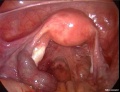 Ectopic molar pregnancy 01.jpg 700 × 535; 60 KB
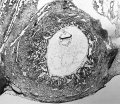
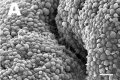 Human - uterine epithelium SEM01.jpg 600 × 400; 37 KB
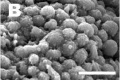 Human - uterine epithelium SEM02.jpg 600 × 400; 26 KB
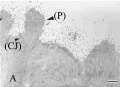 Human - uterine epithelium TEM01.jpg 600 × 433; 25 KB
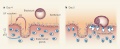
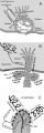
 Implantation cartoon 01.jpg 1,000 × 756; 154 KB
 Implantation cartoon 02.jpg 950 × 1,000; 161 KB
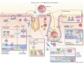
 Implantation LIF.jpg 800 × 325; 48 KB
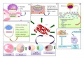
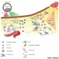 Implantation signaling cartoon.jpg 1,000 × 991; 163 KB
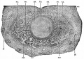
 Keibel Mall 094.jpg 680 × 791; 98 KB
 Keith1921 fig017.jpg 1,095 × 1,000; 236 KB
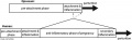
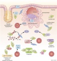
 Model for immune role in implantation - Mucin absence.jpg 600 × 870; 213 KB
 Model for immune role in implantation.jpg 600 × 1,001; 231 KB
 Ovarian ectopic pregnancy 01.jpg 800 × 603; 60 KB
 Pig - uterine epithelium SEM.jpg 800 × 598; 118 KB
 Roles of LIF in embryo implantation.jpeg 600 × 422; 77 KB
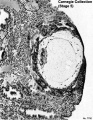
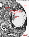
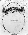
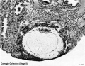 Stage5 bf01.jpg 800 × 618; 149 KB
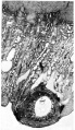
 Stage5 bf02.jpg 800 × 516; 96 KB
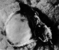
 Stage5 bf03.jpg 800 × 537; 132 KB
 Stage5 bf04.jpg 1,410 × 794; 108 KB
 Stage5 bf05.jpg 1,200 × 692; 256 KB
 Stage5 bf06.jpg 800 × 558; 120 KB
 Stage5 bf07.jpg 304 × 708; 31 KB
 Stage5 bf08.jpg 537 × 800; 133 KB
 Stage5 bf1.jpg 800 × 618; 213 KB
 Stage5 bf11.jpg 618 × 800; 213 KB
 Stage5 bf11a.jpg 464 × 600; 134 KB
 Stage5 bf11b.jpg 464 × 600; 96 KB
 Stage5 bf11L.jpg 618 × 800; 165 KB
 Stage5 bf13.jpg 800 × 537; 142 KB
 Stage5 bf2.jpg 800 × 516; 122 KB
 Stage5 bf22.jpg 516 × 800; 123 KB
 Stage5 bf22b.jpg 516 × 800; 95 KB
 Stage5 bf22L.jpg 516 × 800; 110 KB
 Stage5 bf8004.jpg 1,280 × 1,030; 297 KB
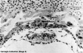
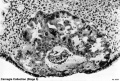
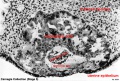
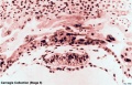
 Stage6 bf01.jpg 646 × 800; 60 KB
 Stage6 bf03.jpg 646 × 800; 65 KB
 Stage7 bf6.jpg 347 × 599; 69 KB
 Stage7 bf7.jpg 400 × 341; 22 KB
 Stage7 bf8.jpg 400 × 341; 18 KB
 Stage7 bf9.jpg 1,200 × 1,039; 501 KB
 Trophoblast hCG function.jpg 600 × 1,798; 229 KB


