Category:Human Embryo
From Embryology
This Embryology category shows pages and media related to human embryonic development occurring in the first 8 weeks of human development.
Subcategories
This category has the following 72 subcategories, out of 72 total.
C
- Carnegie Collection
- Carnegie Embryo 1267
- Carnegie Embryo 1324
- Carnegie Embryo 1332
- Carnegie Embryo 1534
- Carnegie Embryo 175
- Carnegie Embryo 190
- Carnegie Embryo 199
- Carnegie Embryo 2114
- Carnegie Embryo 219
- Carnegie Embryo 2256
- Carnegie Embryo 293
- Carnegie Embryo 368
- Carnegie Embryo 390
- Carnegie Embryo 409
- Carnegie Embryo 423
- Carnegie Embryo 424
- Carnegie Embryo 426
- Carnegie Embryo 431
- Carnegie Embryo 4361
- Carnegie Embryo 437
- Carnegie Embryo 452
- Carnegie Embryo 484
- Carnegie Embryo 490
- Carnegie Embryo 491
- Carnegie Embryo 508
- Carnegie Embryo 509
- Carnegie Embryo 511
- Carnegie Embryo 576
- Carnegie Embryo 6150
- Carnegie Embryo 626
- Carnegie Embryo 657
- Carnegie Embryo 709
- Carnegie Embryo 7900
- Carnegie Embryo 84
- Carnegie Embryo 8913
- Carnegie Stage 1
- Carnegie Stage 10
- Carnegie Stage 11
- Carnegie Stage 12
- Carnegie Stage 13
- Carnegie Stage 15
- Carnegie Stage 16
- Carnegie Stage 17
- Carnegie Stage 18
- Carnegie Stage 19
- Carnegie Stage 20
- Carnegie Stage 21
- Carnegie Stage 22
- Carnegie Stage 23
- Carnegie Stage 3
- Carnegie Stage 6
- Carnegie Stage 7
- Carnegie Stage 8
- Carnegie Stage 9
E
Pages in category 'Human Embryo'
The following 200 pages are in this category, out of 261 total.
(previous page) (next page)A
- Template:Abdominal Wall Muscle Timeline table
- Abnormal Development - Biological Toxins
- Abnormal Development - Chemicals
- Abnormal Development - Drugs
- Abnormal Development - Herbal Drugs
- Abnormal Development - Illegal Drugs
- Abnormal Development - Thalidomide
- Alpha-Fetoprotein
- Amniocentesis
- Assisted Reproductive Technology
- Assisted Reproductive Technology - Glossary
- Australian Drug Categories
- Australian In Vitro Fertilization and Donor Questions
B
C
- Carnegie stage 1
- Carnegie stage 10
- Carnegie stage 10 gallery
- Carnegie stage 11
- Carnegie stage 12
- Carnegie stage 13
- Carnegie stage 14
- Carnegie stage 15
- Carnegie stage 16
- Carnegie stage 17
- Carnegie Stage 17 Movie
- Carnegie stage 18
- Carnegie stage 19
- Carnegie stage 2
- Carnegie stage 20
- Carnegie stage 21
- Carnegie stage 22
- Carnegie stage 22 - selected serial sections
- Carnegie stage 22 - serial sections
- Carnegie stage 23
- Carnegie stage 3
- Carnegie stage 4
- Carnegie stage 5
- Carnegie stage 6
- Carnegie stage 7
- Carnegie stage 8
- Carnegie stage 9
- Carnegie stage 9 gallery
- Template:Carnegie stage table 1
- Template:Carnegie stages
- Carnegie Stages
- Template talk:Carnegie stages
- Chorionic villus sampling
- Comparative Genomic Hybridization
- Template:CRL
- Template:CS1
- Template:CS10
- Template:CS11
- Template:CS12
- Template:CS13
- Template:CS14
- Template:CS15
- Template:CS16
- Template:CS17
- Template:CS18
- Template:CS19
- Template:CS2
- Template:CS20
- Template:CS21
- Template:CS22
- Template:CS23
- Template:CS3
- Template:CS4
- Template:CS5
- Template:CS6
- Template:CS7
- Template:CS8
- Template:CS9
D
E
F
G
H
- Template:Harvard Collection table01
- Human Abnormal Development
- Human Chorionic Gonadotropin
- Template:Human embryo Hertwig G31 table
- Template:Human embryo Klb table
- Template:Human embryo Meyer 300 table
- Template:Human embryo Pfannenstiel III table
- Template:Human embryo Strahl 4mm table
- Template:Human Eyelid timeline table
M
O
P
- Paper - A fifteen-somite human embryo
- Paper - A human embryo of the pre-somite period from the uterine tube
- Paper - A Human Embryo of Thirteen Somites
- Paper - A Human Embryo with Seven Pairs of Somites Measuring about 2 mm in Length
- Paper - A Human Ovum at the Previllous Stage
- Paper - A previllous human embryo (1946)
- Paper - Congenital absence of ventrolateral abdominal musculature (1946)
- Paper - Description of a 4 mm human embryo (1906)
- Special:Badtitle/NS501:Paper - Description of a Human Embryo of 13-14 Mesodermic Somites
- Paper - Development of the human placenta in the first three months of gestation (1960)
- Paper - Development of the trigone of the bladder and the termination of the mesonephric ducts (1946)
- Paper - Growth allometry of the myocardium in human embryos from stages 15 to 23
- Paper - Hydatiform degeneration in an early human embryo (1946)
- Paper - Observations on the neural crest of a ten-somite human embryo (1939)
- Paper - On measuring human embryos
- Paper - On Ossification Centers in Human Embryos
- Paper - On The Age Of Human Embryos
- Paper - Principles of growth and repair in membrane bones (1946)
- Paper - Sequential innervation of the intestinal loop in the human embryo
- Paper - Simple formulae for estimating the age and size of human embryos
- Paper - Structural organization of the human cerebral cortex prior to the appearance of the cortical plate (1983)
- Paper - The Anatomy of a 17.8 mm Human Embryo
- Paper - The Anatomy of the Head End of a 20 mm Human Embryo
- Paper - The chondrocranium of a 20 mm human embryo
- Paper - The development of the human brain stage 12
- Paper - The development of the islands of Langerhans in the human embryo (1903)
- Paper - The Development of the Nose and of the Pharynx and its Derivatives in Man
- Paper - The development of the subcutaneous vascular plexus in the head of the human embryo (1923)
- Paper - The early development of the eye in staged human embryos
- Paper - The early development of the otic vesicle in staged human embryos
- Paper - The first appearance of the neural tube and optic primordium in the human embryo at stage 10
- Paper - The human brain at stages 18-20 including the choroid plexuses and the amygdaloid and septal nuclei (1990)
- Paper - The Nuclei of Origin of the Cranial Nerves in the 10 mm Human Embryo
- Paper - The Origin of the Otic and Optic Primordia in Man
- Paper - The timing and sequence of events in the development of the human endocrine system during the embryonic period proper
- Paper The development of the subcutaneous vascular plexus in the head of the human embryo (1923)
- Template:Pearce1903 table1
- Preimplantation Genetic Diagnosis
- Preimplantation Genetic Screening
- Prenatal Diagnosis
- Prenatal Genetic Diagnosis
R
- Template:Ref-Arey1925
- Template:Ref-Atwell1926b
- Template:Ref-Bartelmez1924
- Template:Ref-Bossy1981
- Template:Ref-Bremer1906
- Template:Ref-Chidester1908
- Template:Ref-FlorianHill1935
- Template:Ref-Hamilton1946b
- Template:Ref-Hamilton1946c
- Template:Ref-HamiltonBoyd1960
- Template:Ref-Ingalls1920
- Template:Ref-Ingalls1929
- Template:Ref-Johnson1914
- Template:Ref-Jordan1909a
Media in category 'Human Embryo'
The following 200 files are in this category, out of 738 total.
(previous page) (next page) Keibel Mall 118.jpg 698 × 1,004; 158 KB
 Keibel Mall 119.jpg 684 × 496; 45 KB
 Keibel Mall 120.jpg 688 × 533; 57 KB
 Keibel Mall 121.jpg 683 × 691; 66 KB
 Keibel Mall 122.jpg 678 × 494; 78 KB
 Keibel Mall 123.jpg 709 × 435; 56 KB
 Keibel Mall 124.jpg 712 × 803; 140 KB
 Keibel Mall 125.jpg 743 × 566; 107 KB
 Keibel Mall 126.jpg 706 × 724; 99 KB
 Keibel Mall 127.jpg 730 × 768; 125 KB
 Keibel Mall 128.jpg 751 × 612; 105 KB
 Keibel Mall 129.jpg 702 × 756; 127 KB
 Keibel Mall 130.jpg 715 × 532; 82 KB
 Keibel Mall 131.jpg 703 × 834; 134 KB
 Keibel Mall 132.jpg 698 × 570; 100 KB
 Keibel Mall 133.jpg 690 × 449; 85 KB
 Keibel Mall 134.jpg 687 × 945; 96 KB
 Keibel Mall 135.jpg 686 × 568; 56 KB
 Keibel Mall 136.jpg 688 × 911; 174 KB
 Keibel Mall 137.jpg 684 × 780; 88 KB
 Keibel Mall 138.jpg 667 × 237; 34 KB
 Keibel Mall 139.jpg 690 × 559; 85 KB
 Keibel Mall 140.jpg 691 × 354; 43 KB
 Keibel Mall 141.jpg 695 × 601; 41 KB
 Keibel Mall 142.jpg 694 × 630; 42 KB
 Keibel Mall 143.jpg 624 × 625; 33 KB
 Keibel Mall 145.jpg 716 × 920; 84 KB
 Keibel Mall 146.jpg 736 × 1,000; 96 KB
 Keibel Mall 147.jpg 1,117 × 851; 157 KB
 Keibel Mall 148.jpg 604 × 909; 93 KB
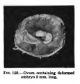 Keibel Mall 150.jpg 300 × 314; 14 KB
 Keibel Mall 151.jpg 690 × 784; 55 KB
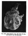 Keibel Mall 152.jpg 400 × 513; 27 KB
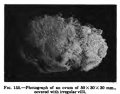 Keibel Mall 155.jpg 400 × 314; 19 KB
 Keibel Mall 156.jpg 400 × 313; 22 KB
 Keibel Mall 157.jpg 300 × 406; 17 KB
 Keibel Mall 158.jpg 630 × 201; 22 KB
 Keibel Mall 159.jpg 639 × 305; 49 KB
 Keibel Mall 160.jpg 337 × 400; 27 KB
 Keibel Mall 161.jpg 570 × 370; 31 KB
 Keibel Mall 163.jpg 369 × 524; 36 KB
 Keibel Mall 166.jpg 706 × 579; 99 KB
 Keibel Mall 167.jpg 702 × 495; 37 KB
 Keibel Mall 168.jpg 677 × 653; 83 KB
 Keibel Mall 308.jpg 677 × 578; 43 KB
 Keibel Mall 309.jpg 687 × 479; 52 KB
 Keibel Mall 310.jpg 686 × 760; 161 KB
 Keibel Mall 311.jpg 700 × 906; 181 KB
 Keibel Mall 314.jpg 701 × 625; 89 KB
 Keibel Mall 315.jpg 736 × 433; 84 KB
 Keibel Mall 316.jpg 716 × 327; 57 KB
 Keibel Mall 317.jpg 688 × 376; 53 KB
 Keibel Mall 318.jpg 687 × 423; 79 KB
 Keibel Mall 319.jpg 300 × 352; 27 KB
 Keibel Mall 320.jpg 693 × 313; 33 KB
 Keibel Mall 321.jpg 746 × 651; 122 KB
 Keibel Mall 322.jpg 735 × 717; 133 KB
 Keibel Mall 323.jpg 700 × 211; 27 KB
 Keibel Mall 324.jpg 722 × 377; 47 KB
 Keibel Mall 421.jpg 410 × 800; 46 KB
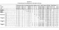
 Keibel Mall Table-embryo and fetal age.jpg 641 × 1,002; 168 KB
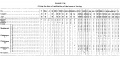
 Keibel Mall Table-embryo length.jpg 694 × 419; 74 KB
 Keibel1908 fig01.jpg 2,880 × 1,876; 614 KB
 Keibel1908 fig02.jpg 1,486 × 800; 164 KB
 Keibel1908 fig03.jpg 904 × 800; 72 KB
 Keibel1908 fig04.jpg 1,408 × 1,000; 265 KB
 Keibel1908 fig05.jpg 1,493 × 1,000; 227 KB
 Keibel1908 fig06.jpg 1,684 × 2,000; 582 KB
 Keibel1908 fig07.jpg 1,246 × 1,000; 199 KB
 Keibel1908 fig08.jpg 1,613 × 1,000; 252 KB
 Keibel1908 fig09.jpg 1,310 × 1,600; 443 KB
 Keibel1908 fig10.jpg 814 × 800; 144 KB
 Keibel1908 fig11.jpg 1,561 × 1,000; 151 KB
 Keibel1908 fig12.jpg 1,658 × 2,000; 537 KB
 Keibel1908 fig13.jpg 1,625 × 2,000; 461 KB
 Keibel1908 fig14.jpg 713 × 600; 65 KB
 Keibel1908 fig15.jpg 1,895 × 2,000; 399 KB
 Keibel1908 fig16a.jpg 2,556 × 2,990; 835 KB
 Keibel1908 fig16b.jpg 2,075 × 2,000; 671 KB
 Keibel1908 fig17.jpg 1,200 × 531; 103 KB
 Keibel1908 fig18.jpg 1,756 × 2,000; 461 KB
 Keibel1908 fig19.jpg 1,251 × 800; 171 KB
 Keibel1908 fig20.jpg 1,160 × 1,200; 153 KB
 Keibel1908 fig21.jpg 1,690 × 1,200; 337 KB
 Keibel1908 fig22.jpg 1,889 × 2,000; 548 KB
 Keibel1908 fig23.jpg 680 × 800; 76 KB
 Keibel1908 fig24.jpg 1,602 × 2,000; 514 KB
 Keibel1908 fig25.jpg 1,777 × 2,000; 552 KB
 Keibel1908 fig26.jpg 649 × 691; 60 KB
 Keibel1908 fig27.jpg 806 × 800; 77 KB
 Keibel1908 fig28.jpg 1,682 × 2,000; 589 KB
 Keibel1908 fig29.jpg 680 × 800; 76 KB
 Keibel1908 fig30a.jpg 1,637 × 2,000; 458 KB
 Keibel1908 fig30b.jpg 1,923 × 1,500; 396 KB
 Keibel1908 fig31.jpg 1,445 × 1,000; 258 KB
 Keibel1908 fig32.jpg 1,211 × 1,000; 167 KB
 Keibel1908 fig33.jpg 1,814 × 2,000; 430 KB
 Keibel1908 fig34a.jpg 1,953 × 1,500; 377 KB
 Keibel1908 fig34b.jpg 1,544 × 1,500; 336 KB
 Keibel1908 fig35.jpg 1,279 × 800; 137 KB
 Keibel1908 fig36.jpg 1,391 × 1,000; 209 KB
 Keibel1908 fig37.jpg 1,618 × 1,500; 358 KB
 Keibel1908 fig38.jpg 767 × 600; 45 KB
 Keibel1908 fig39.jpg 1,684 × 800; 192 KB
 Keibel1908 fig40.jpg 891 × 700; 66 KB
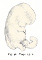
 Keibel1908 fig41.jpg 556 × 800; 33 KB
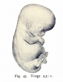
 Keibel1908 fig42.jpg 569 × 800; 51 KB
 Keibel1908 fig43.jpg 615 × 800; 46 KB
 Keibel1908 fig44.jpg 1,527 × 800; 136 KB
 Keibel1908 plate01.jpg 1,447 × 2,000; 437 KB
 Keibel1908 plate02.jpg 1,448 × 2,000; 378 KB
 Keibel1908 plate03.jpg 1,448 × 2,000; 596 KB
 Keibel1908 plate04.jpg 1,448 × 2,000; 514 KB
 Keibel1908 plate05.jpg 1,448 × 2,000; 493 KB
 Keibel1908 plate06.jpg 1,448 × 2,000; 505 KB
 Keibel1908 titlepage.jpg 800 × 1,200; 117 KB
 Kyoto16834 stage17-umbilicus.jpg 1,536 × 1,316; 137 KB
 Kyoto25783 stage18-dorsal.jpg 1,536 × 2,048; 152 KB
 Kyoto25783 stage18-left+yolk.jpg 1,536 × 2,048; 207 KB
 Kyoto25783 stage18-left.jpg 1,536 × 2,048; 226 KB
 Kyoto25783 stage18-right+yolk.jpg 1,536 × 2,048; 219 KB
 Kyoto25783 stage18-right.jpg 1,536 × 2,048; 228 KB
 Kyoto25783 stage18-ventral.jpg 1,536 × 2,048; 169 KB
 Kyoto731 Stage16-01.jpg 908 × 681; 42 KB
 Kyoto731 Stage16-02.jpg 908 × 681; 59 KB
 Kyoto940 stage21-01.jpg 1,288 × 2,048; 356 KB
 Kyoto940 stage21-02.jpg 1,024 × 1,024; 177 KB
 Kyoto940 stage21-03.jpg 1,024 × 1,024; 179 KB
 Kyoto940 stage21-04.jpg 1,288 × 2,048; 276 KB
 Kyoto940 stage21-05.jpg 1,024 × 1,024; 132 KB
 Kyoto940 stage21-06.jpg 1,024 × 1,024; 146 KB
 Kyoto940 stage21-07.jpg 1,288 × 2,048; 370 KB
 Kyoto940 stage21-08.jpg 1,024 × 1,024; 178 KB
 Kyoto940 stage21-09.jpg 1,024 × 1,024; 193 KB
 Kyoto940 stage21-10.jpg 1,288 × 2,048; 349 KB
 Kyoto940 stage21-11.jpg 1,024 × 1,024; 177 KB
 Kyoto940 stage21-12.jpg 1,024 × 1,024; 174 KB
 Mall1891 Fig01.jpg 623 × 576; 99 KB
 Mall1891 Fig02.jpg 600 × 347; 45 KB
 Mall1891 Plate01Fig01.jpg 623 × 831; 70 KB
 Mall1891 Plate01Fig02.jpg 623 × 831; 61 KB
 Mall1891 Plate02Fig01.jpg 623 × 831; 125 KB
 Mall1891 Plate02Fig02.jpg 623 × 831; 76 KB
 Mall1905 fig3.jpg 902 × 1,000; 155 KB
 Mall1906 fig01.jpg 1,158 × 1,569; 133 KB
 Mall1906 fig02.jpg 1,317 × 1,532; 205 KB
 Mall1906 fig03.jpg 797 × 800; 47 KB
 Mall1906 fig04.jpg 1,114 × 1,044; 137 KB
 Mall1906 fig05.jpg 561 × 1,055; 45 KB
 Mall1906 fig06.jpg 522 × 966; 41 KB
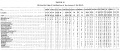
 Mall1906 table01.jpg 1,319 × 1,154; 391 KB
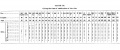
 Mall1906 table02.jpg 2,000 × 858; 404 KB
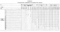
 Mall1906 table03.jpg 2,118 × 878; 295 KB
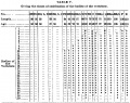
 Mall1906 table04.jpg 2,113 × 1,125; 463 KB
 Mall1906 table05.jpg 1,325 × 1,048; 314 KB
 Mall1906 table06.jpg 2,117 × 1,074; 469 KB
 Mall1906 table07.jpg 2,118 × 1,034; 432 KB
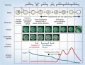 Model human blastocyst development.jpg 946 × 726; 84 KB
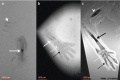 MRI Human Embryo - upper limb 01.jpg 1,418 × 940; 106 KB
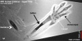 MRI Human Embryo - upper limb 02.jpg 1,000 × 497; 74 KB
 ORahilly1987 fig20-2.jpg 600 × 549; 46 KB
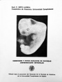 Orts Llorca Madrid embryo catalogue.jpg 439 × 585; 32 KB
 Placental membranes.jpg 600 × 450; 99 KB
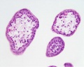 Placental villi 1.jpg 1,280 × 1,024; 77 KB
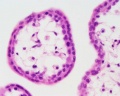 Placental villi 2.jpg 1,280 × 1,024; 70 KB
 Placental villi.jpg 1,280 × 1,024; 199 KB
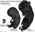 Size comparison embryo-fetus actual.jpg 194 × 178; 14 KB
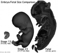 Size comparison embryo-fetus.jpg 327 × 300; 29 KB
 St. John's Wort.jpg 240 × 180; 9 KB
 Stage 19 ear.jpg 1,200 × 786; 116 KB
 Stage 22 image 001.jpg 1,000 × 650; 113 KB
 Stage 22 image 002.jpg 1,000 × 653; 103 KB
 Stage 22 image 003.jpg 1,000 × 667; 150 KB
 Stage 22 image 004.jpg 1,000 × 685; 111 KB
 Stage 22 image 005.jpg 1,000 × 645; 103 KB
 Stage 22 image 006.jpg 1,000 × 637; 107 KB
 Stage 22 image 007.jpg 1,000 × 632; 107 KB
 Stage 22 image 008-eye.jpg 1,200 × 1,059; 555 KB
 Stage 22 image 008.jpg 1,200 × 1,161; 322 KB
 Stage 22 image 009.jpg 1,000 × 638; 106 KB
 Stage 22 image 010.jpg 1,000 × 640; 103 KB
 Stage 22 image 011.jpg 1,200 × 1,109; 312 KB
 Stage 22 image 012.jpg 1,000 × 645; 107 KB
 Stage 22 image 013.jpg 1,000 × 645; 102 KB
 Stage 22 image 014.jpg 1,000 × 635; 88 KB
 Stage 22 image 015.jpg 1,200 × 1,075; 116 KB
 Stage 22 image 016.jpg 1,000 × 637; 75 KB
 Stage 22 image 017.jpg 600 × 493; 75 KB
 Stage 22 image 018.jpg 1,000 × 640; 72 KB
 Stage 22 image 019.jpg 1,000 × 642; 83 KB
 Stage 22 image 020.jpg 1,000 × 637; 92 KB
 Stage 22 image 021.jpg 1,000 × 635; 91 KB
 Stage 22 image 022.jpg 1,000 × 638; 100 KB
 Stage 22 image 023.jpg 1,000 × 633; 106 KB
 Stage 22 image 024.jpg 1,000 × 645; 119 KB
 Stage 22 image 025.jpg 1,200 × 1,079; 236 KB
 Stage 22 image 026.jpg 1,000 × 648; 100 KB
 Stage 22 image 027.jpg 1,000 × 655; 126 KB
 Stage 22 image 028.jpg 1,000 × 662; 121 KB
 Stage 22 image 029.jpg 1,000 × 637; 116 KB


