Category:Head
From Embryology
Subcategories
This category has the following 8 subcategories, out of 8 total.
Pages in category 'Head'
The following 189 pages are in this category, out of 189 total.
2
A
B
- BGD Lecture - Face and Ear Development
- BGD Practical - Face and Ear Quiz
- BGD Practical - Face Quiz
- Template:BGDB Face
- BGDB Face and Ear - Abnormalities
- BGDB Face and Ear - Early Embryo
- BGDB Face and Ear - Fetal
- BGDB Face and Ear - Late Embryo
- BGDB Face and Ear - Postnatal
- BGDB Face and Ear - Trilaminar Embryo
- BGDB Practical - Face and Ear Development
- Talk:BGDB Practical - Face and Ear Development
- BGDB Practical - Face and Ear Development Interactive
- Template:BGDB Practical 6 - Abnormalities Interactive
- Template talk:BGDB Practical 6 - Abnormalities Interactive
- Template:BGDB Practical 6 - Early Embryo Interactive
- Template:BGDB Practical 6 - Fetal Interactive
- Template:BGDB Practical 6 - Late Embryo Interactive
- Template:BGDB Practical 6 - Postnatal Interactive
- Template:BGDB Practical 6 - Trilaminar Embryo Interactive
- Book - Contributions to Embryology Carnegie Institution No.48
- Book - Contributions to Embryology Carnegie Institution No.55
- Book - Human Embryology and Morphology 1
- Book - Text-Book of Embryology 8
- Buccopharyngeal membrane
- Template:Buccopharyngeal membrane
F
H
- Template:Head
- Template:Head abnormalities
- Template:Head and Neck Links
- Head Development
- Head Development - Abnormalities
- Head Development - Carnegie Stage 22
- Template:Head Links
- Template:Holoprosencephaly
- Human Embryology and Morphology 12
- Human Embryology and Morphology 17
- Template:Human Eyelid timeline table
- Human System Development
M
P
- Template:Palate
- Palate Development
- Palate Development 1 Movie
- Palate Development 2 Movie
- Paper - A human foetus exhibiting iniencephaly and other abnormalities (1922)
- Paper - An anencephalic embryo of 25 mm CRL
- Paper - Contribution to the structure and development of the vertebrate head
- Paper - Contribution to the structure and development of the vertebrate head 1
- Paper - Contribution to the structure and development of the vertebrate head 2
- Paper - Contribution to the structure and development of the vertebrate head 3
- Paper - Description of a reconstruction of the head of a thirty-millimetre embryo (1910)
- Paper - Development of olfactory and related structures in staged human embryos
- Paper - Evolutionary factors in the production of pharyngeal diverticula
- Paper - Extroversion of the cerebral hemispheres in a human embryo (1934)
- Paper - Malformations of the human body from a new point of view 1+2
- Paper - Normal facial growth in children (1937)
- Paper - Observations on metopism (1917)
- Paper - On the premature obliteration of sutures in the human skull (1915)
- Paper - On the presence of a series of ectodermal placodes in the head region of a sparrow embryo (1928)
- Paper - On the relation of the chorda dorsalis to the anlage of the pharyngeal bursa or median pharyngeal recess (1912)
- Paper - On the relation of the head chorda to the pharyngeal epithelium in the pig embryo
- Paper - On the so-called ultimobranchial body of the mammalian embryo (1915)
- Paper - Pouches of the pharynx and oesophagus with special reference to the embryological and morphological aspects
- Paper - Preliminary note on the skull of a human fetus of 43 mm greatest length
- Paper - Primary neuromeres and head segmentation (1922)
- Paper - The Anatomy of the Head End of a 20 mm Human Embryo
- Paper - The aortic arch derivatives in human adult (1951)
- Paper - The cartilaginous skull of a human embryo twenty-one millimeters in length (1920)
- Paper - The chondrocranium of a 20 mm human embryo
- Paper - The development of the anterior post-otic somites in the rabbit
- Paper - The development of the cerebrospinal fluid spaces and choroid plexuses in the chick (1937)
- Paper - The development of the cranial arteries in the human embryo
- Paper - The development of the first branchial arch in man and the fate of Meckel's cartilage
- Paper - The development of the human chin (1917)
- Paper - The Development of the Human Mandibular Joint
- Paper - The development of the human pharynx
- Paper - The Development of the Nose and of the Pharynx and its Derivatives in Man
- Paper - The development of the sphenoidal sinus in man and its homology in mammals (1927)
- Paper - The development of the subcutaneous vascular plexus in the head of the human embryo (1923)
- Paper - The developmental alterations in the vascular system of the brain of the human embryo (1921)
- Paper - The genesis, development, and adult anatomy of the nasofrontal region in man
- Paper - The lateral wall of the cavum nasi in man, with especial reference to the various developmental stages
- Paper - The Long Fox lecture - The development of the human skull (1910)
- Paper - The pharyngeal pouches and their derivatives in the mammalia
- Paper - The prenatal development of the human temporomandibular joint
- Paper - The primordial cranium of erinaceus europaeus (1918)
- Paper - The primordial cranium of Erinaceus europaeus (1918)
- Paper - The primordial cranium of microtus amphibius (water-rat), as determined by sections and a model of the 25-mm stage (1917)
- Paper - The primordial cranium of miniopterus schreibersi at the 17 millimetre total length stage (1919)
- Paper - The second visceral arch and groove in the tubo-tympanic region
- Paper - The sinus maxillaris and its relations in the embryo, child, and adult man
- Paper - Three demonstrations on congenital melformations of palate, face, and neck
- Paper - Transformation of the aortic-arch system during the development of the human embryo (1922)
- Paper - Vertebrate cephalogenesis 1 (1890)
- Paper - Vertebrate cephalogenesis 2 (1892)
- Paper - Vertebrate cephalogenesis 4 (1919)
- Paper The development of the subcutaneous vascular plexus in the head of the human embryo (1923)
- Template:Pharyngeal arch
- Template:Pharyngeal Arch table
R
- Template:Ref-Allis1938
- Template:Ref-Ayers1890
- Template:Ref-Ayers1892
- Template:Ref-Bartelmez1924
- Template:Ref-Boulgakow1926
- Template:Ref-Dickie1914
- Template:Ref-Fawcett1910head
- Template:Ref-Fawcett1911
- Template:Ref-Finley1922
- Template:Ref-Finley1923
- Template:Ref-Huber1912
- Template:Ref-JacksonAJ1935
- Template:Ref-Keith1909
- Template:Ref-Keith1932a
- Template:Ref-Keith1932b
- Template:Ref-Kingsbury1915a
- Template:Ref-Kingsbury1915b
- Template:Ref-Locy1895
- Template:Ref-Mann1921
- Template:Ref-Moffatt1957
- Template:Ref-Padget1956
- Template:Ref-Rand1917
- Template:Ref-Raven1933
- Template:Ref-Schaeffer1910
- Template:Ref-Schaeffer1916
- Template:Ref-Stunkard1922
- Template:Ref-Wallis1917
- Template:Ref-Young1937
- Template:Reichert’s cartilage
S
Media in category 'Head'
The following 108 files are in this category, out of 308 total.
(previous page) (next page)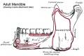 Musculoskeletal- adult mandible.jpg 600 × 402; 58 KB
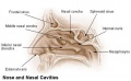 Nasal cavities.jpg 500 × 307; 19 KB
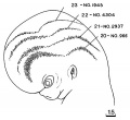 ORahilly1987 fig20-2.jpg 600 × 549; 46 KB
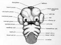 Patten041.jpg 683 × 504; 59 KB
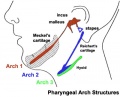 Pharyngeal arch cartilages.jpg 400 × 324; 26 KB
 Pharyngeal arch structure cartoon.gif 480 × 177; 5 KB
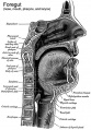 Pharynx cartoon.jpg 418 × 600; 88 KB
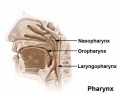 Pharynx.jpg 434 × 340; 13 KB
 PMID28514120-Chen et al-2017.pdf ; 2.87 MB
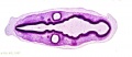
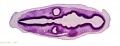
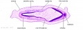
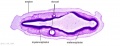
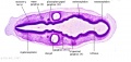
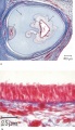 Proboscis histology.jpg 600 × 1,041; 166 KB
 Rugh 135.jpg 1,034 × 800; 190 KB
 Skull - microcephaly 01.jpg 750 × 1,000; 75 KB
 Stage 13 image 001.jpg 1,000 × 357; 44 KB
 Stage 13 image 002.jpg 1,000 × 359; 52 KB
 Stage 13 image 003.jpg 1,000 × 436; 71 KB
 Stage 13 image 004.jpg 1,000 × 386; 65 KB
 Stage 13 image 050.jpg 1,000 × 370; 52 KB
 Stage 13 image 051.jpg 1,000 × 382; 55 KB
 Stage 13 image 052.jpg 1,000 × 476; 81 KB
 Stage 13 image 053.jpg 1,000 × 423; 76 KB
 Stage 22 image 050.jpg 1,000 × 650; 128 KB
 Stage 22 image 051.jpg 1,000 × 653; 113 KB
 Stage 22 image 052.jpg 1,000 × 665; 177 KB
 Stage 22 image 053.jpg 1,000 × 687; 132 KB
 Stage 22 image 054.jpg 1,000 × 636; 119 KB
 Stage 22 image 055.jpg 1,000 × 632; 145 KB
 Stage 22 image 056.jpg 1,000 × 635; 138 KB
 Stage 22 image 065.jpg 1,000 × 637; 100 KB
 Stage 22 image 205.jpg 1,112 × 909; 261 KB
 Stage 22 image 206.jpg 1,200 × 754; 245 KB
 Stage 22 image 207.jpg 1,200 × 753; 208 KB
 Stage 22 image 208.jpg 1,200 × 903; 368 KB
 Stage 22 image 209.jpg 1,200 × 808; 305 KB
 Stage 22 image 211.jpg 1,200 × 760; 242 KB
 Stage 22 image 212.jpg 1,200 × 753; 269 KB
 Stage 22 image 222.jpg 1,180 × 999; 225 KB
 Stage 22 image 322.jpg 1,180 × 999; 254 KB
 Stage 22 vomeronasal organ.jpg 600 × 554; 125 KB
 Stage10 sem11.jpg 1,000 × 636; 76 KB
 Stage11 sem2.jpg 976 × 1,000; 188 KB
 Stage11 sem3.jpg 936 × 1,000; 68 KB
 Stage11 sem3a.jpg 749 × 800; 44 KB
 Stage11 sem3b.gif 468 × 500; 182 KB
 Stage11 sem3b.jpg 468 × 500; 45 KB
 Stage11 sem3c.jpg 375 × 400; 16 KB
 Stage11 sem4.jpg 808 × 1,000; 129 KB
 Stage12 sem6.jpg 1,620 × 1,612; 190 KB
 Stage12 sem6a.jpg 1,000 × 995; 100 KB
 Stage12 sem6b.jpg 800 × 796; 74 KB
 Stage12 sem6c.jpg 600 × 597; 49 KB
 Stage13 face ventral view01.jpg 1,290 × 2,048; 152 KB
 Stage13 oral cavity floor01.jpg 1,315 × 2,048; 234 KB
 Stage13 oral cavity floor02.jpg 1,315 × 2,048; 371 KB
 Stage13 pharyngeal arch excerpts.gif 600 × 300; 86 KB
 Stage13 sem1.jpg 1,000 × 886; 80 KB
 Stage13 sem1a.jpg 800 × 709; 57 KB
 Stage13 sem1b.jpg 600 × 532; 36 KB
 Stage13 sem1c.jpg 400 × 355; 18 KB
 Stage13 sem2.jpg 848 × 1,000; 84 KB
 Stage13 sem2a.jpg 678 × 800; 59 KB
 Stage13 sem2b.jpg 509 × 600; 36 KB
 Stage13 sem2c.jpg 339 × 400; 25 KB
 Stage13 sem2l.jpg 848 × 1,000; 121 KB
 Stage13 spinal cord01.jpg 807 × 637; 51 KB
 Stage13 spinal cord02.jpg 807 × 637; 58 KB
 Stage14 sem2al.jpg 504 × 800; 68 KB
 Stage14 sem2cl.jpg 252 × 400; 22 KB
 Stage14 sem2l.jpg 630 × 1,000; 96 KB
 Stage16 cleft palate.jpg 337 × 400; 14 KB
 Stage16-18 face.jpg 800 × 393; 34 KB
 Stage17 bf10.jpg 1,375 × 2,048; 134 KB
 Stage17 bf11.jpg 1,375 × 2,048; 166 KB
 Streeter-plate01.jpg 1,200 × 950; 138 KB
 Streeter-plate02.jpg 1,200 × 871; 180 KB
 Streeter-plate03.jpg 1,398 × 1,000; 192 KB
 Streeter-plate04.jpg 1,301 × 1,000; 214 KB
 Streeter-plate05.jpg 833 × 1,000; 115 KB
 Streeter1920table5.jpg 1,134 × 569; 74 KB
 Streeter1921 fig01.jpg 981 × 1,000; 103 KB
 Streeter1921 fig02.jpg 1,286 × 1,000; 138 KB
 Streeter1921 fig03.jpg 1,154 × 1,000; 142 KB
 Streeter1921 fig04.jpg 1,030 × 1,000; 101 KB
 Streeter1921 fig05.jpg 600 × 342; 19 KB
 Streeter1921 fig06.jpg 1,007 × 1,000; 106 KB
 Streeter1921 fig07-09.jpg 1,200 × 494; 92 KB
 Streeter1921 fig10.jpg 1,108 × 1,000; 95 KB
 Streeter1921 fig11.jpg 908 × 751; 103 KB
 Streeter1921 fig12.jpg 960 × 745; 117 KB
 Streeter1921 fig13.jpg 800 × 600; 45 KB
 Streeter1921 fig14.jpg 800 × 600; 55 KB
 Streeter1921 fig15.jpg 800 × 600; 54 KB
 Streeter1921 fig16.jpg 800 × 600; 60 KB
 Streeter1921 fig17.jpg 800 × 600; 60 KB
 Streeter1921 fig18.jpg 800 × 600; 48 KB
 Streeter1921 fig19.jpg 800 × 600; 48 KB
 Streeter1921 fig20.jpg 800 × 600; 40 KB
 Streeter1921 fig21.jpg 800 × 600; 40 KB
 Streeter1921 fig22.jpg 917 × 1,000; 157 KB
 Streeter1921 fig23.jpg 917 × 1,000; 152 KB
 Streeter1921 fig24.jpg 921 × 1,000; 130 KB
 Streeter1921 fig25.jpg 921 × 1,000; 118 KB
 Streeter1921 fig26.jpg 1,328 × 1,000; 228 KB
 Streeter1921 fig27.jpg 949 × 1,000; 138 KB
 Streeter1921 table01.jpg 800 × 347; 44 KB
 Unilateral cleft palate.jpg 214 × 300; 12 KB
 Waterston04.jpg 500 × 763; 75 KB
 Waterston06.jpg 720 × 538; 92 KB
 Waterston15.jpg 500 × 674; 65 KB


