Category:Head
From Embryology
Subcategories
This category has the following 8 subcategories, out of 8 total.
Pages in category 'Head'
The following 189 pages are in this category, out of 189 total.
2
A
B
- BGD Lecture - Face and Ear Development
- BGD Practical - Face and Ear Quiz
- BGD Practical - Face Quiz
- Template:BGDB Face
- BGDB Face and Ear - Abnormalities
- BGDB Face and Ear - Early Embryo
- BGDB Face and Ear - Fetal
- BGDB Face and Ear - Late Embryo
- BGDB Face and Ear - Postnatal
- BGDB Face and Ear - Trilaminar Embryo
- BGDB Practical - Face and Ear Development
- Talk:BGDB Practical - Face and Ear Development
- BGDB Practical - Face and Ear Development Interactive
- Template:BGDB Practical 6 - Abnormalities Interactive
- Template talk:BGDB Practical 6 - Abnormalities Interactive
- Template:BGDB Practical 6 - Early Embryo Interactive
- Template:BGDB Practical 6 - Fetal Interactive
- Template:BGDB Practical 6 - Late Embryo Interactive
- Template:BGDB Practical 6 - Postnatal Interactive
- Template:BGDB Practical 6 - Trilaminar Embryo Interactive
- Book - Contributions to Embryology Carnegie Institution No.48
- Book - Contributions to Embryology Carnegie Institution No.55
- Book - Human Embryology and Morphology 1
- Book - Text-Book of Embryology 8
- Buccopharyngeal membrane
- Template:Buccopharyngeal membrane
F
H
- Template:Head
- Template:Head abnormalities
- Template:Head and Neck Links
- Head Development
- Head Development - Abnormalities
- Head Development - Carnegie Stage 22
- Template:Head Links
- Template:Holoprosencephaly
- Human Embryology and Morphology 12
- Human Embryology and Morphology 17
- Template:Human Eyelid timeline table
- Human System Development
M
P
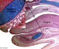
- Template:Palate
- Palate Development
- Palate Development 1 Movie
- Palate Development 2 Movie
- Paper - A human foetus exhibiting iniencephaly and other abnormalities (1922)
- Paper - An anencephalic embryo of 25 mm CRL
- Paper - Contribution to the structure and development of the vertebrate head
- Paper - Contribution to the structure and development of the vertebrate head 1
- Paper - Contribution to the structure and development of the vertebrate head 2
- Paper - Contribution to the structure and development of the vertebrate head 3
- Paper - Description of a reconstruction of the head of a thirty-millimetre embryo (1910)
- Paper - Development of olfactory and related structures in staged human embryos
- Paper - Evolutionary factors in the production of pharyngeal diverticula
- Paper - Extroversion of the cerebral hemispheres in a human embryo (1934)
- Paper - Malformations of the human body from a new point of view 1+2
- Paper - Normal facial growth in children (1937)
- Paper - Observations on metopism (1917)
- Paper - On the premature obliteration of sutures in the human skull (1915)
- Paper - On the presence of a series of ectodermal placodes in the head region of a sparrow embryo (1928)
- Paper - On the relation of the chorda dorsalis to the anlage of the pharyngeal bursa or median pharyngeal recess (1912)
- Paper - On the relation of the head chorda to the pharyngeal epithelium in the pig embryo
- Paper - On the so-called ultimobranchial body of the mammalian embryo (1915)
- Paper - Pouches of the pharynx and oesophagus with special reference to the embryological and morphological aspects
- Paper - Preliminary note on the skull of a human fetus of 43 mm greatest length
- Paper - Primary neuromeres and head segmentation (1922)
- Paper - The Anatomy of the Head End of a 20 mm Human Embryo
- Paper - The aortic arch derivatives in human adult (1951)
- Paper - The cartilaginous skull of a human embryo twenty-one millimeters in length (1920)
- Paper - The chondrocranium of a 20 mm human embryo
- Paper - The development of the anterior post-otic somites in the rabbit
- Paper - The development of the cerebrospinal fluid spaces and choroid plexuses in the chick (1937)
- Paper - The development of the cranial arteries in the human embryo
- Paper - The development of the first branchial arch in man and the fate of Meckel's cartilage
- Paper - The development of the human chin (1917)
- Paper - The Development of the Human Mandibular Joint
- Paper - The development of the human pharynx
- Paper - The Development of the Nose and of the Pharynx and its Derivatives in Man
- Paper - The development of the sphenoidal sinus in man and its homology in mammals (1927)
- Paper - The development of the subcutaneous vascular plexus in the head of the human embryo (1923)
- Paper - The developmental alterations in the vascular system of the brain of the human embryo (1921)
- Paper - The genesis, development, and adult anatomy of the nasofrontal region in man
- Paper - The lateral wall of the cavum nasi in man, with especial reference to the various developmental stages
- Paper - The Long Fox lecture - The development of the human skull (1910)
- Paper - The pharyngeal pouches and their derivatives in the mammalia
- Paper - The prenatal development of the human temporomandibular joint
- Paper - The primordial cranium of erinaceus europaeus (1918)
- Paper - The primordial cranium of Erinaceus europaeus (1918)
- Paper - The primordial cranium of microtus amphibius (water-rat), as determined by sections and a model of the 25-mm stage (1917)
- Paper - The primordial cranium of miniopterus schreibersi at the 17 millimetre total length stage (1919)
- Paper - The second visceral arch and groove in the tubo-tympanic region
- Paper - The sinus maxillaris and its relations in the embryo, child, and adult man
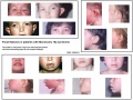
- Paper - Three demonstrations on congenital melformations of palate, face, and neck
- Paper - Transformation of the aortic-arch system during the development of the human embryo (1922)
- Paper - Vertebrate cephalogenesis 1 (1890)
- Paper - Vertebrate cephalogenesis 2 (1892)
- Paper - Vertebrate cephalogenesis 4 (1919)
- Paper The development of the subcutaneous vascular plexus in the head of the human embryo (1923)
- Template:Pharyngeal arch
- Template:Pharyngeal Arch table
R
- Template:Ref-Allis1938
- Template:Ref-Ayers1890
- Template:Ref-Ayers1892
- Template:Ref-Bartelmez1924
- Template:Ref-Boulgakow1926
- Template:Ref-Dickie1914
- Template:Ref-Fawcett1910head
- Template:Ref-Fawcett1911
- Template:Ref-Finley1922
- Template:Ref-Finley1923
- Template:Ref-Huber1912
- Template:Ref-JacksonAJ1935
- Template:Ref-Keith1909
- Template:Ref-Keith1932a
- Template:Ref-Keith1932b
- Template:Ref-Kingsbury1915a
- Template:Ref-Kingsbury1915b
- Template:Ref-Locy1895
- Template:Ref-Mann1921
- Template:Ref-Moffatt1957
- Template:Ref-Padget1956
- Template:Ref-Rand1917
- Template:Ref-Raven1933
- Template:Ref-Schaeffer1910
- Template:Ref-Schaeffer1916
- Template:Ref-Stunkard1922
- Template:Ref-Wallis1917
- Template:Ref-Young1937
- Template:Reichert’s cartilage
S
Media in category 'Head'
The following 200 files are in this category, out of 308 total.
(previous page) (next page) Bailey096.jpg 693 × 501; 59 KB
 Bailey097.jpg 776 × 674; 71 KB
 Bailey098.jpg 680 × 432; 47 KB
 Bailey099.jpg 704 × 464; 52 KB
 Bailey132+133.jpg 940 × 570; 101 KB
 Bailey132.jpg 466 × 413; 43 KB
 Bailey133.jpg 806 × 655; 85 KB
 Bailey135.jpg 940 × 965; 216 KB
 Bailey136.jpg 835 × 566; 114 KB
 Bailey137.jpg 672 × 539; 73 KB
 Bailey138.jpg 831 × 400; 62 KB
 Bailey139.jpg 961 × 671; 96 KB
 Bailey140.jpg 793 × 505; 58 KB
 Bailey141.jpg 761 × 323; 66 KB
 Bailey142.jpg 778 × 479; 72 KB
 Bailey191.jpg 863 × 509; 109 KB
 Bailey192.jpg 960 × 806; 133 KB
 Bailey193.jpg 747 × 848; 94 KB
 Baileytable02.jpg 884 × 1,109; 182 KB
 Bat-craniofacial development.jpg 600 × 720; 140 KB
 BGDB PracManual 2011 Practical 6.pdf ; 426 KB
 Bilateral cleft palate.jpg 214 × 300; 11 KB
 Chicken HH20 MyoR expression 01.jpg 960 × 917; 121 KB
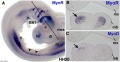 Chicken HH20 MyoR expression 02.jpg 1,200 × 620; 167 KB
 Cleft lip 01.jpg 585 × 438; 34 KB
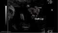 Cleft lip 02.jpg 641 × 362; 22 KB
 Cleft palate.jpg 653 × 776; 124 KB
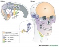 Cranial neural crest skeletal fate 01.jpg 800 × 633; 59 KB
 Cytomegalovirus induced micrognathia and abnormal skeletogenesis.jpg 1,200 × 1,105; 355 KB
 Dickie1914 fig01.jpg 463 × 611; 57 KB
 Dickie1914 fig02.jpg 443 × 526; 58 KB
 Dickie1914 fig03.jpg 672 × 516; 64 KB
 Dickie1914 fig04.jpg 755 × 578; 71 KB
 Dickie1914 fig05.jpg 613 × 567; 51 KB
 Dickie1914 fig06.jpg 504 × 607; 57 KB
 Dickie1914 fig07.jpg 407 × 577; 74 KB
 Dickie1914 fig08.jpg 538 × 523; 93 KB
 Dickie1914 fig09.jpg 351 × 526; 31 KB
 Dickie1914 fig10.jpg 337 × 525; 33 KB
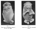
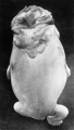
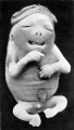
 Fetal facial expression 01.jpg 1,200 × 1,094; 122 KB
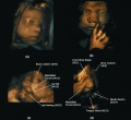 Fetal facial expression 02.jpg 1,914 × 1,762; 205 KB
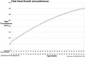 Fetal head growth circumference graph01.jpg 905 × 613; 58 KB
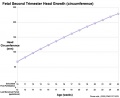 Fetal head growth circumference graph02.jpg 800 × 650; 44 KB
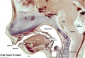
 Fetal head lateral.jpg 632 × 447; 34 KB
 Fetal head medial.jpg 632 × 447; 34 KB
 Fetal head section 01.jpg 1,200 × 821; 186 KB
 Fetal head section.jpg 1,200 × 821; 167 KB
 Fetal mandible and lower lip 01.jpg 1,028 × 557; 57 KB
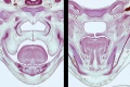
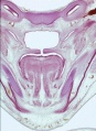
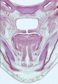
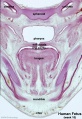
 Fetal week 10 hard palate 01.jpg 800 × 532; 77 KB
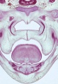
 Fetal week 10 hard palate 02.jpg 398 × 633; 66 KB
 Fetal week 10 hard palate 03.jpg 600 × 450; 122 KB
 Fetal week 10 hard palate 04.jpg 1,198 × 795; 196 KB
 Fetal week 10 hard palate 06.jpg 534 × 778; 88 KB
 Fetal week 10 hard palate 07.jpg 534 × 778; 97 KB
 Fetal week 10 palate 01.gif 534 × 778; 1.14 MB
 Fetal week 10 palate 01.mp4 ; 427 KB
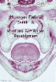
 Fetal week 10 palate icon.jpg 534 × 778; 100 KB
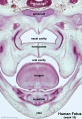
 Fetal week 10 soft palate 01.jpg 571 × 784; 95 KB
 Fetal week 10 soft palate 02.jpg 534 × 778; 87 KB
 Fetal week 10 soft palate 03.jpg 534 × 778; 95 KB
 Fetal week 14 head bone lateral 01.jpg 1,000 × 773; 107 KB
 Fetal week 9 hard palate fusion 01.jpg 661 × 400; 51 KB
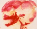
 Fetal week 9 head lateral 01.jpg 700 × 600; 78 KB
 Finley1923 fig01.jpg 494 × 968; 53 KB
 Finley1923 fig02.jpg 700 × 800; 77 KB
 Finley1923 fig03.jpg 600 × 512; 56 KB
 Finley1923 fig04.jpg 700 × 627; 61 KB
 Finley1923 fig05.jpg 674 × 800; 146 KB
 Finley1923 fig06.jpg 729 × 800; 95 KB
 Finley1923 fig07.jpg 314 × 800; 40 KB
 Finley1923 fig08.jpg 296 × 800; 32 KB
 Finley1923 fig09.jpg 456 × 800; 49 KB
 Finley1923 fig10.jpg 221 × 802; 17 KB
 Finley1923 fig11.jpg 432 × 800; 38 KB
 Finley1923 fig12.jpg 593 × 800; 48 KB
 Finley1923 fig13.jpg 594 × 800; 51 KB
 Finley1923 Plate 1.jpg 776 × 1,000; 151 KB
 Finley1923 Plate 2.jpg 864 × 1,200; 153 KB
 Foster113.jpg 927 × 512; 57 KB
 Frazer1926 fig01.jpg 1,200 × 804; 137 KB
 Frazer1926 fig02.jpg 991 × 833; 145 KB
 Frazer1926 fig03.jpg 1,200 × 804; 69 KB
 Frazer1926 fig04.jpg 1,200 × 804; 95 KB
 Frazer1926 fig05.jpg 1,000 × 643; 85 KB
 Frazer1926 fig06.jpg 563 × 811; 35 KB
 Frazer1926 fig07.gif 554 × 600; 134 KB
 Frazer1926 fig07.jpg 1,229 × 996; 95 KB
 Frazer1926 fig08.jpg 616 × 789; 38 KB
 Frazer1926 plate01.jpg 1,914 × 2,681; 469 KB
 Gray0043.jpg 800 × 496; 50 KB
 Gray0048.jpg 500 × 429; 30 KB
 Gray0050.jpg 600 × 332; 46 KB
 Gray0051.jpg 600 × 423; 91 KB
 Gray0379.jpg 713 × 600; 114 KB
 Gray0593.jpg 550 × 437; 66 KB
 Gray0603.jpg 800 × 480; 69 KB
 Gray0604.jpg 716 × 500; 65 KB
 Gray0605.jpg 614 × 600; 98 KB
 Gray0947.jpg 600 × 398; 56 KB
 Gray0994.jpg 600 × 861; 151 KB
 Gray1013.jpg 562 × 500; 56 KB
 Gray1014.jpg 619 × 600; 95 KB
 Gray1015.jpg 598 × 300; 62 KB
 Gray1016.jpg 252 × 400; 18 KB
 Gray1017.jpg 450 × 334; 31 KB
 Gray1018.jpg 390 × 400; 53 KB
 Gray1019.jpg 644 × 650; 87 KB
 Gray1020.jpg 667 × 400; 53 KB
 Gray1022.jpg 629 × 400; 45 KB
 Gray1023.jpg 585 × 400; 47 KB
 Gray1024.jpg 800 × 691; 117 KB
 Gray1027.jpg 598 × 600; 101 KB
 Gray1029.jpg 700 × 435; 75 KB
 Gray1030.jpg 481 × 800; 122 KB
 Gray1202.jpg 563 × 500; 63 KB
 Head - Treacher Collins Syndrome 01.jpg 291 × 391; 16 KB
 Head and heart muscle cartoon.jpg 874 × 800; 129 KB
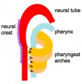 Head arches cartoon.jpg 394 × 402; 29 KB
 Hertwig286.jpg 642 × 800; 126 KB
 Human embryo head week 6 to 8.jpg 540 × 780; 66 KB
 Human fetal temporal bone and mandible 01.jpg 1,200 × 805; 170 KB
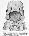
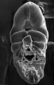
 Human holoprosencephaly cyclopia dissection.jpg 600 × 340; 37 KB
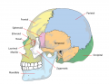 Human skull lateral simplified.png 740 × 576; 138 KB
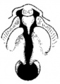
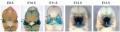
 Human stage16 face 01.jpg 500 × 504; 20 KB
 Human stage17 face 01.jpg 500 × 504; 21 KB
 Human stage18 face 01.jpg 500 × 504; 23 KB
 Human- fetal week 10 cerebellum A.jpg 347 × 284; 24 KB
 Human- fetal week 10 cerebellum B.jpg 347 × 284; 21 KB
 Human- fetal week 10 cerebellum C.jpg 347 × 284; 25 KB
 Human- fetal week 10 cerebellum D.jpg 347 × 284; 23 KB
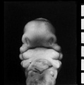
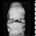
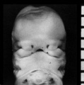
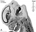 Human- fetal week 10 head A.jpg 600 × 544; 113 KB
 Human- fetal week 10 head A1.jpg 1,200 × 1,088; 159 KB
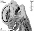
 Human- fetal week 10 head B.jpg 600 × 544; 66 KB
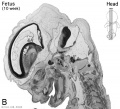
 Human- fetal week 10 head C.jpg 600 × 544; 118 KB
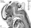
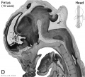 Human- fetal week 10 head D.jpg 600 × 544; 111 KB
 Hunter1934 fig01-02.jpg 1,594 × 1,308; 188 KB
 Hunter1934 fig01.jpg 600 × 1,062; 64 KB
 Hunter1934 fig02.jpg 565 × 1,000; 60 KB
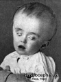 Hydrocephalus.jpg 320 × 432; 29 KB
 Keibel Mall 066-071.jpg 610 × 800; 58 KB
 Keibel Mall 068-069.jpg 1,000 × 358; 35 KB
 Keibel Mall 070-071.jpg 1,000 × 490; 45 KB
 Keibel Mall 2 257.jpg 869 × 800; 120 KB
 Keibel Mall 2 258.jpg 927 × 540; 78 KB
 Keibel Mall 2 334.jpg 1,000 × 332; 50 KB
 Keibel Mall 2 335.jpg 793 × 800; 61 KB
 Keibel Mall 2 336.jpg 777 × 800; 34 KB
 Keibel Mall 231.jpg 450 × 511; 21 KB
 Keibel Mall 232.jpg 450 × 511; 35 KB
 Keibel Mall 266.jpg 740 × 608; 77 KB
 Keibel Mall 367.jpg 857 × 850; 112 KB
 Keibel Mall 368.jpg 800 × 599; 63 KB
 Keibel Mall 370.jpg 900 × 740; 111 KB
 Keith1902 fig001.jpg 823 × 750; 75 KB
 Keith1902 fig002.jpg 932 × 800; 69 KB
 Keith1902 fig003.jpg 1,000 × 700; 113 KB
 Keith1902 fig004.jpg 580 × 314; 27 KB
 Keith1902 fig005.jpg 650 × 557; 36 KB
 Keith1902 fig006.jpg 1,000 × 689; 112 KB
 Keith1902 fig007.jpg 946 × 700; 80 KB
 Keith1902 fig008.jpg 925 × 700; 122 KB
 Keith1902 fig009.jpg 1,000 × 568; 78 KB
 Keith1902 fig010a-c.jpg 800 × 1,000; 112 KB
 Keith1902 fig010d.jpg 800 × 581; 66 KB
 Keith1902 fig011.jpg 954 × 500; 66 KB
 Keith1902 fig012.jpg 875 × 500; 75 KB
 Keith1902 fig013.jpg 662 × 800; 67 KB
 Keith1902 fig014.jpg 707 × 1,000; 98 KB
 Keith1902 fig015a.jpg 971 × 600; 74 KB
 Keith1902 fig015b.jpg 1,000 × 719; 97 KB
 Keith1902 fig021a.jpg 923 × 700; 0 bytes
 Keith1902 fig021b.jpg 800 × 727; 75 KB
 Keith1902 fig022.jpg 807 × 800; 92 KB
 Keith1902 fig023.jpg 800 × 411; 85 KB
 Keith1902 fig024.jpg 600 × 433; 47 KB
 Keith1902 fig025.jpg 876 × 800; 61 KB
 Keith1902 fig026.jpg 1,000 × 728; 92 KB
 Keith1902 fig027.jpg 881 × 800; 93 KB
 Keith1902 fig028.jpg 707 × 800; 77 KB
 Keith1902 fig029.jpg 872 × 600; 81 KB
 Keith1902 fig030.jpg 803 × 700; 87 KB
 Keith1902 fig031.jpg 1,000 × 557; 151 KB
 Keith1902 fig032.jpg 1,000 × 496; 81 KB
 Keith1902 fig033.jpg 971 × 800; 139 KB
 Keith1902 fig034.jpg 921 × 700; 110 KB
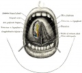
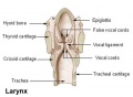 Larynx.jpg 473 × 345; 16 KB
 Lizard embryo 11.jpg 1,200 × 900; 122 KB
 Low 14.jpg 508 × 554; 60 KB
 Mall1906 fig04.jpg 1,114 × 1,044; 137 KB
 ME16 001.jpg 1,740 × 2,500; 557 KB
 ME16 002.jpg 1,037 × 1,500; 272 KB
 ME18 001.jpg 773 × 1,200; 176 KB
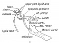
 Meckel.jpg 800 × 667; 181 KB
 Monosomy 18p syndrome facial features.jpg 967 × 727; 106 KB
 Mouse Bmp4 expression face 01.jpg 1,200 × 322; 58 KB
 Mouse Bmp4 expression limb and face 01.jpg 1,200 × 513; 91 KB
- Mouse face Bmp4.mp4 ; 480 KB
 Mouse head E11.5 microCT 01.jpg 1,409 × 1,200; 308 KB
 Mouse head TFII-I expression 01.jpg 1,000 × 862; 298 KB
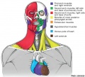
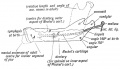
 Musculoskeletal- adult hyoid.jpg 450 × 309; 45 KB


