Category:Gle Embryo: Difference between revisions
(Created page with "This {{Embryology}} category shows pages, media, papers related to the historic Graf Spee's Gläveke Embryo (Gle). Gle. , or Gläveke or Glaevecke (von Spee, 1889 and 1896)....") |
mNo edit summary |
||
| Line 1: | Line 1: | ||
This {{Embryology}} category shows pages, media, papers related to the historic Graf Spee's Gläveke Embryo (Gle). | This {{Embryology}} category shows pages, media, papers related to the historic Graf Spee's Gläveke Embryo (Gle). | ||
Gle. , or Gläveke or Glaevecke (von Spee, 1889 and 1896). Illustrated as Normentafel No. 2 by Keibel and Elze (1908). See also Kollmann (1907) and Keibel and Mall (1910, 1912). | Gle. , or Gläveke or Glaevecke (von Spee, 1889 and 1896). Illustrated as Normentafel No. 2 by Keibel and Elze (1908). See also Kollmann (1907) and Keibel and Mall (1910, 1912). | ||
{{Carnegie stage | |||
{{Carnegie stage 8 links}} | |||
{{Carnegie_stage_table_1}} | {{Carnegie_stage_table_1}} | ||
| Line 28: | Line 30: | ||
[[Category:Human Embryo]] | [[Category:Human Embryo]] | ||
[[Category:Carnegie Stage | [[Category:Carnegie Stage 8]] | ||
[[Category:Historic Embryology]][[Category:1800's]] | [[Category:Historic Embryology]][[Category:1800's]] | ||
Revision as of 11:00, 8 August 2017
This Embryology category shows pages, media, papers related to the historic Graf Spee's Gläveke Embryo (Gle).
Gle. , or Gläveke or Glaevecke (von Spee, 1889 and 1896). Illustrated as Normentafel No. 2 by Keibel and Elze (1908). See also Kollmann (1907) and Keibel and Mall (1910, 1912).
| Week: | 1 | 2 | 3 | 4 | 5 | 6 | 7 | 8 |
| Carnegie stage: | 1 2 3 4 | 5 6 | 7 8 9 | 10 11 12 13 | 14 15 | 16 17 | 18 19 | 20 21 22 23 |
Description
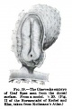
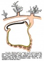
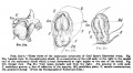
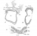
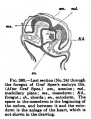
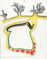
Chorion, 6 x 4.5 mm. Chorionic cavity, 5.3 x 3.8 mm. Embryonic disc, 1.54 mm. Primitive streak and node, notochordal plate, and neurenteric canal (Van Beneden, 1899) identified (Keibel and Mall, 1912, fig. 231).
Neural groove. Indication of foregut and pericardial cavities.
Anlage of endocardium.
No somites detected, although a small cavity on the left side (Keibel and Elze, 1908, fig. 4f) could be considered as the first Anlage of a myocoele. May belong to stage 9.
References
Pohlman AG. The development of the cloaca in human embryos. (1911) Amer. J Anat. 12: 1-26.
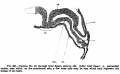
- "One of the earliest stages in the formation of the tail fold is found in the Spee reconstruction ('96) of embryo Gle (2.0 mm.). A schematic sagittal section is presented in his fig. 1, and emphasis is laid on the point that the primitive streak is composed of all three germ layers. No. 1 of the present tabulation was reconstructed by Mall ('97), and while a slightly older stage in the development, shows the cloacal sac limited ventrally by all three germ layers."
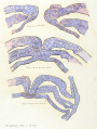
- Early Tract Development: Fig 223 | Fig 224 | Fig 225 | Fig 226 | Fig 227 | Fig 228 | Fig 229 | Fig 230 | Fig 231 | Fig 232 | Fig 233 | Fig 234 | Fig 235 | Fig 236 | Fig 237 | Fig 238 | Fig 239 | Fig 240 | Fig 241 | Fig 242 | Fig 243 | Fig 244 | Fig 245 | Fig 246 | Fig 247 | All Figures
Media in category 'Gle Embryo'
The following 9 files are in this category, out of 9 total.
 Keibel Mall 019.jpg 360 × 541; 40 KB
 Keibel Mall 020.jpg 581 × 791; 80 KB
 Keibel Mall 021.jpg 1,000 × 553; 113 KB
 Keibel Mall 2 231.jpg 1,019 × 1,000; 132 KB
 Keibel Mall 380.jpg 389 × 510; 46 KB
 Keibel Mall 381.jpg 800 × 491; 42 KB
 Keibel1908 fig02.jpg 1,486 × 800; 164 KB
 Keiller Glaevecke embryo drawing 1.jpg 1,280 × 1,695; 484 KB
 Keiller Glaevecke embryo drawing 2.jpg 1,280 × 1,602; 403 KB


