Category:Gastrointestinal Tract
From Embryology
This Embryology category is a link to resource pages, images, podcasts and movies that relate to gastrointestinal tract development.
Subcategories
This category has the following 16 subcategories, out of 16 total.
Pages in category 'Gastrointestinal Tract'
The following 133 pages are in this category, out of 334 total.
(previous page) (next page)P
- Paper - Sequential innervation of the intestinal loop in the human embryo
- Paper - Some factors influencing the position of the small intestine (1915)
- Paper - Studies of the intestine and peritoneum in the human foetus - part 1
- Paper - Studies of the intestine and peritoneum in the human foetus - part 2
- Paper - Studies of the intestine and peritoneum in the human foetus - part 3
- Paper - Studies of the intestine and peritoneum in the human foetus - part 4
- Paper - Studies of the intestine and peritoneum in the human foetus - part 5
- Paper - Studies of the intestine and peritoneum in the human foetus - part 6
- Paper - The angiology, angiogenesis, and organogenesis of the submaxillary gland
- Paper - The bi-lobed form of the ventral pancreas in mammals
- Paper - The comparative anatomy of the lips and labial villi of vertebrates
- Paper - The critical period in the development of the intestines (1914)
- Paper - The development of the cloaca in human embryos
- Paper - The development of the form of the gastrointestinal canal in humans 1
- Paper - The development of the form of the gastrointestinal canal in humans 2
- Paper - The development of the great omentum and transverse mesocolon
- Paper - The development of the human pharynx
- Paper - The Development of the Infra-Umbilical Portion of the Abdominal Wall, with Remarks on the Aetiology of Ectopia Vesicae
- Paper - The development of the lobule of the pig's liver (1919)
- Paper - The development of the lobus quadratus of the liver with special reference to an unusual anomaly of this lobe in the adult (1914)
- Paper - The development of the mucous membrane oesophagus stomach and small intestine in human embryo
- Paper - The development of the mucous membrane of the large intestine and vermiform process in the human embryo
- Paper - The development of the rectum in the human embryo
- Paper - The development of the serous glands (von Ebner's) of the vallate papillae in man (1917)
- Paper - The development of the spiral coil in the large intestine of the pig
- Paper - The early looping of the alimentary canal in the mammalian and human foetus and the mechanisms assumed to be active in this process
- Paper - The early stages of the development of the ileo-colic sphincter (1924)
- Paper - The embryogenesis of human bile capillaries and ducts
- Paper - The form of the stomach in human embryos with notes upon the nomenclature of the stomach
- Paper - The formation of the duodenal curve
- Paper - The formation of the duodenal curve (1919)
- Paper - The formation of the umbilical cord and the umbilical region of the anterior abdominal wall
- Paper - The gall bladder and the extrahepatic biliary passages in late embryonic and early fetal life
- Paper - The genesis of Jackson's membrane (1914)
- Paper - The genesis of Jackson's membrane: notes on the genito-mesenteric fold of peritoneum and the supra-adhesion foramen
- Paper - The later development of the bursa pharyngea - Homo
- Paper - The morphology and development of intestinal folds and villi in vertebrates
- Paper - The nature of the malformations of the rectum and urogenital passages
- Paper - The regular occurrence of intestinal diverticula in embryos of the pig, rabbit and man
- Paper - The regular occurrence of intestinal diverticula in embryos of the pig, rabbit, and man
- Paper - The relations of endogenous and exogenous factors in bone and tooth development (1937)
- Paper - The relative frequency of the various positions of the vermiform appendix (1924)
- Paper - The role of the primitive mesothelium in the development of the mammalian spleen (1936)
- Paper - Transposition of Abdominal Viscera (1926)
- Paper - V. Meckel's diverticulum patent at the navel (1902)
- Template:Parotid gland
- Template:Phenylketonuria
R
- Template:Ref-Beattie1924
- Template:Ref-Bossy1981
- Template:Ref-BurnsOgryzlo1938
- Template:Ref-Carey1920a
- Template:Ref-Carey1920b
- Template:Ref-Cave1936
- Template:Ref-Crymble1915
- Template:Ref-deVries1974
- Template:Ref-Elze1909
- Template:Ref-Enbom1939
- Template:Ref-Flint1903
- Template:Ref-Franklin1948
- Template:Ref-Fraser1919b
- Template:Ref-Frazer1915
- Template:Ref-Frazer1919
- Template:Ref-Gladstone1913
- Template:Ref-Gladstone1924
- Template:Ref-Guthrie1945
- Template:Ref-Hilton1902
- Template:Ref-Hubbard1902
- Template:Ref-Hunter1927
- Template:Ref-Hunter1928
- Template:Ref-Jackson1909b
- Template:Ref-Johnson1910
- Template:Ref-Johnson1913
- Template:Ref-Johnson1914
- Template:Ref-Johnson1914b
- Template:Ref-Kirk1910
- Template:Ref-Latta1921
- Template:Ref-Lewis1912b
- Template:Ref-Lineback1916
- Template:Ref-Lineback1920
- Template:Ref-Lockwood1882
- Template:Ref-Lockwood1884
- Template:Ref-Mall1898a
- Template:Ref-Mall1898b
- Template:Ref-Mall1899
- Template:Ref-McGill1910
- Template:Ref-Minot1900a
- Template:Ref-Pernkoof1922
- Template:Ref-Pernkoof1925
- Template:Ref-Reid1908
- Template:Ref-Reid1911a
- Template:Ref-Reid1911b
- Template:Ref-Reid1912
- Template:Ref-Reid1913a
- Template:Ref-Reid1913b
- Template:Ref-Reid1913c
- Template:Ref-Reid1914a
- Template:Ref-Reid1914b
- Template:Ref-Snook1934b
- Template:Ref-Tandler1900
- Template:Ref-Wagstaffe1924
- Template:Ref-Wakeley1923
- Template:Ref-Wakeley1930
- Template:Ref-Woods-Jones1904
- Template:Reid DG.
S
- Template:Salivary gland
- Salivary Gland Development
- Site Map
- Talk:Site Map
- Template:Small intestine
- Template:Small Intestine Length table1
- Template:Splanchnic mesoderm
- Template:Stage 23 MRI movie 7
- Stage 23 MRI Movie 7
- Template:Stomach
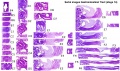
- Template:Stomach Histology
- Template:Sublingual gland
- Template:Submandibular gland
- Template:Submaxillary gland
V
Media in category 'Gastrointestinal Tract'
The following 123 files are in this category, out of 523 total.
(previous page) (next page) Stage 11 historic-Atwell1930-1b.jpg 323 × 600; 26 KB
 Stage 11 historic-Atwell1930-1c.jpg 215 × 400; 14 KB
 Stage 11 historic-Atwell1930-2.jpg 800 × 639; 87 KB
 Stage 11 historic-Atwell1930-2a.jpg 800 × 639; 87 KB
 Stage 11 historic-Atwell1930-2b.jpg 600 × 479; 50 KB
 Stage 11 historic-Atwell1930-2c.jpg 400 × 319; 23 KB
 Stage 11 historic-Atwell1930-3.jpg 1,000 × 679; 87 KB
 Stage 11 historic-Atwell1930-3a.jpg 800 × 543; 57 KB
 Stage 11 historic-Atwell1930-3b.jpg 600 × 407; 32 KB
 Stage 11 historic-Atwell1930-3c.jpg 400 × 271; 15 KB
 Stage 11 historic-Atwell1930-4.jpg 1,000 × 1,121; 114 KB
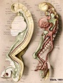
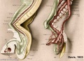
 Stage 11 historic-Davis1923-1.jpg 804 × 1,000; 87 KB
 Stage 11 historic-Davis1923-1a.jpg 643 × 800; 61 KB
 Stage 11 historic-Davis1923-1b.jpg 482 × 600; 41 KB
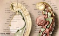
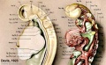
 Stage 11 historic-Davis1923-1c.jpg 321 × 400; 20 KB
 Stage 11 historic-Davis1923-2.jpg 768 × 1,000; 128 KB
 Stage 11 historic-Davis1923-2a.jpg 614 × 800; 83 KB
 Stage 11 historic-Davis1923-2b.jpg 461 × 600; 48 KB
 Stage 11 historic-Davis1923-2c.jpg 307 × 400; 23 KB
 Stage 11 historic-Davis1923-3.jpg 1,000 × 615; 84 KB
 Stage 11 historic-Davis1923-3a.jpg 800 × 492; 59 KB
 Stage 11 historic-Davis1923-3b.jpg 600 × 369; 41 KB
 Stage 11 historic-Davis1923-3c.jpg 400 × 246; 22 KB
 Stage 11 historic-Davis1923-4.jpg 1,000 × 722; 90 KB
 Stage 11 historic-Davis1923-4a.jpg 800 × 578; 63 KB
 Stage 11 historic-Davis1923-4b.jpg 600 × 434; 39 KB
 Stage 11 historic-Davis1923-4c.jpg 400 × 289; 21 KB
 Stage 11 historic-Heuser1930-1.jpg 521 × 1,000; 94 KB
 Stage 11 historic-Heuser1930-1a.jpg 417 × 800; 57 KB
 Stage 11 historic-Heuser1930-1b.jpg 313 × 600; 30 KB
 Stage 11 historic-Heuser1930-1c.jpg 209 × 400; 15 KB
 Stage 13 image 022.jpg 1,000 × 473; 101 KB
 Stage 13 image 023.jpg 1,000 × 544; 110 KB
 Stage 13 image 056.jpg 1,000 × 516; 102 KB
 Stage 13 image 057.jpg 1,000 × 511; 99 KB
 Stage 13 image 058.jpg 1,000 × 481; 94 KB
 Stage 13 image 059.jpg 1,000 × 513; 92 KB
 Stage 13 image 060.jpg 1,000 × 486; 96 KB
 Stage 13 image 061.jpg 1,000 × 600; 101 KB
 Stage 13 image 068.jpg 1,000 × 557; 97 KB
 Stage 13 image 074.jpg 1,000 × 549; 113 KB
 Stage 13 image 075.jpg 1,000 × 567; 116 KB
 Stage 13 image 076.jpg 1,000 × 667; 120 KB
 Stage 13 image 077.jpg 1,000 × 612; 127 KB
 Stage 13 image 078.jpg 1,000 × 486; 81 KB
 Stage 13 image 098.jpg 1,000 × 623; 144 KB
 Stage 22 image 167.jpg 1,000 × 662; 194 KB
 Stage 22 image 180.jpg 1,000 × 668; 145 KB
 Stage 22 image 181.jpg 1,000 × 653; 263 KB
 Stage 22 image 182.jpg 1,000 × 653; 211 KB
 Stage 22 image 183.jpg 1,000 × 662; 147 KB
 Stage 22 image 184.jpg 1,000 × 655; 256 KB
 Stage 22 image 185.jpg 1,000 × 656; 109 KB
 Stage 22 image 186.jpg 1,000 × 658; 209 KB
 Stage 22 image 197.jpg 1,000 × 668; 135 KB
 Stage 22 image 198.jpg 1,000 × 664; 192 KB
 Stage 22 image 202.jpg 1,455 × 920; 617 KB
 Stage 22 image 203.jpg 1,200 × 754; 334 KB
 Stage 22 image 210.jpg 1,101 × 794; 447 KB
 Stage 22 image 210a.jpg 1,000 × 644; 310 KB
 Stage 22 image 210b.jpg 800 × 515; 207 KB
 Stage 22 image 210c.jpg 400 × 257; 52 KB
 Stage 22 image 213.jpg 738 × 554; 198 KB
 Stage 22 image 214.jpg 738 × 554; 203 KB
 Stage 22 image 215.jpg 600 × 450; 105 KB
 Stage 22 image 216.jpg 600 × 450; 110 KB
 Stage 22 image 302.jpg 1,455 × 920; 625 KB
 Stage11 historic-Atwell1930-2.jpg 1,000 × 799; 131 KB
 Stage11 sem2.jpg 976 × 1,000; 188 KB
 Stage11 sem3.jpg 936 × 1,000; 68 KB
 Stage11 sem3a.jpg 749 × 800; 44 KB
 Stage11 sem3b.gif 468 × 500; 182 KB
 Stage11 sem3b.jpg 468 × 500; 45 KB
 Stage11 sem3c.jpg 375 × 400; 16 KB
 Stage11 sem4.jpg 808 × 1,000; 129 KB
 Stage12 sem9 cloacal membrane.jpg 1,220 × 1,696; 239 KB
 Stage12 sem9.jpg 1,220 × 1,696; 229 KB
 Stage12 sem9a.jpg 719 × 1,000; 110 KB
 Stage12 sem9b.jpg 575 × 800; 81 KB
 Stage12 sem9c.jpg 431 × 600; 53 KB
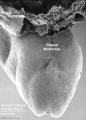
 Stage14 stomach.jpg 240 × 240; 26 KB
 Stage14-git.jpg 600 × 357; 96 KB
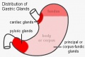 Stomach gastric gland distribution.jpg 500 × 334; 28 KB
 Stomach histology 001.jpg 400 × 533; 87 KB
 Stomach histology 002.jpg 400 × 532; 53 KB
 Stomach histology 003.jpg 599 × 400; 53 KB
 Stomach histology 004.jpg 1,280 × 1,024; 435 KB
 Stomach histology 005.jpg 1,280 × 1,024; 432 KB
 Stomach histology 006.jpg 1,280 × 1,024; 227 KB
 Stomach histology 007.jpg 1,280 × 1,024; 308 KB
 Stomach histology 008.jpg 1,280 × 1,024; 452 KB
 Thyng1914 fig02.jpg 1,189 × 800; 112 KB
 Tongue1.png 373 × 277; 9 KB
 Tongue2.png 327 × 318; 6 KB
 Tongue3.png 249 × 269; 6 KB
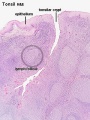 Tonsil histology 01.jpg 450 × 600; 106 KB
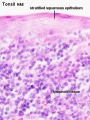 Tonsil histology 02.jpg 450 × 600; 62 KB
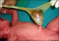 Umbilical cord hernia 01.jpg 846 × 600; 68 KB
 Waterston01.jpg 1,094 × 618; 184 KB
 Waterston02.jpg 661 × 729; 130 KB
 Waterston03.jpg 800 × 547; 100 KB
 Waterston04.jpg 500 × 763; 75 KB
 Waterston05.jpg 414 × 678; 81 KB
 Waterston06.jpg 720 × 538; 92 KB
 Waterston07.jpg 562 × 655; 79 KB
 Waterston08.jpg 600 × 672; 89 KB
 Waterston09.jpg 600 × 774; 105 KB
 Waterston10.jpg 600 × 717; 83 KB
 Waterston11.jpg 500 × 728; 75 KB
 Waterston12.jpg 300 × 420; 18 KB
 Waterston13.jpg 429 × 681; 63 KB
 Waterston14.jpg 438 × 680; 66 KB
 Waterston15.jpg 500 × 674; 65 KB
 Waterston16.jpg 593 × 675; 73 KB
 Waterston17.jpg 500 × 710; 74 KB
 Waterston18.jpg 500 × 662; 71 KB
 Waterston19.jpg 500 × 642; 66 KB
 Waterston1914 fig01.jpg 1,200 × 671; 195 KB
 Waterston1914 figure animation.gif 489 × 752; 325 KB
 Waterston20.jpg 500 × 720; 75 KB
 West02.jpg 619 × 549; 34 KB
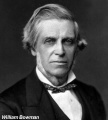 William Bowman.jpg 600 × 665; 49 KB
 Zorn2008 fig01.jpg 1,200 × 1,158; 110 KB


