Category:Carnegie Stage 16: Difference between revisions
From Embryology
mNo edit summary |
|||
| Line 16: | Line 16: | ||
* '''No. BR, 9.75 mm G. L.''', 8.8 mm. Summarized by Tandler (1907)<ref>Tandler, J. 1907. ''Ueber einen menschlichen Embryo vom 38. Tage.'' Anat. Ariz., 31. 49-56.</ref>, who, on the basis of the coital history, gave the age as the “38th day.” About stage 16. | * '''No. BR, 9.75 mm G. L.''', 8.8 mm. Summarized by Tandler (1907)<ref>Tandler, J. 1907. ''Ueber einen menschlichen Embryo vom 38. Tage.'' Anat. Ariz., 31. 49-56.</ref>, who, on the basis of the coital history, gave the age as the “38th day.” About stage 16. | ||
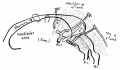
* '''9-mm embryo'''. The peripheral nervous system was described by Masy (1955)<ref>Masy, S. 1955. ''Le systeme nerveux peripherique cranien de l'embryon humain de 9 mm.'' J. Embryol. Exp. Morphol, 3, 30-43.</ref> in this embryo of stage 16. | * '''9-mm embryo'''. The peripheral nervous system was described by Masy (1955)<ref>Masy, S. 1955. ''Le systeme nerveux peripherique cranien de l'embryon humain de 9 mm.'' J. Embryol. Exp. Morphol, 3, 30-43.</ref> in this embryo of stage 16. | ||
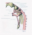
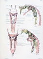
* '''Huber No. 3''', 10 mm. The nuclei of origin of the cranial nerves and the peripheral nervous system were described by Streeter (1908a,b).<ref>Streeter, G. L. 1908a. The nuclei of origin of the cranial nerves in the 10 mm human embryo. Anat. Rec, 2, 111-115.</ref><ref>Streeter, G. L. 1908b. The peripheral nervous system in the human embryo at the end of the first month (10 mm). Amer. J. Anat., 8, 285-301.</ref> An advanced example of stage 16. | * '''Huber No. 3''', 10 mm. The nuclei of origin of the cranial nerves and the peripheral nervous system were described by Streeter (1908a,b).<ref>Streeter, G. L. 1908a. The nuclei of origin of the cranial nerves in the 10 mm human embryo. Anat. Rec, 2, 111-115.</ref><ref>Streeter, G. L. 1908b. [[Paper_-_The Peripheral Nervous System in the Human Embryo at the End of the First Month (10 mm)|The peripheral nervous system in the human embryo at the end of the first month (10 mm)]]. Amer. J. Anat., 8, 285-301.</ref> An advanced example of stage 16. | ||
* '''Wetzel (We) embryo''', 10 mm. Discussed by v. Hayek (1934)<ref>v. Hayek, H. 1934. ''Ein menschlicher Embryo vom 40''. Tage. Anat. Anz., 78, 315-320.</ref> in regard to age, which, based on the coital history, was given as the “40th day.” Probably about stage 16. | * '''Wetzel (We) embryo''', 10 mm. Discussed by v. Hayek (1934)<ref>v. Hayek, H. 1934. ''Ein menschlicher Embryo vom 40''. Tage. Anat. Anz., 78, 315-320.</ref> in regard to age, which, based on the coital history, was given as the “40th day.” Probably about stage 16. | ||
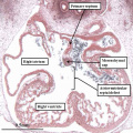
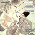
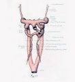
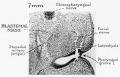
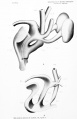
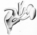
* '''Carnegie No. 6516''', 10.5 mm (corrected). Double ureters described by Wharton (1949)<ref name=“PIMD18130385”><pubmed>18130385</pubmed></ref>. | * '''Carnegie No. 6516''', 10.5 mm (corrected). Double ureters described by Wharton (1949)<ref name=“PIMD18130385”><pubmed>18130385</pubmed></ref>. | ||
* '''No. H60''', University of Missouri, 11 mm. Described briefly by Bonnot and Severs (1906).<ref>Bonnot, E., and Severs, R. 1906. On the structure of a human embryo eleven millimeters in length. Anat. Anz., 29, 452-459.</ref> Probably belongs to stage 16. | * '''No. H60''', University of Missouri, 11 mm. Described briefly by Bonnot and Severs (1906).<ref>Bonnot, E., and Severs, R. 1906. On the structure of a human embryo eleven millimeters in length. Anat. Anz., 29, 452-459.</ref> Probably belongs to stage 16. | ||
===References=== | ===References=== | ||
Revision as of 09:27, 15 September 2015
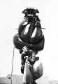
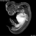
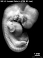
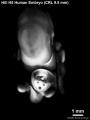
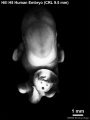
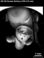
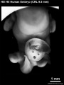
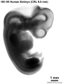
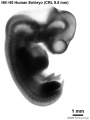
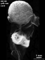
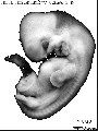
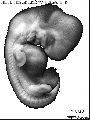
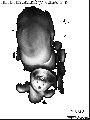
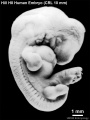
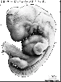
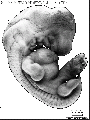
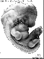
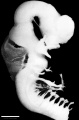
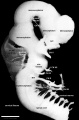
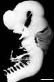
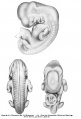
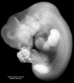
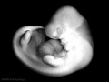
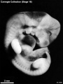
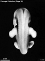
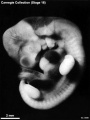
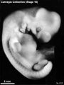
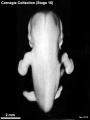
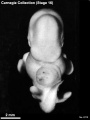
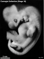
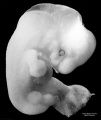
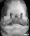
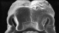
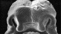
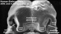
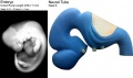
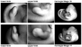
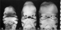
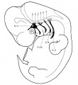
This Embryology category shows pages and media related to embryonic development in week 6, 37 - 42 days, GA week 8. The embryos have a crown rump length (CRL) of 8 - 11 mm. Stage 16 and 17 are the only two stages occurring in week 6 of the historic 23 Carnegie stages of embryonic development.
There is also a specific Carnegie stage 16 resource page.
| Carnegie Collection Embryos - Stage 16 | ||||||||||
|---|---|---|---|---|---|---|---|---|---|---|
| Serial No. | Size (mm) | Grade | Fixative | Embedding Medium | Plane | Thinness (µm) | Stain | Year | Notes | |
| 163 | E., 9.0 Ch., 35x35x20 | Good | Formol | P | Transverse | 20 | Al. coch. | 1899 | Used by Bardeen and Lewis | |
| 221 | E., 7.5 Ch., 40x33x33 | Poor | Formalin | P | Sagittal | 20 | Al. coch. | 1903 | Macerated | |
| 383 | E., 7.0 Ch., 15x15x15 | Poor | Formalin | P | Transverse | 10 | Al. carm., H. & Congo red | 1904 | ||
| 397 | E., 8.0 Ch., 15x15x15 | Poor | Formalin | P | Transverse | 10 | (Stain - Haematoxylin Eosin) | 1907 | ||
| 422 | E., 9.0 Ch., 30x30x30 | Poor | Alc. | P | Transverse | 40 | Al. coch. | 1910 | Tubal. Partly macerated | |
| 559 | E., 8.6 Ch., 20x15x12 | Good | Formalin | P | Transverse | 20 | H. & Congo red | 1911 | Cyclopia. Formerly listed as stage 17 | |
| 589 | E., 11 Ch., 30x13x13 | Poor | p | P | Sagittal | 50 | Al. coch. | 1912 | ||
| 617 | E., 7.0 Ch., 18x14x12 | Good | Formalin | P | Transverse | 15 | Al. coch. | 1912 | Median in group | |
| 636 | E., 10 Ch.,28x28x22 | Poor | Formalin | P | Transverse | 50 | Al. coch. | 1913 | Macerated | |
| 651f | E., 7 Ch., 25x20x15 | Poor | p | p | p | p | p | 1913 | Spina bifida | |
| 675 | E., 10 Ch., 50x30x25 | Poor | Formalin | P | Sagittal | 100 | Carmine | 1915 | Abnormal head and limbs | |
| 792 | E., 8.0 Ch., 40x30x30 | Good | Formalin | P | Transverse | 20 | Al. coch. | 1913 | Advanced | |
| 887 | E, 9.0 Ch., 31x28x17 | Good | Formalin | P | Transverse | 40 | Al. coch. | 1914 | Near next stage | |
| 1121 | E., 11.8 | Good | Corros. acetic | P | Coronal | 40 | Al. coch. | 1915 | Operative. Median in group | |
| 1197 | E., 10.0 Ch., 23x19x15 | Good | Formalin | C | Sagittal | 20 | (Stain - Haematoxylin Eosin), or. G. | 1915 | Advanced | |
| 1544 | E., 7.2 | Good | Zenker | P | Sagittal | 20 | Al. coch. | 1916 | Tubal. Mechanical injury | |
| 1836 | E., 11.0 | Good | Formalin | P | Transverse | 20 | (Stain - Haematoxylin Eosin) | 1917 | Most-advanced third | |
| 4677 | E., 9.5 Ch., 48x36x30 | Good | Formalin | P | Transverse | 10 | Al. coch. | 1924 | Median in group | |
| 5515 | E., 12.0 Ch., 47x37x25 | Good | Formalin | C-P | Transverse | 10 | (Stain - Haematoxylin Eosin) | 1927 | Near next stage | |
| 6054 | E., 7,0 Ch., 21x17x12 | Good | Formalin | C-P | Transverse | 8 | (Stain - Haematoxylin Eosin) | 1930 | Least-advanced third | |
| 6507 | E., 9.0* | Excellent | Corros. acetic | C-P | Coronal | 10 & 8 | Al. coch. | p | Middle or most-advanced third | |
| 6509 | E. 8.1* | Excellent | Corros. acetic | C-P | Coronal | 10 | Al. coch. | p | Least-adianced or middle third | |
| 6510 | E., 10.1* | Excellent | Corros. acetic | C-P | Coronal | 10 | Al. coch. | p | Close to No. 6507. Ag added | |
| 6511 | E, 8.1* | Good | Corros. acetic | C-P | Sagittal | 10 | Al. coch., iron H. | p | Surface injured by fixative. Most-advanced third | |
| 6512 | E., 7.0* | Excellent | Corros. acetic | C-P | Transverse | 10 | Al. coch. | p | Least-advanced third. Borderline | |
| 6513 | E., 7.2* | Good | Corros. acetic | C-P | Coronal | 10 | Al. coch. | p | Surface injured by fixative. Least advanced in group | |
| 6514 | E, 9 0* | Poor | Corros. acetic | C-P | Sagittal | 10 | Al. coch. | p | Most-advanced third | |
| 6516 | E., 10 5* | Good | Corros acetic | C-P | Sagittal | 8 | Al. coch. | p | Most-advanced third. Double left kidney and ureter | |
| 6517 | E., 10.5* | Excellent | Corros. acetic | C-P | Transverse | 8 | Al. coch. | ? | Close to No. 6516 | |
| 6686 | E., 11.0 Ch., l7x17xP | Poor | Formalin | C-P | Coronal | 20 | Al. coch. | 1933 | Tubal. Partly macerated | |
| 6750 | E., 10.0 | Good | Formalin | C-P | Transverse | 10 | H. & phlox. | 1933 | Tubal. Advanced | |
| 6909 | E., 11.0 | Good | Bouin | C-P | Coronal | 10 | (Stain - Haematoxylin Eosin) | 1934 | Tubal. Advanced | |
| 6931 | E., 8.8 Ch., 3""x 33x16 | Good | Formalin | C-P | Coronal | 10 | Al coch., phlox. | 1934 | Least-advanced third. Type specimen | |
| 6950 | E.. 9 0 Ch., 3lx20x18 | Good | Formalin | C-P | Transverse | 10 | (Stain - Haematoxylin Eosin) | 1934 | Tubal. Partly fragmented | |
| 7?15 | E., 9.7 | Exc | Bouin | C-P | Coronal | 10 | H. & phlox | 1935 | Operative. Less advanced | |
| 7629 | E., 11.5 Ch., 31x31 | Good | Formalin | C-P | Coronal | 10 | Al. coch., phlox | 1939 | Hysterectomy. Most advanced in group | |
| 7804 | E., 9.5 Ch., 26x21x16 | Good | Formalin | C-P | Transverse | 10 | (Stain - Haematoxylin Eosin) | 1940 | Least-advanced third | |
| 7897 | E., 12.2 Ch., 31x24x23 | Good | Formalin | C-P | Transverse | 10 | (Stain - Haematoxylin Eosin) | 1941 | Tubal. Advanced | |
| 8098 | E., 10.0 Ch., 30 | Good | Formalin | C-P | Coronal | 6 | (Stain - Haematoxylin Eosin) | 1942 | Tubal. Median in group | |
| 8112 | E., 10.9 | Excellent | Bouin | C-P | Coronal | 8 | (Stain - Haematoxylin Eosin) | 1943 | Most-advanced third | |
| 8179 | E., 11.9 Ch., 23x18x17 | Good | Formalin | C-P | Coronal | 10 | (Stain - Haematoxylin Eosin) | 1943 | Tubal | |
| 8436 | E., 10.9 Ch., 13x15x1? | Good | Formalin | P | Coronal | 10 | Azan | 1946 | Advanced | |
| 8692 | E., 10 | Good | Bouin | P | Transverse | 10 | (Stain - Haematoxylin Eosin) | 1949 | Rubella. Medical abortion. Mechanically damaged | |
| 8697 | E., 11.3 | Poor | Formalin | C-P | Transverse | 10 | (Stain - Haematoxylin Eosin) | 1949 | Perhaps stage 17 | |
| 8773 | E, 11 | Excellent | Bouin | P | Coronal | 10 | Azan | 1950 | ||
| 8971 | E., 10 Ch., 20.5x14.5x13.7 | Poor | Formalin | Transverse | 15 | (Stain - Haematoxylin Eosin) | 1932 | Synophthalmia. Univ. Chicago No. H 1439 | ||
| Template:CE9055 | E., ca. 10 | Excellent | Bouin | P | Transverse | 20 | Azan & Ag | 1953 | Damaged | |
| 9229 | E, 9.5 | Excellent | Formalin | P | Transverse | 6 | Ag & (Stain - Haematoxylin Eosin) | 1954 | Stage 15, 16, or 17? Mislaid | |
Abbreviations
| ||||||||||
| Week: | 1 | 2 | 3 | 4 | 5 | 6 | 7 | 8 |
| Carnegie stage: | 1 2 3 4 | 5 6 | 7 8 9 | 10 11 12 13 | 14 15 | 16 17 | 18 19 | 20 21 22 23 |
- Carnegie Stages: 1 | 2 | 3 | 4 | 5 | 6 | 7 | 8 | 9 | 10 | 11 | 12 | 13 | 14 | 15 | 16 | 17 | 18 | 19 | 20 | 21 | 22 | 23 | About Stages | Timeline
Embryo Examples
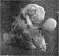
- No. BR, 9.75 mm G. L., 8.8 mm. Summarized by Tandler (1907)[1], who, on the basis of the coital history, gave the age as the “38th day.” About stage 16.
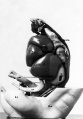
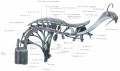
- 9-mm embryo. The peripheral nervous system was described by Masy (1955)[2] in this embryo of stage 16.
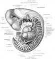
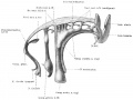
- Huber No. 3, 10 mm. The nuclei of origin of the cranial nerves and the peripheral nervous system were described by Streeter (1908a,b).[3][4] An advanced example of stage 16.
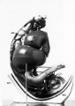
- Wetzel (We) embryo, 10 mm. Discussed by v. Hayek (1934)[5] in regard to age, which, based on the coital history, was given as the “40th day.” Probably about stage 16.
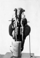
- Carnegie No. 6516, 10.5 mm (corrected). Double ureters described by Wharton (1949)[6].
- No. H60, University of Missouri, 11 mm. Described briefly by Bonnot and Severs (1906).[7] Probably belongs to stage 16.
References
- ↑ Tandler, J. 1907. Ueber einen menschlichen Embryo vom 38. Tage. Anat. Ariz., 31. 49-56.
- ↑ Masy, S. 1955. Le systeme nerveux peripherique cranien de l'embryon humain de 9 mm. J. Embryol. Exp. Morphol, 3, 30-43.
- ↑ Streeter, G. L. 1908a. The nuclei of origin of the cranial nerves in the 10 mm human embryo. Anat. Rec, 2, 111-115.
- ↑ Streeter, G. L. 1908b. The peripheral nervous system in the human embryo at the end of the first month (10 mm). Amer. J. Anat., 8, 285-301.
- ↑ v. Hayek, H. 1934. Ein menschlicher Embryo vom 40. Tage. Anat. Anz., 78, 315-320.
- ↑ <pubmed>18130385</pubmed>
- ↑ Bonnot, E., and Severs, R. 1906. On the structure of a human embryo eleven millimeters in length. Anat. Anz., 29, 452-459.
Subcategories
This category has the following 49 subcategories, out of 49 total.
C
- Carnegie Embryo 1121
- Carnegie Embryo 1197
- Carnegie Embryo 1544
- Carnegie Embryo 163
- Carnegie Embryo 1836
- Carnegie Embryo 221
- Carnegie Embryo 383
- Carnegie Embryo 397
- Carnegie Embryo 422
- Carnegie Embryo 4677
- Carnegie Embryo 5515
- Carnegie Embryo 559
- Carnegie Embryo 589
- Carnegie Embryo 6054
- Carnegie Embryo 617
- Carnegie Embryo 636
- Carnegie Embryo 6507
- Carnegie Embryo 6509
- Carnegie Embryo 6510
- Carnegie Embryo 6511
- Carnegie Embryo 6512
- Carnegie Embryo 6513
- Carnegie Embryo 6514
- Carnegie Embryo 6516
- Carnegie Embryo 6517
- Carnegie Embryo 6527
- Carnegie Embryo 6686
- Carnegie Embryo 675
- Carnegie Embryo 6750
- Carnegie Embryo 6909
- Carnegie Embryo 6931
- Carnegie Embryo 6950
- Carnegie Embryo 7629
- Carnegie Embryo 7804
- Carnegie Embryo 7897
- Carnegie Embryo 792
- Carnegie Embryo 8098
- Carnegie Embryo 8112
- Carnegie Embryo 8179
- Carnegie Embryo 8436
- Carnegie Embryo 852
- Carnegie Embryo 8692
- Carnegie Embryo 8697
- Carnegie Embryo 8773
- Carnegie Embryo 887
- Carnegie Embryo 8971
- Carnegie Embryo 9229
Pages in category 'Carnegie Stage 16'
The following 72 pages are in this category, out of 72 total.
C
- Template:Carnegie Embryo Stage16
- Carnegie stage 16
- Template:Carnegie stage 16 links
- Template:CE1121
- Template:CE1197
- Template:CE1544
- Template:CE163
- Template:CE1836
- Template:CE221
- Template:CE383
- Template:CE397
- Template:CE422
- Template:CE4677
- Template:CE5515
- Template:CE559
- Template:CE589
- Template:CE6054
- Template:CE617
- Template:CE636
- Template:CE6507
- Template:CE6509
- Template:CE6510
- Template:CE6511
- Template:CE6512
- Template:CE6513
- Template:CE6514
- Template:CE6516
- Template:CE6517
- Template:CE6686
- Template:CE675
- Template:CE6750
- Template:CE6909
- Template:CE6931
- Template:CE6950
- Template:CE7629
- Template:CE7804
- Template:CE7897
- Template:CE792
- Template:CE8098
- Template:CE8112
- Template:CE8179
- Template:CE8436
- Template:CE8692
- Template:CE8697
- Template:CE8773
- Template:CE887
- Template:CE8971
- Template:CE9229
- Template:CS16
P
- Paper - Developmental horizons in human embryos stages 15-18
- Paper - Growth allometry of the myocardium in human embryos from stages 15 to 23
- Paper - On the structure of a human embryo eleven millimeters in length
- Paper - The Peripheral Nervous System in the Human Embryo at the End of the First Month (10 mm)
S
- Stage 15 to 22 Head Movie
- Stage 16 EFIC Movie 1
- Stage 16 EFIC Movie 2
- Stage 16 EFIC Movie 3
- Template:Stage 16 movies gallery
- Stage 16 MRI Movie 1
- Stage 16 MRI Movie 2
- Stage 16 MRI Movie 3
- Stage 16 MRI Movie 4
- Stage 16 MRI Movie 5
- Template:Stage 16 MRI movie links
- Stage 16 to 18 Face Movie
- Template:Streeter1908 figures
Media in category 'Carnegie Stage 16'
The following 104 files are in this category, out of 104 total.
 Anderson2016-fig12b.jpg 800 × 800; 138 KB
 Anderson2016-fig14.jpg 800 × 800; 168 KB
 Anderson2016-fig15a.jpg 800 × 800; 142 KB
 Anderson2016-fig15b.jpg 800 × 800; 148 KB
 Bonnot1906 fig01.jpg 658 × 630; 61 KB
 Bonnot1906 fig02.jpg 1,176 × 1,689; 138 KB
 Bonnot1906 fig03.jpg 1,192 × 1,689; 154 KB
 Bonnot1906 plate03.jpg 1,179 × 1,692; 144 KB
 Bonnot1906 plate04.jpg 1,161 × 1,675; 122 KB
 Carnegie stage 16 OPT.jpg 800 × 801; 51 KB
 Congdon1922-35.jpg 920 × 1,000; 97 KB
 Congdon1922-36.jpg 920 × 1,000; 113 KB
 Congdon1922-plate02.jpg 877 × 1,200; 191 KB
 Crowder1957 fig03.jpg 621 × 632; 189 KB
 Crowder1957 fig04.jpg 996 × 891; 291 KB
 Frazer1910 fig09.jpg 1,000 × 582; 80 KB
 Gilbert1957 fig02.jpg 1,280 × 824; 130 KB
 HillH5 Stage 16 bf01.jpg 1,125 × 1,500; 142 KB
 HillH5 Stage 16 bf02.jpg 1,125 × 1,500; 142 KB
 HillH5 Stage 16 bf03.jpg 1,125 × 1,500; 148 KB
 HillH5 Stage 16 bf04.jpg 1,125 × 1,500; 149 KB
 HillH5 Stage 16 bf05.jpg 1,125 × 1,500; 116 KB
 HillH5 Stage 16 bf06.jpg 1,125 × 1,500; 111 KB
 HillH5 Stage 16 bf07.jpg 1,125 × 1,500; 151 KB
 HillH5 Stage 16 bf08.jpg 1,125 × 1,500; 149 KB
 HillH5 Stage 16 bf09.jpg 1,125 × 1,500; 109 KB
 HillH5 Stage 16 bf10.jpg 1,125 × 1,500; 110 KB
 HillH5 Stage 16 bf11.jpg 1,125 × 1,500; 147 KB
 HillH5 Stage 16 bf12.jpg 1,125 × 1,500; 154 KB
 HillH5 Stage 16 bf13.gif 450 × 600; 296 KB
 HillH5 Stage 16 bf14.gif 450 × 600; 300 KB
 HillH5 Stage 16 bf15.gif 450 × 600; 184 KB
 HillH5 Stage 16 bf16.gif 450 × 600; 257 KB
 HillH5 Stage 16 bf17.gif 450 × 600; 238 KB
 HillH5 Stage 16 bf18.gif 450 × 600; 314 KB
 HillH8 Stage 16 bf01.jpg 1,125 × 1,500; 136 KB
 HillH8 Stage 16 bf02.jpg 1,125 × 1,500; 145 KB
 HillH8 Stage 16 bf03.jpg 1,125 × 1,500; 170 KB
 HillH8 Stage 16 bf04.jpg 1,125 × 1,500; 171 KB
 HillH8 Stage 16 bf05.jpg 1,125 × 1,500; 176 KB
 HillH8 Stage 16 bf06.jpg 1,125 × 1,500; 169 KB
 HillH8 Stage 16 bf07.gif 600 × 800; 490 KB
 HillH8 Stage 16 bf08.gif 450 × 600; 295 KB
 HillH8 Stage 16 bf09.gif 600 × 800; 571 KB
 HillH8 Stage 16 bf10.gif 450 × 600; 338 KB
 HillH8 Stage 16 bf11.gif 600 × 800; 585 KB
 HillH8 Stage 16 bf12.gif 450 × 600; 348 KB
 Human embryonic renal branching 1.jpg 1,280 × 779; 236 KB
 Human embryonic tongue 02.jpg 650 × 850; 220 KB
 Human embryonic tongue 06.jpg 650 × 470; 99 KB
 Human Stage14-16 CN5-01.jpg 1,028 × 681; 44 KB
 Human stage16 face 01.jpg 500 × 504; 20 KB
 Human Stage16 neural01.jpg 1,352 × 2,048; 247 KB
 Human Stage16 neural02.jpg 1,352 × 2,048; 286 KB
 Human Stage16 neural03.jpg 1,352 × 2,048; 245 KB
 Keibel Mall 049-051.jpg 680 × 1,000; 66 KB
 Keibel Mall 2 428.jpg 1,280 × 620; 110 KB
 Keith1921 fig044.jpg 1,027 × 1,079; 144 KB
 Kyoto731 Stage16-01.jpg 908 × 681; 42 KB
 Kyoto731 Stage16-02.jpg 908 × 681; 59 KB
 Mall1908a plate03.jpg 1,743 × 2,738; 289 KB
 Mall1908a plate03fig09.jpg 1,743 × 1,306; 186 KB
 ME34 001.jpg 2,153 × 1,500; 186 KB
 ME34 002.jpg 1,280 × 871; 216 KB
 Pohlman1911 plate3.jpg 2,171 × 3,361; 454 KB
 Pohlman1911 plate3E.jpg 1,988 × 1,895; 198 KB
 Stage 16 MRI 3D01.mp4 ; 601 KB
- Stage 16 MRI 3D02.mp4 ; 2.51 MB
- Stage 16 MRI 3D03.mp4 ; 1.2 MB
 Stage16 bf1.jpg 1,000 × 750; 23 KB
 Stage16 bf10.jpg 1,778 × 2,000; 319 KB
 Stage16 bf1a.jpg 800 × 600; 17 KB
 Stage16 bf1b.jpg 600 × 450; 11 KB
 Stage16 bf1c.jpg 400 × 300; 6 KB
 Stage16 bf2.jpg 525 × 700; 38 KB
 Stage16 bf3.jpg 525 × 700; 29 KB
 Stage16 bf4.jpg 525 × 700; 37 KB
 Stage16 bf5.jpg 450 × 600; 31 KB
 Stage16 bf6.jpg 450 × 600; 23 KB
 Stage16 bf7.jpg 450 × 600; 24 KB
 Stage16 bf8.jpg 450 × 600; 30 KB
 Stage16 bf9.jpg 1,677 × 2,000; 319 KB
 Stage16 cleft palate.jpg 337 × 400; 14 KB
- Stage16 EFIC C01.mp4 ; 1.09 MB
- Stage16 EFIC C02.mp4 ; 1.34 MB
- Stage16 EFIC S01.mp4 ; 1,018 KB
- Stage16 EFIC T01.mp4 ; 430 KB
 Stage16 em01.jpg 800 × 456; 44 KB
 Stage16 em11.gif 800 × 456; 336 KB
 Stage16 em11.jpg 800 × 456; 63 KB
- Stage16 em11.mp4 ; 70 KB
 Stage16 embryo and brain 01.jpg 1,280 × 752; 91 KB
- Stage16 MRI C01.mp4 ; 577 KB
- Stage16 MRI S01.mp4 ; 718 KB
- Stage16 MRI T01.mp4 ; 1.18 MB
 Stage16-17-limbs01.jpg 1,000 × 572; 77 KB
 Stage16-18 face.jpg 800 × 393; 34 KB
 Streeter1908-fig01.jpg 1,112 × 1,212; 132 KB
 Streeter1908-plate01.jpg 1,600 × 1,732; 360 KB
 Streeter1908-plate02.jpg 2,031 × 1,200; 238 KB
 Streeter1908-plate03.jpg 1,600 × 1,198; 167 KB
 Streeter1922-fig10.jpg 667 × 1,000; 86 KB
 Streeter1957 fig01.jpg 1,292 × 1,500; 218 KB
 Sudler1902-fig08.jpg 1,000 × 753; 114 KB


