Category:Carnegie Stage 15
From Embryology
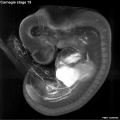
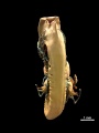
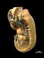
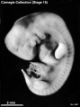
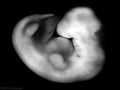
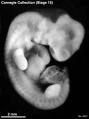
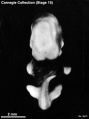
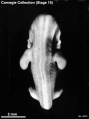
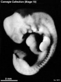
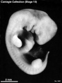
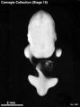
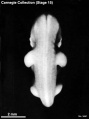
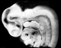
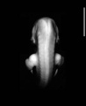
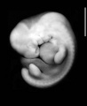
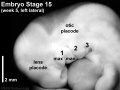
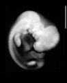
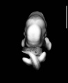
This Embryology category shows pages and media related to Carnegie stage 15 of embryonic development occurring during post fertilisation Week 5 or GA week 7.
There is also a specific Carnegie stage 15 resource page. See also Carnegie Stage 15 - Events
| Week: | 1 | 2 | 3 | 4 | 5 | 6 | 7 | 8 |
| Carnegie stage: | 1 2 3 4 | 5 6 | 7 8 9 | 10 11 12 13 | 14 15 | 16 17 | 18 19 | 20 21 22 23 |
| Carnegie Collection Embryos - Stage 15 | ||||||||||
|---|---|---|---|---|---|---|---|---|---|---|
| Serial No. | Size (mm) | Grade | Fixative | Embedding Medium | Plane | Thinness (µm) | Stain | Year | Notes | |
| 2 | E., 7.0 Ch., 25x25x25 | Good | Alc. | P | Transverse | 15 | Al. carm. | 1888 | Least-advanced third | |
| 88 | E.,8 Ch., 30x28x15 | Poor | Alc. | P | Coronal | P | Al. coch. | 1897 | ||
| 113 | E.,8 241 E,6.0 | Poor | P | p | Sagittal | 10 | Borax carmine | ? | ||
| 241 | E,6.0 | Good | Formalin | P | Transverse | 10 | H. & Congo red | 1904 | ||
| 371 | E,6.6 | Good | Formalin | P | Sagittal | 10 | Al. coch. | 1913 | Shrunken and cracked | |
| 389 | E., 9 | Poor | p | p | Sagittal | 20 | (Stain - Haematoxylin Eosin) | 1907 | Tubal | |
| 721 | E., 9.0 Ch., 30x20x10 | Exc. | Zenker formol | P | Transverse | 15 | (Stain - Haematoxylin Eosin) | 1913 | Median in group | |
| 810 | E., 7.0 Ch, 30x25x15 | Good | Alc. | P | Sagittal | 20 | Al. coch | 1913 | ||
| 855 | E.,7.5 | Poor | Formalin | P | Transverse | 100 | Al. coch. | 1914 | Pathological between limbs | |
| 1006 | E,9.0 Ch., 37x26x22 | Poor | Formalin | P | Coronal | 20 | (Stain - Haematoxylin Eosin) or. G. | 1914 | Operative. Most-advanced third | |
| 1091 | E,7.2 Ch., 28x26x20 | Poor | P | P | Coronal | 20 | Al. coch. | 1915 | Macerated | |
| 1354 | E,7.8 Ch, 35x30x25 | Good | Formalin | P | Sagittal | 20 | Al. coch. | 1916 | Least-advanced third | |
| 1767 | E , 11.0 Ch, 41x23x5 | Good | Formalin | P | Sagittal | 40 | (Stain - Haematoxylin Eosin) or. G. | 1917 | Most-advanced third | |
| 2743 | E., 7.2 Ch., 19xl8x14 | Poor | Formalin | P | Transverse | 20 | Al. coch. | 1919 | Macerated. Least-advanced third | |
| 3216 | E, 6.5 Ch, 30x30x5 | Good | Formalin | P | Transverse | 20 | Al. coch. | 1920 | Hysterectomy. Least-advanced third | |
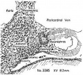
| 3385 | E,83 Ch., 25x20x16 | Exc. | Corros. acetic | P | Trans. | 20 | (Stain - Haematoxylin Eosin) or. G. | 1921 | Some sections lost. Most-advanced third. Ag added | |
| 3441 | E,8.0 Ch., 25x24x20 | Good | Formalin | P | Sag. | 10 | Al. coch. | 1921 | ||
| 3512 | E,8,5 Ch., 33x28x25 | Good | Formalin | P | Trans. | 10 | Al. coch. | 1921 | ||
| 3952 | E,6,7 Ch., 30x25x15 | Good | Formalin | P | Cor. | 15 | Al. coch. | 1922 | Median in group | |
| 4602 | E,9.3 Ch,, 33x30x26 | Good | Formalin | P | Sag. | 15 | Al. coch. | 1924 | Medical abortion | |
| 4782 | E,9.0 Ch., I4xl3x11 | Poor | Formalin | P | Cor. | 20 | Al. coch. | 1924 | ||
| 5772 | E, 8 | Poor | ? | P | Cor. | 15 | Al. coch. eosin | 1928 | ||
| Template:CE5?92 | E, 3 | Good | Corros. acetic | C-P | Cor. | 10 | Al. coch. phlox | 1929 | Transitional to next stage | |
| 6223 | E ? | Poor | Alc. | C-P | Sag. | 8 | Or. G. | 1930 | Fragmented sections. Not saved | |
| 6504 | ||||||||||
| 6506 | ||||||||||
| 6508 | ||||||||||
| 6595 | ||||||||||
| ???? | ||||||||||
Abbreviations
| ||||||||||
| Madrid Collection - Stage 15 | ||||||
|---|---|---|---|---|---|---|
| Carnegie Stage |
Embryo | Days | CRL (mm) | Section thickness |
Staining | Section plane |
| 15 | GV4 | 36 | 7 | 8 | (Stain - Haematoxylin Eosin) | transverse |
Embryo Examples
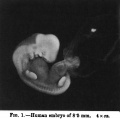
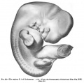
- Carnegie No. 2, 7 mm. Described in detail by Mall (1891).[1]
- Hochstetter's embryo I, 7 mm. ( (No. 10 of Hochstetter's series) Described in monographic form by Elze (1907).[2]Includes illustrations of reconstructions. Also shown in Fig. 52 of Keibel and Mall (1910).[3]
- Legg embryo, 7 mm. Described in detail by Thompson (1915).[4]Includes illustrations of reconstructions.
- 8-mm embryo. The peripheral nervous system was described by Volcher (1963)[5] in this embryo of stage 15.
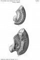
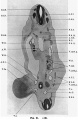
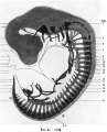
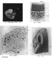
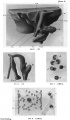
- Keibel No. 1495, 8.5 mm. This advanced example of stage 15 was well described in detail by Barniville (1914)[6].
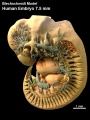
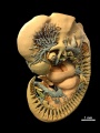
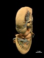
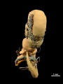
- Blechschmidt 7.5 mm Blechschmidt Model generated from serial sections of this embryo.
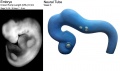
- Newcastle N340 Optical Projection Tomography of this embryo described by J. Kerwin etal., (2004).[7]
References
- ↑ Mall, F. P. 1891. A human embryo twenty-six days old. J. Morph, 5, 459-480.
- ↑ Elze, C. 1907. Beschreibung eines menschlichen Embryo von zirka 7 mm grosster Lange unter besonderer Beriicksichtigung der Frage nach der Enrwickelung der Extremitatenarterien und nach der morphologischen Bedeutung der lateralen Schilddriisenanlage. (Description of a human embryo of about 7 mm greatest length with special reference to the question of the development of the extremities arteries and to the morphological importance of lateral shield primordia). Anat. Hefte, 106, 411-492.
- ↑ Keibel F. and Mall FP. Manual of Human Embryology I. (1910) J. B. Lippincott Company, Philadelphia.
- ↑ Thompson, P. 1915. Description of a human embryo, 7 mm. greatest length. Studies in Anatomy. University of Birmingham, pp. 1-50.
- ↑ Volcher, R. 1963. Le systeme nerveux périphérique d'un embryon humain de 8 mm.(The peripheral nervous system of an 8 mm. human embryo) Arch. Biol. (Liege), 74, 95-127. PMID 13997757
- ↑ Barniville HL. The morphology and histology of a human embryo of 8.5 mm. (1914) J Anat. Physiol. 49(1):1-71. PMID 17233012
- ↑ <pubmed>15298700</pubmed>| PMC514604 | BMC Neurosci.
Subcategories
This category has the following 38 subcategories, out of 38 total.
C
- Carnegie Embryo 1006
- Carnegie Embryo 1091
- Carnegie Embryo 113
- Carnegie Embryo 1354
- Carnegie Embryo 1767
- Carnegie Embryo 2
- Carnegie Embryo 241
- Carnegie Embryo 2743
- Carnegie Embryo 3216
- Carnegie Embryo 3385
- Carnegie Embryo 3441
- Carnegie Embryo 3512
- Carnegie Embryo 371
- Carnegie Embryo 3805
- Carnegie Embryo 389
- Carnegie Embryo 3952
- Carnegie Embryo 4602
- Carnegie Embryo 4782
- Carnegie Embryo 5772
- Carnegie Embryo 6223
- Carnegie Embryo 6504
- Carnegie Embryo 6506
- Carnegie Embryo 6508
- Carnegie Embryo 6595
- Carnegie Embryo 7199
- Carnegie Embryo 721
- Carnegie Embryo 7324
- Carnegie Embryo 7364
- Carnegie Embryo 7370
- Carnegie Embryo 810
- Carnegie Embryo 855
- Carnegie Embryo 88
- Carnegie Embryo 8929
- Carnegie Embryo 8997
- Carnegie Embryo 9140
- Carnegy Embryo 2
W
Pages in category 'Carnegie Stage 15'
The following 57 pages are in this category, out of 57 total.
C
- Template:Carnegie Embryo Stage15
- Carnegie stage 15
- Template:Carnegie stage 15 links
- Template:CE1006
- Template:CE1091
- Template:CE113
- Template:CE1354
- Template:CE143
- Template:CE1767
- Template:CE2
- Template:CE241
- Template:CE2743
- Template:CE3216
- Template:CE3385
- Template:CE3441
- Template:CE3512
- Template:CE371
- Template:CE389
- Template:CE3952
- Template:CE4602
- Template:CE4782
- Template:CE5772
- Template:CE6223
- Template:CE6504
- Template:CE6506
- Template:CE6508
- Template:CE6595
- Template:CE7199
- Template:CE721
- Template:CE7324
- Template:CE7364
- Template:CE76
- Template:CE810
- Template:CE855
- Template:CE88
- Template:CE8929
- Template:CE8997
- Template:CE9140
- Template:CS15
P
- Paper - A Human Embryo Twenty-Six Days Old
- Paper - Developmental horizons in human embryos stages 15-18
- Paper - Growth allometry of the myocardium in human embryos from stages 15 to 23
- Paper - The Disappearance of the Precervical Sinus
- Paper - The Morphology and Histology of a Human Embryo of 8.5 mm
Media in category 'Carnegie Stage 15'
The following 67 files are in this category, out of 67 total.
 7.5mm Embryo movie 1 icon.jpg 299 × 400; 38 KB
 Bardeen1906-plate01.jpg 1,565 × 2,322; 238 KB
 Barniville1914 fig01.jpg 765 × 758; 59 KB
 Barniville1914 fig21.jpg 1,023 × 1,569; 301 KB
 Barniville1914 figA.jpg 1,000 × 1,237; 269 KB
 Barniville1914 plate01.jpg 2,042 × 2,271; 817 KB
 Barniville1914 plate02.jpg 1,458 × 2,509; 791 KB
 Carnegie stage 15 OPT.jpg 800 × 801; 56 KB
 Congdon1922-27-28.jpg 997 × 612; 68 KB
 Crowder1957 fig01.jpg 515 × 463; 91 KB
 Crowder1957 fig02.jpg 497 × 651; 132 KB
 Crowder1957 plate01.jpg 1,280 × 1,457; 548 KB
 Crowder1957 plate02.jpg 1,280 × 1,673; 645 KB
 Embryo 7.5mm model 01.gif 448 × 600; 903 KB
 Frazer1926 fig04.jpg 1,200 × 804; 95 KB
 Frazer1926 plate01.jpg 1,914 × 2,681; 469 KB
 Gilbert1957 fig02.jpg 1,280 × 824; 130 KB
 Human 7.5mm embryo model 01.jpg 747 × 1,000; 149 KB
 Human 7.5mm embryo model 02.jpg 747 × 1,000; 130 KB
 Human 7.5mm embryo model 03.jpg 747 × 1,000; 72 KB
 Human 7.5mm embryo model 04.jpg 747 × 1,000; 56 KB
 Human 7.5mm embryo model 05.jpg 747 × 1,000; 87 KB
 Human 7.5mm embryo model 06.jpg 747 × 1,000; 139 KB
 Human 7.5mm embryo model 07.jpg 747 × 1,000; 118 KB
 Human 7.5mm embryo model 08.jpg 747 × 1,000; 69 KB
 Human 7.5mm embryo model 09.jpg 747 × 1,000; 63 KB
 Human 7.5mm embryo model 10.jpg 747 × 1,000; 106 KB
 Human Carnegie stage 15 HOXC5 expression.jpg 670 × 504; 90 KB
 Human CS13-15 otic vesicle 01.jpg 1,574 × 1,779; 364 KB
 Human embryo olfactory 01.jpg 2,044 × 1,552; 986 KB
 Keibel Mall 052.jpg 604 × 600; 37 KB
 Mall1891 Fig01.jpg 623 × 576; 99 KB
 Mall1891 Fig02.jpg 600 × 347; 45 KB
 Mall1891 Plate01Fig01.jpg 623 × 831; 70 KB
 Mall1891 Plate01Fig02.jpg 623 × 831; 61 KB
 Mall1891 Plate02Fig01.jpg 623 × 831; 125 KB
 Mall1891 Plate02Fig02.jpg 623 × 831; 76 KB
 Pohlman1911 plate2.jpg 2,050 × 3,230; 331 KB
 Pohlman1911 plate2D.jpg 1,822 × 1,397; 166 KB
 Stage15 bf1.jpg 1,000 × 750; 25 KB
 Stage15 bf10.jpg 448 × 600; 28 KB
 Stage15 bf1a.jpg 800 × 600; 17 KB
 Stage15 bf1b.jpg 600 × 450; 11 KB
 Stage15 bf1c.jpg 400 × 300; 6 KB
 Stage15 bf2.jpg 1,874 × 2,000; 1.36 MB
 Stage15 bf21.jpg 600 × 450; 27 KB
 Stage15 bf2a.jpg 959 × 1,024; 344 KB
 Stage15 bf2b.jpg 468 × 500; 124 KB
 Stage15 bf3.jpg 448 × 600; 29 KB
 Stage15 bf4.jpg 448 × 600; 22 KB
 Stage15 bf5.jpg 448 × 600; 23 KB
 Stage15 bf6.jpg 448 × 600; 28 KB
 Stage15 bf7.jpg 448 × 600; 28 KB
 Stage15 bf8.jpg 448 × 600; 22 KB
 Stage15 bf9.jpg 448 × 600; 22 KB
 Stage15 embryo and brain 01.jpg 1,280 × 752; 81 KB
 Stage15 sagittal section upper half 01.jpg 1,500 × 1,206; 211 KB
 Stage15dorsal7207.jpg 1,252 × 1,536; 68 KB
 Stage15left7207.jpg 1,252 × 1,536; 106 KB
 Stage15lefthead7207.jpg 1,280 × 961; 128 KB
 Stage15right7207.jpg 1,252 × 1,536; 100 KB
 Stage15ventral7207.jpg 1,252 × 1,536; 65 KB
 Streeter1922-fig14.jpg 617 × 672; 70 KB
 Streeter1957 fig01.jpg 1,292 × 1,500; 218 KB
 Sudler1902-fig05.jpg 1,000 × 769; 182 KB
 Sudler1902-fig06.jpg 600 × 865; 146 KB
 Sudler1902-fig07.jpg 1,000 × 1,089; 148 KB


