Category:Carnegie Stage 13: Difference between revisions
From Embryology
mNo edit summary |
|||
| Line 15: | Line 15: | ||
* [[:File:Keibel Mall 034a.jpg|His embryo (a)]], 4 mm. Described in detail by His (1880).<ref name="His1880">His, W. 1880-1885. [[Book - Anatomy Of Human Embryos|'''Anatomie menscblicher Embryonen''']]. Vogel, Leipzig.</ref> | * [[:File:Keibel Mall 034a.jpg|His embryo (a)]], 4 mm. Described in detail by His (1880).<ref name="His1880">His, W. 1880-1885. [[Book - Anatomy Of Human Embryos|'''Anatomie menscblicher Embryonen''']]. Vogel, Leipzig.</ref> | ||
* Fischel embryo, 4.2 mm, Hochstetter Atlas. This embryo is sharply and spirally curved, and the length given is approximate. Its place in stage 13 is verified by the form of the limb buds. | * Fischel embryo, 4.2 mm, Hochstetter Atlas.<ref>Hochstetter, F. 1907. Atlas. Munich. Cited by Streeter (1945).</ref> This embryo is sharply and spirally curved, and the length given is approximate. Its place in stage 13 is verified by the form of the limb buds. | ||
* Keibel embryo No. 112, 5.3 mm. Described in the Keibel and Elze Normentafeln (1908). The lens is a flat, slightly thickened disc. The otic vesicle is detached, but a remnant of the stalk is still present; there is some indication of the endolymphatic appendage. There are 36 pairs of somites. | * Keibel embryo No. 112, 5.3 mm. Described in the Keibel and Elze Normentafeln (1908).<ref name="KeibelElze1908">Keibel, F., and Elze, C. 1908. Normentafeln zur Entwicklungsgeschichte der Wirbeltiere.(Normal Plates for Evolution of Vertebrates) [[Book - Normal Plates of the Development of Vertebrates 8|'''8. Heft ''Normentafeln zur Entwicklungsgeschichte des Menschen''''']] (Vol. 8. Normal Plates of the Development of the Human Embryo) Fisher, Jena., Germany.</ref> The lens is a flat, slightly thickened disc. The otic vesicle is detached, but a remnant of the stalk is still present; there is some indication of the endolymphatic appendage. There are 36 pairs of somites. | ||
* R. Meyer, 5-mm embryo, No. 318, Anatomisches Institut, Zurich. This embryo is a more advanced example of stage 13. The epithelium of the lens disc begins to indent. There are 38 pairs of somites. The embryo is referred to in the Keibel and Elze Normentafeln. | * R. Meyer, 5-mm embryo, No. 318, Anatomisches Institut, Zurich. This embryo is a more advanced example of stage 13. The epithelium of the lens disc begins to indent. There are 38 pairs of somites. The embryo is referred to in the Keibel and Elze Normentafeln.<ref name="KeibelElze1908"/> | ||
* C. Rabl, 4-mm embryo. The specimen closely resembles the Fol 5.6-mm embryo and the Hertwig G 31 embryo. It was used by Rabl in his study of the face (1902). | * C. Rabl, 4-mm embryo. The specimen closely resembles the Fol 5.6-mm embryo and the Hertwig G 31 embryo. It was used by Rabl in his study of the face (1902). | ||
* Broman, embryo Lf., 3 mm, Anatomisches Institut, Lund. This embryo was described systematically by Broman (1896). He made revisions for the Keibel and Elze Normentafeln. Broman reports that it has 30 pairs of somites. Regarding its relatively small size, it is to be noted that it was fixed in absolute alcohol and then preserved for two years in weak spirits. This embryo is a less advanced example of stage 13. | * Broman, embryo Lf., 3 mm, Anatomisches Institut, Lund. This embryo was described systematically by Broman (1896). He made revisions for the Keibel and Elze Normentafeln.<ref name="KeibelElze1908"/> Broman reports that it has 30 pairs of somites. Regarding its relatively small size, it is to be noted that it was fixed in absolute alcohol and then preserved for two years in weak spirits. This embryo is a less advanced example of stage 13. | ||
* Carnegie No. 148, 4.3 mm. A monographic description of this embryo was published by Gage (1905). | * Carnegie No. 148, 4.3 mm. A monographic description of this embryo was published by Gage (1905). | ||
* Fol, 5.6-mm embryo. This well-preserved embryo is a more advanced example. It was carefully described and illustrated by Fol (1884). | * Fol, 5.6-mm embryo. This well-preserved embryo is a more advanced example. It was carefully described and illustrated by Fol (1884). | ||
* Hertwig, 4.9-mm embryo, G31 Anatomisches-biologisches Institut, Berlin. This well-preserved embryo, a typical representative of stage 13, was first described in a study of the development of the pancreas by Jankelowitz (1895). An excellent systematic study followed later (Ingalis, 1907, 1908). The embryo was considered also in the Keibel and Elze Normentafeln. There are 35 pairs of somites. | * Hertwig, 4.9-mm embryo, G31 Anatomisches-biologisches Institut, Berlin. This well-preserved embryo, a typical representative of stage 13, was first described in a study of the development of the pancreas by Jankelowitz (1895). An excellent systematic study followed later (Ingalis, 1907, 1908). The embryo was considered also in the Keibel and Elze Normentafeln.<ref name="KeibelElze1908"/> There are 35 pairs of somites. | ||
* 4-mm and 5-mm twin embryos, University of Basel. A graphic reconstruction of the normal specimen was Issued by Müller and O'Rahilly (1980a), and reconstructions, with particular reference to the nervous system, were published by Müller and O'Rahilly (1984), who described the cerebral dysraphia (future anencephaly) present in one twin. | * 4-mm and 5-mm twin embryos, University of Basel. A graphic reconstruction of the normal specimen was Issued by Müller and O'Rahilly (1980a), and reconstructions, with particular reference to the nervous system, were published by Müller and O'Rahilly (1984), who described the cerebral dysraphia (future anencephaly) present in one twin. | ||
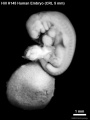
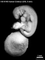
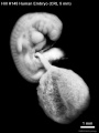
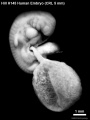
* Free Hospital for Women, Brookline, Massachusetts, No. 5, 5 mm. The histochemistry of this embryo was studied by McKay et al. (1955). | * Free Hospital for Women, Brookline, Massachusetts, No. 5, 5 mm. The histochemistry of this embryo was studied by McKay et al. (1955). | ||
Revision as of 21:16, 20 July 2015
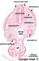
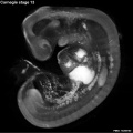
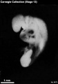
Carnegie Stage 13
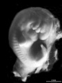
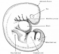
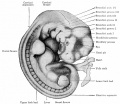
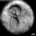
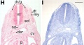
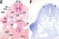
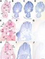
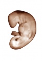
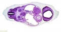
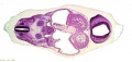
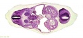
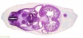
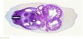
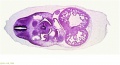
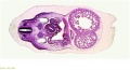
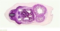
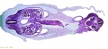
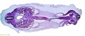
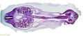
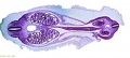
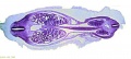
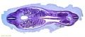
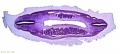
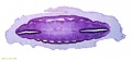
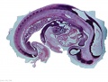
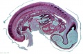
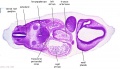
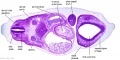
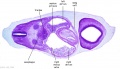
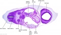
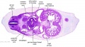
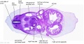
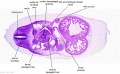
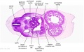
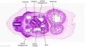
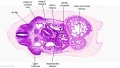
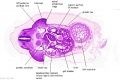
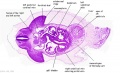
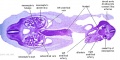
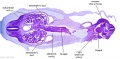
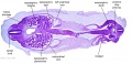
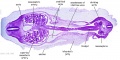
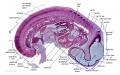
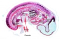
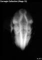
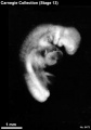
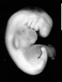
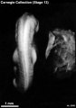
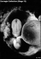
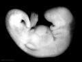
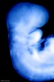
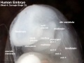
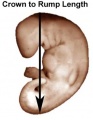
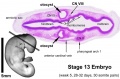
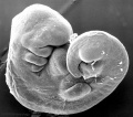
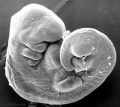
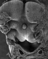
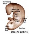
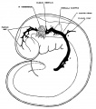
This Embryology category shows pages and media related to embryonic development in week 4 to week 5, 28 - 32 days, GA week 6-7. The embryos have a crown rump length (CRL) of 4 - 6 mm and somite number 30 pairs.
There is also a specific Carnegie stage 13 resource page.
| Week: | 1 | 2 | 3 | 4 | 5 | 6 | 7 | 8 |
| Carnegie stage: | 1 2 3 4 | 5 6 | 7 8 9 | 10 11 12 13 | 14 15 | 16 17 | 18 19 | 20 21 22 23 |
- Carnegie Stages: 1 | 2 | 3 | 4 | 5 | 6 | 7 | 8 | 9 | 10 | 11 | 12 | 13 | 14 | 15 | 16 | 17 | 18 | 19 | 20 | 21 | 22 | 23 | About Stages | Timeline
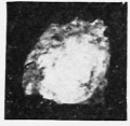
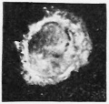
Embryo Examples
- His embryo (a), 4 mm. Described in detail by His (1880).[1]
- Fischel embryo, 4.2 mm, Hochstetter Atlas.[2] This embryo is sharply and spirally curved, and the length given is approximate. Its place in stage 13 is verified by the form of the limb buds.
- Keibel embryo No. 112, 5.3 mm. Described in the Keibel and Elze Normentafeln (1908).[3] The lens is a flat, slightly thickened disc. The otic vesicle is detached, but a remnant of the stalk is still present; there is some indication of the endolymphatic appendage. There are 36 pairs of somites.
- R. Meyer, 5-mm embryo, No. 318, Anatomisches Institut, Zurich. This embryo is a more advanced example of stage 13. The epithelium of the lens disc begins to indent. There are 38 pairs of somites. The embryo is referred to in the Keibel and Elze Normentafeln.[3]
- C. Rabl, 4-mm embryo. The specimen closely resembles the Fol 5.6-mm embryo and the Hertwig G 31 embryo. It was used by Rabl in his study of the face (1902).
- Broman, embryo Lf., 3 mm, Anatomisches Institut, Lund. This embryo was described systematically by Broman (1896). He made revisions for the Keibel and Elze Normentafeln.[3] Broman reports that it has 30 pairs of somites. Regarding its relatively small size, it is to be noted that it was fixed in absolute alcohol and then preserved for two years in weak spirits. This embryo is a less advanced example of stage 13.
- Carnegie No. 148, 4.3 mm. A monographic description of this embryo was published by Gage (1905).
- Fol, 5.6-mm embryo. This well-preserved embryo is a more advanced example. It was carefully described and illustrated by Fol (1884).
- Hertwig, 4.9-mm embryo, G31 Anatomisches-biologisches Institut, Berlin. This well-preserved embryo, a typical representative of stage 13, was first described in a study of the development of the pancreas by Jankelowitz (1895). An excellent systematic study followed later (Ingalis, 1907, 1908). The embryo was considered also in the Keibel and Elze Normentafeln.[3] There are 35 pairs of somites.
- 4-mm and 5-mm twin embryos, University of Basel. A graphic reconstruction of the normal specimen was Issued by Müller and O'Rahilly (1980a), and reconstructions, with particular reference to the nervous system, were published by Müller and O'Rahilly (1984), who described the cerebral dysraphia (future anencephaly) present in one twin.
- Free Hospital for Women, Brookline, Massachusetts, No. 5, 5 mm. The histochemistry of this embryo was studied by McKay et al. (1955).
References
- ↑ His, W. 1880-1885. Anatomie menscblicher Embryonen. Vogel, Leipzig.
- ↑ Hochstetter, F. 1907. Atlas. Munich. Cited by Streeter (1945).
- ↑ 3.0 3.1 3.2 3.3 Keibel, F., and Elze, C. 1908. Normentafeln zur Entwicklungsgeschichte der Wirbeltiere.(Normal Plates for Evolution of Vertebrates) 8. Heft Normentafeln zur Entwicklungsgeschichte des Menschen (Vol. 8. Normal Plates of the Development of the Human Embryo) Fisher, Jena., Germany.
Subcategories
This category has the following 42 subcategories, out of 42 total.
C
- Carnegie Embryo 1
- Carnegie Embryo 112
- Carnegie Embryo 116
- Carnegie Embryo 148
- Carnegie Embryo 1787
- Carnegie Embryo 186
- Carnegie Embryo 19
- Carnegie Embryo 239
- Carnegie Embryo 248
- Carnegie Embryo 3956
- Carnegie Embryo 4046
- Carnegie Embryo 407
- Carnegie Embryo 463
- Carnegie Embryo 523
- Carnegie Embryo 5541
- Carnegie Embryo 5682
- Carnegie Embryo 5874
- Carnegie Embryo 588
- Carnegie Embryo 6032
- Carnegie Embryo 6469
- Carnegie Embryo 6473
- Carnegie Embryo 7433
- Carnegie Embryo 7618
- Carnegie Embryo 7669
- Carnegie Embryo 786
- Carnegie Embryo 7889
- Carnegie Embryo 800
- Carnegie Embryo 8066
- Carnegie Embryo 808
- Carnegie Embryo 8119
- Carnegie Embryo 8147
- Carnegie Embryo 8239
- Carnegie Embryo 836
- Carnegie Embryo 8372
- Carnegie Embryo 8581
- Carnegie Embryo 8967
- Carnegie Embryo 9296
- Carnegie Embryo 9297
- Carnegie Embryo 963
- Carnegie Embryo 9697
W
Pages in category 'Carnegie Stage 13'
The following 81 pages are in this category, out of 81 total.
C
- Cardiovascular 3D stage 13 Movie
- Template:Carnegie Collection stage 13 table
- Template:Carnegie Embryo Stage13
- Carnegie stage 13
- Carnegie stage 13 - serial sections
- Template:Carnegie stage 13 links
- Template:CE1
- Template:CE1075
- Template:CE112
- Template:CE116
- Template:CE136
- Template:CE148
- Template:CE186
- Template:CE239
- Template:CE248
- Template:CE3956
- Template:CE4046
- Template:CE407
- Template:CE523
- Template:CE5541
- Template:CE5682
- Template:CE5874
- Template:CE588
- Template:CE6032
- Template:CE6469
- Template:CE6473
- Template:CE7433
- Template:CE7618
- Template:CE7669
- Template:CE786
- Template:CE7889
- Template:CE800
- Template:CE8066
- Template:CE808
- Template:CE8119
- Template:CE8147
- Template:CE8239
- Template:CE826
- Template:CE836
- Template:CE8372
- Template:CE8581
- Template:CE87
- Template:CE8967
- Template:CE9296
- Template:CE9297
- Template:CE963
- Template:CE9697
- Template:CS13
E
P
- Paper - Developmental horizons in human embryos. Description of age group XIII
- Paper - Histochemical horizons in human embryos - Stage 13
- Paper - Normal development of early human embryos: Observation of 90 specimens at Carnegie stages 7 to 13
- Paper - The Disappearance of the Precervical Sinus
- Placenta - Stage 13
R
Media in category 'Carnegie Stage 13'
The following 189 files are in this category, out of 189 total.
 050653-01.jpg 1,122 × 1,490; 124 KB
 Anderson2016-fig37.jpg 800 × 800; 69 KB
 Bailey086.jpg 720 × 662; 85 KB
 Bailey087.jpg 928 × 803; 132 KB
 Bailey088.jpg 888 × 701; 102 KB
 Bailey246.jpg 806 × 681; 89 KB
 Bailey256.jpg 894 × 545; 75 KB
 Beverley and Mark 1997.jpg 800 × 542; 88 KB
 Boyd1950 fig07.jpg 884 × 1,000; 159 KB
 Carnegie stage 13 caudal trunk.jpg 400 × 625; 53 KB
 Carnegie stage 13 OPT.jpg 800 × 801; 53 KB
 Congdon1922-32.jpg 1,133 × 1,000; 132 KB
 Congdon1922-plate01.jpg 877 × 1,200; 145 KB
 Frazer1910 fig02.jpg 750 × 804; 144 KB
 Frazer1926 fig03.jpg 1,200 × 804; 69 KB
 Frazer1926 plate01.jpg 1,914 × 2,681; 469 KB
 Gilbert1957 fig01.jpg 1,280 × 1,579; 156 KB
 HillH145 Stage 13 bf01.jpg 908 × 1,210; 81 KB
 HillH145 Stage 13 bf02.jpg 908 × 1,210; 83 KB
 HillH145 Stage 13 bf03.jpg 908 × 1,210; 86 KB
 HillH145 Stage 13 bf04.jpg 908 × 1,210; 84 KB
 HillH145 Stage 13 bf05.jpg 1,257 × 1,257; 240 KB
 HillH145 Stage 13 bf06.jpg 1,257 × 1,257; 244 KB
 HillH145 Stage 13 bf07.jpg 1,257 × 1,257; 244 KB
 HillH145 Stage 13 bf08.jpg 1,257 × 1,257; 224 KB
 Human Carnegie stage 13 GJA1 expression 2.jpg 705 × 384; 76 KB
 Human Carnegie stage 13 GJA1 expression.jpg 706 × 470; 96 KB
 Human Carnegie stage 13 SOX11 MAZ GJA1 expression.jpg 777 × 1,000; 192 KB
 Human Carnegie stage 13 stem cell markers.jpg 657 × 585; 78 KB
 Human CS13 otic vesicle 01.jpg 1,028 × 774; 112 KB
 Human CS13-15 otic vesicle 01.jpg 1,574 × 1,779; 364 KB
 Human embryonic tongue 03.jpg 650 × 470; 159 KB
 Human embryonic tongue 04.jpg 650 × 470; 131 KB
 Human neural crest cell migration-in vitro.jpg 1,280 × 959; 163 KB
 Human Stage13 sagittal upper half01.jpg 1,518 × 2,048; 269 KB
 Human Stage13 sagittal upper half02.jpg 1,518 × 2,048; 289 KB
 ILP2006 Carnegie stage 13.jpg 337 × 490; 63 KB
 Mall1908a fig136a.jpg 269 × 262; 8 KB
 Mall1908a fig136b.jpg 263 × 250; 9 KB
 McClure1925 fig01.jpg 1,000 × 1,585; 116 KB
 McClure1925 fig02.jpg 1,000 × 1,364; 159 KB
 ME18 001.jpg 773 × 1,200; 176 KB
 Pohlman1911 plate1.jpg 1,851 × 2,888; 372 KB
 Pohlman1911 plate1B.jpg 1,226 × 1,449; 180 KB
 Stage 13 image 001.jpg 1,000 × 357; 44 KB
 Stage 13 image 002.jpg 1,000 × 359; 52 KB
 Stage 13 image 003.jpg 1,000 × 436; 71 KB
 Stage 13 image 004.jpg 1,000 × 386; 65 KB
 Stage 13 image 005.jpg 1,000 × 451; 81 KB
 Stage 13 image 006.jpg 1,000 × 439; 83 KB
 Stage 13 image 007.jpg 1,000 × 514; 93 KB
 Stage 13 image 008.jpg 1,000 × 436; 78 KB
 Stage 13 image 009.jpg 1,000 × 449; 86 KB
 Stage 13 image 010.jpg 1,000 × 463; 85 KB
 Stage 13 image 011.jpg 1,000 × 454; 91 KB
 Stage 13 image 012.jpg 1,000 × 524; 98 KB
 Stage 13 image 013.jpg 1,000 × 507; 98 KB
 Stage 13 image 014.jpg 1,000 × 459; 95 KB
 Stage 13 image 015.jpg 1,000 × 519; 80 KB
 Stage 13 image 016.jpg 1,000 × 525; 82 KB
 Stage 13 image 017.jpg 1,000 × 522; 87 KB
 Stage 13 image 018.jpg 1,000 × 473; 87 KB
 Stage 13 image 019.jpg 1,000 × 457; 85 KB
 Stage 13 image 020.jpg 1,000 × 504; 100 KB
 Stage 13 image 021.jpg 1,000 × 461; 97 KB
 Stage 13 image 022.jpg 1,000 × 473; 101 KB
 Stage 13 image 023.jpg 1,000 × 544; 110 KB
 Stage 13 image 024.jpg 1,000 × 538; 116 KB
 Stage 13 image 025.jpg 1,000 × 516; 128 KB
 Stage 13 image 026.jpg 1,000 × 526; 120 KB
 Stage 13 image 027.jpg 1,000 × 590; 123 KB
 Stage 13 image 028.jpg 1,000 × 537; 120 KB
 Stage 13 image 029.jpg 1,000 × 486; 76 KB
 Stage 13 image 030.jpg 1,000 × 365; 57 KB
 Stage 13 image 031.jpg 1,000 × 453; 79 KB
 Stage 13 image 032.jpg 1,000 × 449; 81 KB
 Stage 13 image 033.jpg 1,000 × 451; 87 KB
 Stage 13 image 034.jpg 1,000 × 441; 87 KB
 Stage 13 image 035.jpg 1,000 × 425; 88 KB
 Stage 13 image 036.jpg 1,000 × 436; 82 KB
 Stage 13 image 037.jpg 1,000 × 423; 81 KB
 Stage 13 image 038.jpg 1,000 × 461; 90 KB
 Stage 13 image 039.jpg 1,000 × 425; 92 KB
 Stage 13 image 040.jpg 1,000 × 433; 104 KB
 Stage 13 image 041.jpg 1,000 × 446; 99 KB
 Stage 13 image 042.jpg 1,000 × 452; 99 KB
 Stage 13 image 043.jpg 1,000 × 444; 91 KB
 Stage 13 image 044.jpg 1,000 × 454; 93 KB
 Stage 13 image 045.jpg 1,000 × 451; 87 KB
 Stage 13 image 046.jpg 1,000 × 449; 82 KB
 Stage 13 image 047.jpg 1,000 × 469; 71 KB
 Stage 13 image 048.jpg 1,000 × 756; 136 KB
 Stage 13 image 049.jpg 1,000 × 649; 129 KB
 Stage 13 image 050.jpg 1,000 × 370; 52 KB
 Stage 13 image 051.jpg 1,000 × 382; 55 KB
 Stage 13 image 052.jpg 1,000 × 476; 81 KB
 Stage 13 image 053.jpg 1,000 × 423; 76 KB
 Stage 13 image 054.jpg 1,000 × 453; 86 KB
 Stage 13 image 055.jpg 1,000 × 466; 98 KB
 Stage 13 image 056.jpg 1,000 × 516; 102 KB
 Stage 13 image 057.jpg 1,000 × 511; 99 KB
 Stage 13 image 058.jpg 1,000 × 481; 94 KB
 Stage 13 image 059.jpg 1,000 × 513; 92 KB
 Stage 13 image 060.jpg 1,000 × 486; 96 KB
 Stage 13 image 061.jpg 1,000 × 600; 101 KB
 Stage 13 image 062.jpg 1,000 × 573; 97 KB
 Stage 13 image 063.jpg 1,000 × 496; 100 KB
 Stage 13 image 064.jpg 1,000 × 577; 78 KB
 Stage 13 image 065.jpg 1,000 × 576; 85 KB
 Stage 13 image 066.jpg 1,000 × 579; 93 KB
 Stage 13 image 067.jpg 1,000 × 577; 93 KB
 Stage 13 image 068.jpg 1,000 × 557; 97 KB
 Stage 13 image 069.jpg 1,000 × 581; 100 KB
 Stage 13 image 070.jpg 1,000 × 554; 101 KB
 Stage 13 image 071.jpg 1,000 × 526; 100 KB
 Stage 13 image 072.jpg 1,000 × 614; 116 KB
 Stage 13 image 073.jpg 1,000 × 619; 118 KB
 Stage 13 image 074.jpg 1,000 × 549; 113 KB
 Stage 13 image 075.jpg 1,000 × 567; 116 KB
 Stage 13 image 076.jpg 1,000 × 667; 120 KB
 Stage 13 image 077.jpg 1,000 × 612; 127 KB
 Stage 13 image 078.jpg 1,000 × 486; 81 KB
 Stage 13 image 079.jpg 1,000 × 409; 80 KB
 Stage 13 image 080.jpg 1,000 × 533; 91 KB
 Stage 13 image 081.jpg 1,000 × 449; 90 KB
 Stage 13 image 082.jpg 1,000 × 451; 90 KB
 Stage 13 image 083.jpg 1,000 × 440; 98 KB
 Stage 13 image 084.jpg 1,000 × 420; 101 KB
 Stage 13 image 085.jpg 1,000 × 434; 99 KB
 Stage 13 image 086.jpg 1,000 × 499; 97 KB
 Stage 13 image 087.jpg 1,000 × 493; 95 KB
 Stage 13 image 088.jpg 1,000 × 484; 97 KB
 Stage 13 image 089.jpg 1,000 × 498; 104 KB
 Stage 13 image 090.jpg 1,000 × 512; 103 KB
 Stage 13 image 091.jpg 1,000 × 470; 93 KB
 Stage 13 image 092.jpg 1,000 × 481; 86 KB
 Stage 13 image 093.jpg 1,000 × 454; 92 KB
 Stage 13 image 094.jpg 1,000 × 469; 90 KB
 Stage 13 image 095.jpg 1,000 × 452; 76 KB
 Stage 13 image 096.jpg 1,000 × 469; 75 KB
 Stage 13 image 097.jpg 1,000 × 720; 162 KB
 Stage 13 image 098.jpg 1,000 × 623; 144 KB
 Stage 13 image 099.jpg 350 × 500; 56 KB
 Stage 13 image 100.jpg 350 × 500; 56 KB
 Stage 13 image 101.jpg 1,000 × 649; 65 KB
 Stage 13 kidney sections 2.jpg 600 × 400; 53 KB
 Stage 13 MRI 3D02.mp4 ; 440 KB
- Stage 13 MRI 3D03.mp4 ; 1.38 MB
 Stage13 and 22 thyroid development a.jpg 800 × 640; 94 KB
 Stage13 and 22 thyroid development.jpg 1,000 × 800; 283 KB
 Stage13 bf1.jpg 1,000 × 868; 31 KB
 Stage13 bf13.jpg 412 × 590; 19 KB
 Stage13 bf14.jpg 412 × 590; 20 KB
 Stage13 bf15.jpg 412 × 590; 20 KB
 Stage13 bf16.jpg 1,536 × 2,048; 188 KB
 Stage13 bf17.jpg 412 × 590; 23 KB
 Stage13 bf18.jpg 412 × 590; 28 KB
 Stage13 bf19.jpg 412 × 590; 25 KB
 Stage13 bf1a.jpg 800 × 694; 23 KB
 Stage13 bf1c.jpg 400 × 347; 8 KB
 Stage13 bf2.jpg 1,000 × 750; 61 KB
 Stage13 bf20.jpg 412 × 590; 23 KB
 Stage13 bf21.jpg 412 × 590; 32 KB
 Stage13 bf2a.jpg 800 × 600; 40 KB
 Stage13 bf2b.jpg 600 × 450; 24 KB
 Stage13 bf2c.jpg 400 × 300; 12 KB
 Stage13 bf3.jpg 1,317 × 2,000; 312 KB
 Stage13 bf3a.jpg 659 × 1,000; 99 KB
 Stage13 bf3b.jpg 527 × 800; 64 KB
 Stage13 bf8.jpg 600 × 451; 32 KB
 Stage13 crown rump length.jpg 288 × 371; 10 KB
 Stage13 face ventral view01.jpg 1,290 × 2,048; 152 KB
 Stage13 oral cavity floor01.jpg 1,315 × 2,048; 234 KB
 Stage13 oral cavity floor02.jpg 1,315 × 2,048; 371 KB
 Stage13 otocyst.jpg 1,000 × 655; 55 KB
 Stage13 pharyngeal arch excerpts.gif 600 × 300; 86 KB
 Stage13 sem1.jpg 1,000 × 886; 80 KB
 Stage13 sem1a.jpg 800 × 709; 57 KB
 Stage13 sem1b.jpg 600 × 532; 36 KB
 Stage13 sem1c.jpg 400 × 355; 18 KB
 Stage13 sem3.jpg 818 × 1,000; 81 KB
 Stage13 sem3c.jpg 327 × 400; 20 KB
 Stage13 spinal cord01.jpg 807 × 637; 51 KB
 Stage13 spinal cord02.jpg 807 × 637; 58 KB
 Stage13 surface bulges.jpg 337 × 389; 14 KB
 Streeter-plate02.jpg 1,200 × 871; 180 KB
 Streeter1915 fig01.jpg 1,000 × 1,133; 124 KB
 Streeter1921 fig13.jpg 800 × 600; 45 KB
 Streeter1957 fig01.jpg 1,292 × 1,500; 218 KB


