Category:Carnegie Embryo 22: Difference between revisions
mNo edit summary |
mNo edit summary |
||
| (9 intermediate revisions by the same user not shown) | |||
| Line 1: | Line 1: | ||
[[File:Keibel Mall 143.jpg|thumb|Carnegie Embryo No. 22]] | |||
This {{Embryology}} category shows pages and images that relate to the Carnegie Collection Embryo No. {{CE22}}. This embryo was classified as [[Carnegie stage 21|Stage 21]] in [[Week 8]]. | This {{Embryology}} category shows pages and images that relate to the Carnegie Collection Embryo No. {{CE22}}. This embryo was classified as [[Carnegie stage 21|Stage 21]] in [[Week 8]]. | ||
{{Carnegie stage 21 links}} | |||
<br> | |||
{| class="wikitable" | {| class="wikitable" | ||
|- | |- | ||
! Serial No. !! Size (mm) !! Grade !! Fixative !! Embedding Medium !! Plane !! Thinness (µm) !! Stain !! Point Score !! Sex !! Year !! Notes | ! Serial No. !! Size (mm) !! Grade !! Fixative !! Embedding Medium !! Plane !! Thinness (µm) !! Stain !! Point Score !! Sex !! Year !! Notes | ||
|- | |- | ||
| | | {{CE22}} || E, 20 Ch, 35x30x30 || Good || Alc. || P || {{Transverse}} || 50 || Al. coch. || 34.5 || F || 1895 || | ||
|} | |} | ||
{{Carnegie Collection stage 21 table}} | {{Carnegie Collection stage 21 table}} | ||
<br> | |||
{{Carnegie_stage_table_1}} | |||
{{Carnegie_stages}} | |||
===References=== | ===References=== | ||
| Line 15: | Line 23: | ||
{{Ref-BardeenLewis1901}} | {{Ref-BardeenLewis1901}} | ||
{{Ref-Lewis1902}} | |||
{{Ref-Pearce1903}} | |||
{{Ref-Bardeen1905}} | {{Ref-Bardeen1905}} | ||
{{Ref-KeibelMall1910}} [[Book - Manual of Human Embryology 8|VIII. Determination of the Age of Human Embryos and Fetuses]] | |||
{{Ref-Lisser1911}} | {{Ref-Lisser1911}} | ||
{{Ref-Streeter1906ear}} | {{Ref-Streeter1906ear}} | ||
{{Ref-Streeter1957}} | |||
{{Carnegie numbered embryo links}} | {{Carnegie numbered embryo links}} | ||
[[Category:Carnegie Stage 21]][[Category:Week 8]] | ==Limb== | ||
{{Ref-Lewis1902}} | |||
Embryo [[:Category:Carnegie Embryo 22|'''XXII''']] measures 20 mm. V. B. and 18 mm. X. B. It is about seven weeks old. The entire arm has a more posterior position. The lower angle of the scapula is at the level of the sixth rib, its anterior limit is about at the seventh cervical vertebra. The entire arm as well as its various parts have increased in size. The muscles are sharper and better developed than the preceding stage. Every muscle that the adult arm presents can now be recognized and each one now contains muscle fibers. The tendons are better formed and can be traced farther towards their final insertions. The ligaments and fasciae are also more distinct. The process of ligament and tendon formation from the condensed mesenchyma is still in progress at the distal ends of the digits. The skeletal elements are for the most part fairly well formed in cartilage except the distal row of phalanges. | |||
===The Skeletal System=== | |||
====The vertebral column==== | |||
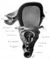
The intervertebral discs are reduced in thickness, while the bodies of the vertebra have increased and occnpy about four-fifths of each segment. The neural and transverse processes are larger and for the most part of cartilage. At the tip of the neural processes, which reach about one-half way around the spinal cord, is a small mass of condensed tissue at what juay be considered the growing point. These processes arise entirely from the body and not from the disc. So the body has probably grown at the expense of the disc. The perichondrium, wliich surrounds the body and its processes, is thickened along the ventral side of the bodies into the anterior common ligament. | |||
The {{rib}}s are of cartilage surrounded by thick perichondrium, wliicli is continuous with the condensed tissue anlage of the one-half the sternum. The distance between the two halves of the sternum is not as great as in the preceding stage and at the anterior end they ai'e just beginning to come in contact with each other. There are no joint cavities between the ribs and vertebrae. | |||
The {{clavicle}} is composed of cartilage somewhat different in appear ance from that in the other bones. It is continuous with the acromion and sternum by an area of condensed tissue. It is surrounded by a typical perichondrium. There are distinct coraco-clavicular, costoclavicular, and interclavicular ligaments. | |||
The cartilaginous scapula is very much larger than in the precedingstage and contains no large areas of condensed tissue. It has moved posteriorly and lies in the region from the last cervical to the fifth thoracic vertebrae. Its dorsal border also extends farther dorsal than in any of the preceding stages. The acromion and coracoid processes are large and of cartilage with only the ordinary thickness of perichondrium which is continuous with that surrounding the rest of the scapula. The spine has not yet appeared but the thickened anterior border from which the supraspinatus muscle arises probably represents by its lateral lip the spine and by its median lip the future anterior border. The acromion arise partially from the lateral side of the anterior border. The head seems to have enlarged at the expense of part of the base of the coracoid process as the long head of the biceps now arises from the junction of the coracoid and the head, and the head of the humerus does not rest against such a large proportional area of the coracoid. There is a distinct suprascapular and a coraco-acromial ligament. At the posterior angle of the scapula there is small mass of condensed tissue which gives attachment to a portion of the serratus, latissimus, and teres major muscles. | |||
The humerus is much hirger than in embryo XLIII, and has much the adult shape, though of course it is thicker in proportion to its length. It is composed of cartilage. There is a capsular and a coraco-humeral ligament. No joint cavity exists between the scapula and humerus. The tuberosities and condyles are fairly well formed in cartilage and •condensed tissue. The bicipital groove is present. | |||
The ulna and radius are larger and longer than in the preceding stage 3ind are well formed in cartilage. The olecranon, coranoid and styloid processes are partially formed in cartilage and condensed tissue. The perichondrium about the ulna and radius is quite thick. The capsular .and orbicular ligaments are present. No joint cavities exist and the cartilages are separated by condensed tissue continuous with the peri•ciiondrium. | |||
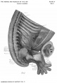
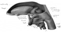
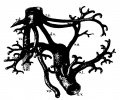
'''Fig. 13.''' Cartilaginous skeleton of the arm of embrj-o XXII, lateral view. X 12 diameters. | |||
All the bones of the carpus are represented by cartilage, and in about their relative positions. The amount of condensed tissue matrix is much less than in the preceding stage. The condensed tissue matrix is continuous with the ulna and radius and the five metacarpals without joint cavities. Indications of ligaments of the wrist are present. | |||
The five metacarpals are present in cartilage surrounded by thick perichondrium. The first is the shortest. | |||
The first two rows of phalanges are present in all the digits. They are of cartilage surrounded by a very thick perichondrium, which is continuous with the condensed tissue between them and the metacarpals and between the phalanges them.selves. It is also continuous with the enlarged condensed tissue tip of each digit. There are thickenings for the various ligaments connecting the metacarpals and phalanges and the phalanges with each other. | |||
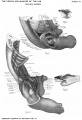
===The Muscular System=== | |||
(Plate II, Fig. C.) The trapezius muscle fibers extend from the occiput to the level of the sixth rib. There is a considerable interval of fascia connecting them to the neural processes of the lower cervical and the thoracic vertebrae. There is a tendonous attachment to the clavicle and acromion and into fascia or condensed tissne on the surface of the infraspinatus between the trapezius and the deltoid. | |||
The rliomhoid muscle lies in the region of the seventh cervical to the fourth thoracic vertebrae. It is inserted into the condensed tissue along the dorsal border of the scapula. | |||
The latissimus dorsi muscle fibers extend from the humerus to the level of the ninth rib. There is a considerable interval of fascia between them and the neural processes of the lower thoracic and the first two or three lumbar vertebrae. This dorsal fascia is not very well marked. The latissimus also has fibers attached to the condensed tissue at the inferior angle of the scapula. | |||
The serratus anterior muscle is separate from the levator scapulae except near its attachment to the scapula. It is a broad, thin sheet, having digitations to the first eight ribs. | |||
The pedoralis major muscle is well developed. The separation between the clavicular and the sterno-costal portions is less marked than in the preceding stage.' The muscle is attached as low as the sixth rib. | |||
The pedoralis minor muscle is quite distinct from the major, as a considerable layer of loose mesenchymal tissue lies between them. It arises from the second, third and fourth ribs and passes to the coracoid process. | |||
The siibclavius muscle is inserted into the clavicle at an angle of 45°. As the scapula and clavicle sink down towards the level of the first rib the angle at which this muscle is inserted into the clavicle decreases. | |||
The teres major muscle arises from the lower angle of the scapula and passes to the humerus. It is interesting to note that at this stage tendon of the latissimus dorsi twists around the lower l^order of the teres to be inserted with it into the humerus. | |||
The deltoid muscle is large and well developed. | |||
The suprasfinatus muscle arises from the thickened anterior border of the scapula. It cannot be said to take origin more from the lateral surface than from the median surface of the scapula. | |||
The infraspinatus muscle occupies the middle of the lateral surface of the scapula and passes beneath the deltoid to the great tuberosity of the humerus. | |||
The suhscapularis muscle arises from most of the median surface of the scapula. Its tendon of insertion is broad and thin and closely applied to the capsular ligament. | |||
The three heads of the triceps muscle are easily distinguished. The long and external heads are of about the same size. The anconeus muscle is continuous with the triceps but arises from the external condyle and passes to the side of the olecranon and adjoining surface of the shaft of the ulna. | |||
The long head of the biceps muscle arises from the junction of the coracoid process and the head of the scapula and passes through the bicipital groove. The two heads are inserted together into the condensed tissue swelling on the radius. | |||
The corncobracJtviUs muscle and short head of the biceps are intimately connected for most of the length of the former. | |||
The brachialis muscle has spread out over more of the distal surface of the humerus than in the preceding stage. | |||
The flexor muscles of the forearm are easier to distinguish than in the preceding stage. | |||
The tendon of the palinaris longus is narrower in proportion than in embryo XLIII. | |||
The tendon of the flexor carpi raclialis muscle can be traced farther towards its insertion into the base of the second metacarpal than in embryo XLIII. | |||
The muscle fibers of the flexor sublimis digitorum still run to the carpus before the ^\ide tendon begins. This tendon soon splits into four tendons which go to the four ulnar digits. These tendons are better formed than in the preceding stage and split to surround the tendons of the deep flexor. Their ends fuse with the thick perichondrium about the phalanges. | |||
The flexor carpi uhiaris muscle shows distinctly its two heads of origin. It has a well-formed tendon of insertion. | |||
The deep flexor muscles can be separated into the flexor polUcis longus and the flexor profundus digitormn muscles. The muscle fibers of the profundus continue into the carpal region and end in a broad tendon which divides at the base of the metacarpus into four well-formed tendons. These fuse with the condensed tissue at the tips of the digits. There is a slight split in each of these tendons near its end. The tendon of the flexor longus pollieis behaves similarly. | |||
The pronator quadratns muscle is oval in cross section, and connects the distal ends of the radius and ulna. | |||
The lumbricales are quite well developed and their fairly well-formed tendons end in the perichondrium on the radial side of the digit. | |||
The interossei mnscles and the small mnscles of the thumb and little finger are now fairly well developed. Muscle fibers are present. | |||
The extensor muscles of the forearm show considerable advance over the preceding stage. The tendons of the extensor communis digitoriim are longer and narrower. The muscle fibers continue to the base of the metacarpus, where the splitting into the four tendons takes place. The tendons are inserted into the condensed tissue tips of the digits. The edge of the tendons near their insertions are more or less continuous with the perichondrium about the digit. | |||
The tendon of the extensor carpi ulnaris is beginning to. form. One branch of it seems to join the communis tendon. This may be the tendon of the extensor minimi digiti. | |||
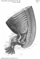
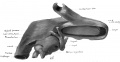
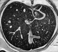
'''Fig. 14.''' Lateral view of the arm of embryo XXII. from Plate VIII. | |||
The extensor carpi radialis longior et hrevior are not to be separated. | |||
The supinator muscle is well developed and has the posterior interosseus nerve passing through it. | |||
The abductor pollicis longus and extensor pollicis brevis muscles are only to be separated where the muscle fibres pass into tendons, which fuse with the perichondrium of the first digit. The separation occurs at the lower end of the radius. These two muscles are fairly distinct from the supinator and the extensor pollicis longus and extensor indicis projiriiis muscles. The last two muscles are inseparable for part of their course and shortly after dividing each forms a round tendon. The extensor pollicis longus then spreads out into the sheath about the first digit. The extensor indicis muscle joins the nlnar side of the tendon of the commimis to the second digit. | |||
===The Nerves=== | |||
The drachial plexus has a decided posterior inclination and seems to have been pulled down against the first rib. The three cords are so close together that it was impossible to separate them satisfactorily though indications of the cords are present. There is nothing especially peculiar about the distribution of the nerves from the plexus either motor or sensory, which is not present in the adult. | |||
[[Category:Carnegie Stage 21]][[Category:Week 8]][[Category:Transverse]] | |||
[[Category:1800's]] | [[Category:1800's]] | ||
Latest revision as of 09:48, 20 October 2020

This Embryology category shows pages and images that relate to the Carnegie Collection Embryo No. 22. This embryo was classified as Stage 21 in Week 8.
| Serial No. | Size (mm) | Grade | Fixative | Embedding Medium | Plane | Thinness (µm) | Stain | Point Score | Sex | Year | Notes |
|---|---|---|---|---|---|---|---|---|---|---|---|
| 22 | E, 20 Ch, 35x30x30 | Good | Alc. | P | Transverse | 50 | Al. coch. | 34.5 | F | 1895 |
| Carnegie Collection - Stage 21 | |||||||||||
|---|---|---|---|---|---|---|---|---|---|---|---|
| Serial No. | Size (mm) | Grade | Fixative | Embedding Medium | Plane | Thinness (µm) | Stain | Point Score | Sex | Year | Notes |
| 22 | E, 20 Ch, 35x30x30 | Good | Alc. | P | Transverse | 50 | Al. coch. | 34.5 | Female | 1895 | |
| 57 | E, 23 Ch., ca. 30 | Poor | Alc. | P | Sagittal | 50 | Al. coch. | 36 | Male | 1896 | |
| 128 | E, 20 Ch., 50x43 | Good | Formalin | P | Coronal | 50 | Al. coch. | 33 | Female | 1898 | |
| 229 | E, 19 | Poor | Alc. | P | Sagittal | 50 | Al. coch. | 33 | Female | 1903 | |
| 349 | E, 24 | Good | Zenker | C | Coronal | 250 | Unstained | 36 | ? | 1905 | Double vascular injection |
| 455 | E, 24 Ch., 42x34x20 | Good | Alc. | P | Transverse | 30 | (Stain - Haematoxylin Eosin) | 36.5 | Male | 1910 | |
| 632 | E, 24 Ch., 60x50x30 | Good | Bichlor. acetic | P | Sagittal | 40, 100, 250 | Al. coch. | 33 | Female | 1913 | Injected |
| 903C | E, 23.5 | Good | Formalin | P | Transverse | 40 | Al. coch. | 38.5 | Female | 1914 | |
| 1008 | E, 26,4 | Good | Formalin | P | Sagittal | 40 | Al. coch. | 39 | ?? | 1914 | |
| 1358F | E, 23 | Good | Formalin | P | Sagittal | 40 | Al. coch. | 37.5 | Female | 1916 | |
| 2937 | E,, 24.2 | Good | Bouin | P | Transverse | 50 | (Stain - Haematoxylin Eosin) aur., or. G. | 39 | Female | 1920 | |
| 3167 | E., 24.5 Ch., 60x50x40 | Poor | Bichlor, acetic, formol | P | Transverse | 20 | Al. coch. | 32 | Male | 1920 | |
| 4090 | E, 22.2 Ch.. 66x46x30 | Good | Formalin | P | Transverse | 40 | Al. coch. | 30 | Female | 1922 | |
| 4160 | E,25 | Poor | Formalin | P | Sagittal | 25 | (Stain - Haematoxylin Eosin) | 39 | Male | 1923 | Tubal |
| 4960 | E.22 Ch,, 47x42x28 | Good | Formalin | P | Transverse | 15 | Al. coch., Mallory | 31.5 | Female | 1925 | |
| 5??6 | E. 215 | Good | Formalin | P | Sagittal | 20 | (Stain - Haematoxylin Eosin) | 34 | Female | 1927 | |
| 6531 | E,22 | Poor | Glacial acetic, | C-P | Transverse | 10 | (Stain - Haematoxylin Eosin) | 31.5 | Female | 1931 | Leitz Collection |
| 7254 | E,225 | Exc | Bouin | C-P | Transverse | 20 | (Stain - Haematoxylin Eosin) | 33.5 | Male | 1936 | |
| 7592 | E,22-> | Exc. | Bouin | C-P | Transverse | 20 | (Stain - Haematoxylin Eosin) | 36 | Female | 1937 | |
| 7864 | E., 24 | Exc, | Formalin | C-P | Frontal | 20 | (Stain - Haematoxylin Eosin) | 32.5 | Male | 1941 | |
| 8553 | E., 22 | Exc | Bouin | C-P | Transverse | 12 | (Stain - Haematoxylin Eosin) | 38 | Female | 1947 | |
| 9614 | E,,22 5 | Exc | Bouin | P | Coronal | 10 &15 | Azan | ? | ? | 1958 | Rubella. Hysterectomy |
Abbreviations
| |||||||||||
| Week: | 1 | 2 | 3 | 4 | 5 | 6 | 7 | 8 |
| Carnegie stage: | 1 2 3 4 | 5 6 | 7 8 9 | 10 11 12 13 | 14 15 | 16 17 | 18 19 | 20 21 22 23 |
- Carnegie Stages: 1 | 2 | 3 | 4 | 5 | 6 | 7 | 8 | 9 | 10 | 11 | 12 | 13 | 14 | 15 | 16 | 17 | 18 | 19 | 20 | 21 | 22 | 23 | About Stages | Timeline
References
Mall FP. Development of the human coelom. (1897) J Morphol. 12: 395-453.
Bardeen CR. and Lewis WH. The development of the limbs, body-wall and back. (1901) Amer. J Anat. 1: 1-36.
Lewis WH. The development of the arm in man. (1902) Amer. J Anat. 1(2): 145-184.
Pearce RM. The development of the islands of Langerhans in the human embryo. (1903) Amer. J Anat. : 446-455.
Bardeen CR. Studies of the development of the human skeleton. (1905) Amer. J Anat. 4:265-302.
Keibel F. and Mall FP. Manual of Human Embryology I. (1910) J. B. Lippincott Company, Philadelphia. VIII. Determination of the Age of Human Embryos and Fetuses
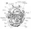
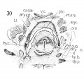
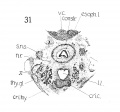
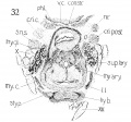
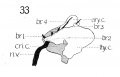
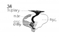
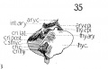
Lisser H. Studies on the development of the human larynx. (1911) Amer. J Anat. 12: 27-66.
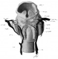
Streeter GL. On the development of the membranous labyrinth and the acoustic and facial nerves in the human embryo. (1906) Amer. J Anat. 6:139-165.
Streeter GL. Developmental Horizons In Human Embryos Description Or Age Groups XIX, XX, XXI, XXII, And XXIII, Being The Fifth Issue Of A Survey Of The Carnegie Collection. (1957) Carnegie Instn. Wash. Publ. 611, Contrib. Embryol., 36: 167-196.
| Carnegie Collection - Carnegie Embryos Sorted by Stage | |||||||||||||||||||||||||||||||||||||||||||||||||||||||||||||||||||||||||||||||||||||||||||||||||||||||||||||||||||||||||||||||||||||||||||||||||||||||||||||||||||||||||||||||||||||||||||||||||||||||||||||||||||||||||||||||||||||||||||||||||||||||||||||||||||||||||||||||||||||||||||||||||||||||||||||||||||||||||||||||||||||||||||||||||||||||||||||||||||||||||||||||||||||||||||||||||||||||||||||||||||||||||||||||||||||||||||||||||||||||||||||||||||||||||||||||||||||||||||||||||||||||||||||||||||||||||||||||||||||||||||||||||||||||||||||||||||||||||||||||||||||||||||||||||||||||||||||||||||||||||||||||||||||||||||||||||||||||||||||||||||||||||||||||||||||||||||||||||||||||||||||||||||||||||||||||||||||||||||||||||||||||||||||||||||||||||||||||||||||||||||||||||||||||||||||||||||||||||||||||||||||||||||||||||||||||||||||||||||||||||||||||||||||||||||||||||||||||||||||||||||||||||||||||||||||||||||||||||||||||||||||||||||||||||||||||||||||||||||||||||||||||||||||||||||||||||||||||||||||||
|---|---|---|---|---|---|---|---|---|---|---|---|---|---|---|---|---|---|---|---|---|---|---|---|---|---|---|---|---|---|---|---|---|---|---|---|---|---|---|---|---|---|---|---|---|---|---|---|---|---|---|---|---|---|---|---|---|---|---|---|---|---|---|---|---|---|---|---|---|---|---|---|---|---|---|---|---|---|---|---|---|---|---|---|---|---|---|---|---|---|---|---|---|---|---|---|---|---|---|---|---|---|---|---|---|---|---|---|---|---|---|---|---|---|---|---|---|---|---|---|---|---|---|---|---|---|---|---|---|---|---|---|---|---|---|---|---|---|---|---|---|---|---|---|---|---|---|---|---|---|---|---|---|---|---|---|---|---|---|---|---|---|---|---|---|---|---|---|---|---|---|---|---|---|---|---|---|---|---|---|---|---|---|---|---|---|---|---|---|---|---|---|---|---|---|---|---|---|---|---|---|---|---|---|---|---|---|---|---|---|---|---|---|---|---|---|---|---|---|---|---|---|---|---|---|---|---|---|---|---|---|---|---|---|---|---|---|---|---|---|---|---|---|---|---|---|---|---|---|---|---|---|---|---|---|---|---|---|---|---|---|---|---|---|---|---|---|---|---|---|---|---|---|---|---|---|---|---|---|---|---|---|---|---|---|---|---|---|---|---|---|---|---|---|---|---|---|---|---|---|---|---|---|---|---|---|---|---|---|---|---|---|---|---|---|---|---|---|---|---|---|---|---|---|---|---|---|---|---|---|---|---|---|---|---|---|---|---|---|---|---|---|---|---|---|---|---|---|---|---|---|---|---|---|---|---|---|---|---|---|---|---|---|---|---|---|---|---|---|---|---|---|---|---|---|---|---|---|---|---|---|---|---|---|---|---|---|---|---|---|---|---|---|---|---|---|---|---|---|---|---|---|---|---|---|---|---|---|---|---|---|---|---|---|---|---|---|---|---|---|---|---|---|---|---|---|---|---|---|---|---|---|---|---|---|---|---|---|---|---|---|---|---|---|---|---|---|---|---|---|---|---|---|---|---|---|---|---|---|---|---|---|---|---|---|---|---|---|---|---|---|---|---|---|---|---|---|---|---|---|---|---|---|---|---|---|---|---|---|---|---|---|---|---|---|---|---|---|---|---|---|---|---|---|---|---|---|---|---|---|---|---|---|---|---|---|---|---|---|---|---|---|---|---|---|---|---|---|---|---|---|---|---|---|---|---|---|---|---|---|---|---|---|---|---|---|---|---|---|---|---|---|---|---|---|---|---|---|---|---|---|---|---|---|---|---|---|---|---|---|---|---|---|---|---|---|---|---|---|---|---|---|---|---|---|---|---|---|---|---|---|---|---|---|---|---|---|---|---|---|---|---|---|---|---|---|---|---|---|---|---|---|---|---|---|---|---|---|---|---|---|---|---|---|---|---|---|---|---|---|---|---|---|---|---|---|---|---|---|---|---|---|---|---|---|---|---|---|---|---|---|---|---|---|---|---|---|---|---|---|---|---|---|---|---|---|---|---|---|---|---|---|---|---|---|---|---|---|---|---|---|---|---|---|---|---|---|---|---|---|---|---|---|---|---|---|---|---|---|---|---|---|---|---|---|---|---|---|---|---|---|---|---|---|---|---|---|---|---|---|---|---|---|---|---|---|---|---|---|---|---|---|---|---|---|---|---|---|---|---|---|---|---|---|---|---|---|---|---|---|---|---|---|---|---|---|---|---|---|---|---|---|---|---|---|---|---|---|---|---|---|---|---|---|---|---|---|---|---|---|---|---|---|---|---|---|---|---|---|---|---|---|---|---|---|---|---|---|---|---|---|---|---|---|---|---|---|---|---|---|---|---|---|---|---|---|---|---|---|---|---|---|---|---|---|---|---|---|---|---|---|---|---|---|---|---|---|---|---|---|---|---|---|---|---|---|---|---|---|---|---|---|---|---|---|---|---|---|---|---|---|---|---|---|---|---|---|---|---|---|---|---|---|---|---|---|---|---|---|---|---|---|---|---|---|---|---|---|---|---|---|---|---|---|---|---|---|---|---|---|---|---|---|---|---|---|---|---|---|---|---|---|---|---|---|---|---|---|---|---|---|---|---|---|---|---|---|---|---|---|---|---|---|---|---|---|---|---|---|---|---|---|---|---|---|---|---|---|---|---|---|---|---|---|---|---|---|---|---|---|---|---|---|---|---|---|---|---|---|---|---|---|---|---|---|---|---|---|---|---|---|---|---|---|---|---|---|---|---|---|---|---|---|---|---|---|---|---|---|---|
| |||||||||||||||||||||||||||||||||||||||||||||||||||||||||||||||||||||||||||||||||||||||||||||||||||||||||||||||||||||||||||||||||||||||||||||||||||||||||||||||||||||||||||||||||||||||||||||||||||||||||||||||||||||||||||||||||||||||||||||||||||||||||||||||||||||||||||||||||||||||||||||||||||||||||||||||||||||||||||||||||||||||||||||||||||||||||||||||||||||||||||||||||||||||||||||||||||||||||||||||||||||||||||||||||||||||||||||||||||||||||||||||||||||||||||||||||||||||||||||||||||||||||||||||||||||||||||||||||||||||||||||||||||||||||||||||||||||||||||||||||||||||||||||||||||||||||||||||||||||||||||||||||||||||||||||||||||||||||||||||||||||||||||||||||||||||||||||||||||||||||||||||||||||||||||||||||||||||||||||||||||||||||||||||||||||||||||||||||||||||||||||||||||||||||||||||||||||||||||||||||||||||||||||||||||||||||||||||||||||||||||||||||||||||||||||||||||||||||||||||||||||||||||||||||||||||||||||||||||||||||||||||||||||||||||||||||||||||||||||||||||||||||||||||||||||||||||||||||||||||
| Week: | 1 | 2 | 3 | 4 | 5 | 6 | 7 | 8 |
| Carnegie stage: | 1 2 3 4 | 5 6 | 7 8 9 | 10 11 12 13 | 14 15 | 16 17 | 18 19 | 20 21 22 23 |
Limb
Lewis WH. The development of the arm in man. (1902) Amer. J Anat. 1(2): 145-184.
Embryo XXII measures 20 mm. V. B. and 18 mm. X. B. It is about seven weeks old. The entire arm has a more posterior position. The lower angle of the scapula is at the level of the sixth rib, its anterior limit is about at the seventh cervical vertebra. The entire arm as well as its various parts have increased in size. The muscles are sharper and better developed than the preceding stage. Every muscle that the adult arm presents can now be recognized and each one now contains muscle fibers. The tendons are better formed and can be traced farther towards their final insertions. The ligaments and fasciae are also more distinct. The process of ligament and tendon formation from the condensed mesenchyma is still in progress at the distal ends of the digits. The skeletal elements are for the most part fairly well formed in cartilage except the distal row of phalanges.
The Skeletal System
The vertebral column
The intervertebral discs are reduced in thickness, while the bodies of the vertebra have increased and occnpy about four-fifths of each segment. The neural and transverse processes are larger and for the most part of cartilage. At the tip of the neural processes, which reach about one-half way around the spinal cord, is a small mass of condensed tissue at what juay be considered the growing point. These processes arise entirely from the body and not from the disc. So the body has probably grown at the expense of the disc. The perichondrium, wliich surrounds the body and its processes, is thickened along the ventral side of the bodies into the anterior common ligament.
The ribs are of cartilage surrounded by thick perichondrium, wliicli is continuous with the condensed tissue anlage of the one-half the sternum. The distance between the two halves of the sternum is not as great as in the preceding stage and at the anterior end they ai'e just beginning to come in contact with each other. There are no joint cavities between the ribs and vertebrae.
The clavicle is composed of cartilage somewhat different in appear ance from that in the other bones. It is continuous with the acromion and sternum by an area of condensed tissue. It is surrounded by a typical perichondrium. There are distinct coraco-clavicular, costoclavicular, and interclavicular ligaments.
The cartilaginous scapula is very much larger than in the precedingstage and contains no large areas of condensed tissue. It has moved posteriorly and lies in the region from the last cervical to the fifth thoracic vertebrae. Its dorsal border also extends farther dorsal than in any of the preceding stages. The acromion and coracoid processes are large and of cartilage with only the ordinary thickness of perichondrium which is continuous with that surrounding the rest of the scapula. The spine has not yet appeared but the thickened anterior border from which the supraspinatus muscle arises probably represents by its lateral lip the spine and by its median lip the future anterior border. The acromion arise partially from the lateral side of the anterior border. The head seems to have enlarged at the expense of part of the base of the coracoid process as the long head of the biceps now arises from the junction of the coracoid and the head, and the head of the humerus does not rest against such a large proportional area of the coracoid. There is a distinct suprascapular and a coraco-acromial ligament. At the posterior angle of the scapula there is small mass of condensed tissue which gives attachment to a portion of the serratus, latissimus, and teres major muscles.
The humerus is much hirger than in embryo XLIII, and has much the adult shape, though of course it is thicker in proportion to its length. It is composed of cartilage. There is a capsular and a coraco-humeral ligament. No joint cavity exists between the scapula and humerus. The tuberosities and condyles are fairly well formed in cartilage and •condensed tissue. The bicipital groove is present.
The ulna and radius are larger and longer than in the preceding stage 3ind are well formed in cartilage. The olecranon, coranoid and styloid processes are partially formed in cartilage and condensed tissue. The perichondrium about the ulna and radius is quite thick. The capsular .and orbicular ligaments are present. No joint cavities exist and the cartilages are separated by condensed tissue continuous with the peri•ciiondrium.
Fig. 13. Cartilaginous skeleton of the arm of embrj-o XXII, lateral view. X 12 diameters.
All the bones of the carpus are represented by cartilage, and in about their relative positions. The amount of condensed tissue matrix is much less than in the preceding stage. The condensed tissue matrix is continuous with the ulna and radius and the five metacarpals without joint cavities. Indications of ligaments of the wrist are present.
The five metacarpals are present in cartilage surrounded by thick perichondrium. The first is the shortest.
The first two rows of phalanges are present in all the digits. They are of cartilage surrounded by a very thick perichondrium, which is continuous with the condensed tissue between them and the metacarpals and between the phalanges them.selves. It is also continuous with the enlarged condensed tissue tip of each digit. There are thickenings for the various ligaments connecting the metacarpals and phalanges and the phalanges with each other.
The Muscular System
(Plate II, Fig. C.) The trapezius muscle fibers extend from the occiput to the level of the sixth rib. There is a considerable interval of fascia connecting them to the neural processes of the lower cervical and the thoracic vertebrae. There is a tendonous attachment to the clavicle and acromion and into fascia or condensed tissne on the surface of the infraspinatus between the trapezius and the deltoid.
The rliomhoid muscle lies in the region of the seventh cervical to the fourth thoracic vertebrae. It is inserted into the condensed tissue along the dorsal border of the scapula.
The latissimus dorsi muscle fibers extend from the humerus to the level of the ninth rib. There is a considerable interval of fascia between them and the neural processes of the lower thoracic and the first two or three lumbar vertebrae. This dorsal fascia is not very well marked. The latissimus also has fibers attached to the condensed tissue at the inferior angle of the scapula.
The serratus anterior muscle is separate from the levator scapulae except near its attachment to the scapula. It is a broad, thin sheet, having digitations to the first eight ribs.
The pedoralis major muscle is well developed. The separation between the clavicular and the sterno-costal portions is less marked than in the preceding stage.' The muscle is attached as low as the sixth rib.
The pedoralis minor muscle is quite distinct from the major, as a considerable layer of loose mesenchymal tissue lies between them. It arises from the second, third and fourth ribs and passes to the coracoid process.
The siibclavius muscle is inserted into the clavicle at an angle of 45°. As the scapula and clavicle sink down towards the level of the first rib the angle at which this muscle is inserted into the clavicle decreases.
The teres major muscle arises from the lower angle of the scapula and passes to the humerus. It is interesting to note that at this stage tendon of the latissimus dorsi twists around the lower l^order of the teres to be inserted with it into the humerus.
The deltoid muscle is large and well developed.
The suprasfinatus muscle arises from the thickened anterior border of the scapula. It cannot be said to take origin more from the lateral surface than from the median surface of the scapula.
The infraspinatus muscle occupies the middle of the lateral surface of the scapula and passes beneath the deltoid to the great tuberosity of the humerus.
The suhscapularis muscle arises from most of the median surface of the scapula. Its tendon of insertion is broad and thin and closely applied to the capsular ligament.
The three heads of the triceps muscle are easily distinguished. The long and external heads are of about the same size. The anconeus muscle is continuous with the triceps but arises from the external condyle and passes to the side of the olecranon and adjoining surface of the shaft of the ulna.
The long head of the biceps muscle arises from the junction of the coracoid process and the head of the scapula and passes through the bicipital groove. The two heads are inserted together into the condensed tissue swelling on the radius.
The corncobracJtviUs muscle and short head of the biceps are intimately connected for most of the length of the former.
The brachialis muscle has spread out over more of the distal surface of the humerus than in the preceding stage.
The flexor muscles of the forearm are easier to distinguish than in the preceding stage.
The tendon of the palinaris longus is narrower in proportion than in embryo XLIII.
The tendon of the flexor carpi raclialis muscle can be traced farther towards its insertion into the base of the second metacarpal than in embryo XLIII.
The muscle fibers of the flexor sublimis digitorum still run to the carpus before the ^\ide tendon begins. This tendon soon splits into four tendons which go to the four ulnar digits. These tendons are better formed than in the preceding stage and split to surround the tendons of the deep flexor. Their ends fuse with the thick perichondrium about the phalanges.
The flexor carpi uhiaris muscle shows distinctly its two heads of origin. It has a well-formed tendon of insertion.
The deep flexor muscles can be separated into the flexor polUcis longus and the flexor profundus digitormn muscles. The muscle fibers of the profundus continue into the carpal region and end in a broad tendon which divides at the base of the metacarpus into four well-formed tendons. These fuse with the condensed tissue at the tips of the digits. There is a slight split in each of these tendons near its end. The tendon of the flexor longus pollieis behaves similarly.
The pronator quadratns muscle is oval in cross section, and connects the distal ends of the radius and ulna.
The lumbricales are quite well developed and their fairly well-formed tendons end in the perichondrium on the radial side of the digit.
The interossei mnscles and the small mnscles of the thumb and little finger are now fairly well developed. Muscle fibers are present.
The extensor muscles of the forearm show considerable advance over the preceding stage. The tendons of the extensor communis digitoriim are longer and narrower. The muscle fibers continue to the base of the metacarpus, where the splitting into the four tendons takes place. The tendons are inserted into the condensed tissue tips of the digits. The edge of the tendons near their insertions are more or less continuous with the perichondrium about the digit.
The tendon of the extensor carpi ulnaris is beginning to. form. One branch of it seems to join the communis tendon. This may be the tendon of the extensor minimi digiti.
Fig. 14. Lateral view of the arm of embryo XXII. from Plate VIII.
The extensor carpi radialis longior et hrevior are not to be separated.
The supinator muscle is well developed and has the posterior interosseus nerve passing through it.
The abductor pollicis longus and extensor pollicis brevis muscles are only to be separated where the muscle fibres pass into tendons, which fuse with the perichondrium of the first digit. The separation occurs at the lower end of the radius. These two muscles are fairly distinct from the supinator and the extensor pollicis longus and extensor indicis projiriiis muscles. The last two muscles are inseparable for part of their course and shortly after dividing each forms a round tendon. The extensor pollicis longus then spreads out into the sheath about the first digit. The extensor indicis muscle joins the nlnar side of the tendon of the commimis to the second digit.
The Nerves
The drachial plexus has a decided posterior inclination and seems to have been pulled down against the first rib. The three cords are so close together that it was impossible to separate them satisfactorily though indications of the cords are present. There is nothing especially peculiar about the distribution of the nerves from the plexus either motor or sensory, which is not present in the adult.
Pages in category 'Carnegie Embryo 22'
The following 14 pages are in this category, out of 14 total.
P
- Paper - Development and variation of the nerves and the musculature of the inferior extremity and of the neighboring regions of the trunk in man
- Paper - Development of the human coelom (1897)
- Paper - On the development of the membranous labyrinth and the acoustic and facial nerves in the human embryo
- Paper - On the embryology of the corpus ponto-bulbare and its relation to the development of the pons (1909)
- Paper - Studies of the development of the human skeleton (1905)
- Paper - Studies on the development of the human larynx (1911)
- Paper - The development of the arm in man (1902)
- Paper - The development of the islands of Langerhans in the human embryo (1903)
- Paper - The development of the limbs, body-wall and back
- Paper - The development of the limbs, body-wall and back (1901)
- Paper - The development of the nuclei pontis and the nucleus arcuatus in man (1912)
- Template:Pearce1903 table1
Media in category 'Carnegie Embryo 22'
The following 26 files are in this category, out of 26 total.
 Bardeen1906-plate06.jpg 1,568 × 2,299; 379 KB
 Bardeen1906-plate51.jpg 1,570 × 2,330; 392 KB
 Bardeen1906-plate52.jpg 1,596 × 2,350; 404 KB
 Keibel Mall 143.jpg 624 × 625; 33 KB
 Lisser1911 fig24.jpg 826 × 705; 36 KB
 Lisser1911 fig25.jpg 826 × 705; 28 KB
 Lisser1911 fig26.jpg 826 × 705; 29 KB
 Lisser1911 fig27.jpg 826 × 705; 38 KB
 Lisser1911 fig28.jpg 826 × 705; 60 KB
 Lisser1911 fig29.jpg 1,070 × 998; 252 KB
 Lisser1911 fig30.jpg 1,070 × 998; 249 KB
 Lisser1911 fig31.jpg 1,070 × 998; 165 KB
 Lisser1911 fig32.jpg 1,070 × 998; 242 KB
 Lisser1911 fig33.jpg 846 × 542; 42 KB
 Lisser1911 fig34.jpg 846 × 542; 41 KB
 Lisser1911 fig35.jpg 846 × 542; 62 KB
 Lisser1911 fig36.jpg 1,531 × 1,543; 186 KB
 Lisser1911 fig37.jpg 3,354 × 1,729; 349 KB
 Lisser1911 fig38.jpg 3,041 × 1,586; 351 KB
 Lisser1911 fig39.jpg 2,340 × 2,746; 375 KB
 Mall1897 fig52.jpg 907 × 1,000; 113 KB
 Mall1906-fig26.jpg 766 × 639; 78 KB
 Mall1906-fig27.jpg 890 × 800; 168 KB
 Streeter1906 plate01.jpg 2,708 × 1,786; 701 KB
 Streeter1957 fig07.jpg 1,280 × 1,201; 117 KB
 Streeter1957 fig7-21.jpg 680 × 800; 25 KB


