Category:Carnegie Embryo 1656
This Embryology category shows pages and images that relate to the Carnegie Collection Embryo No. 1656. This embryo would be early fetal development Week 10 based upon the CRL 67 mm.
References
Kunitomo K. The development and reduction of the tail and of the caudal end of the spinal cord (1920) Contrib. Embryol., Carnegie Inst. Wash. Publ. 272, 9: 163-198.
Embryo No. 1656, 67 mm Crown-Rump Length
Kunitomo K. The development and reduction of the tail and of the caudal end of the spinal cord (1920) Contrib. Embryol., Carnegie Inst. Wash. Publ. 272, 9: 163-198.
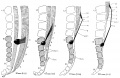
There are 34 vertebrae in embryo No. 1656, the last being the smallest. At the thirty-first and thirty-second the vertebral column shows a ventral curve, the angle being sharper than in the younger specimens. The vertebrae are separated by embryonic tissue which is to develop at a later stage into intervertebral fibro-cartilage. This separation becomes progressively more marked above the thirtieth vertebra. Between the vertebrae which still lie close together is a small space where the chorda dorsalis coils as it emerges from the vertebral bodies in the median line. Several of these coils can be seen in figure 46, which is a profile reconstruction through the caudal end of the embryo. The blood-vessels enter the vertebral bodies from the ventral and dorsal side.
In the conus medullaris there are two medullary ventricles. The more cranially situated one is somewhat smaller tnan the other, measuring 0.55 by 0.25 by 0.33 mm. Its form, as seen in the sagittal plane, can be recognized in figure 46 {vent. t. cran.). The lower cavity is oblong in shape, measures 1.1 by 0.3 by 0.36 mm., and presents a canallike appendage 1.7 mm. in length, as seen in figure 46 {Append.). This appendage tapers to a point and continues as a cell-strand. Toward the caudal end of the strand, in the path of the filum terminale, are two small groups of cells which represent the remnants of the ependymal cells of the medullary tube (fig. 46, Ee. epend.).
The phenomenon of dedifferentiation at the caudal end of the spinal cord is well shown in this specimen. The appendage of the lower cavity was a complete ventriculus terminalis at the first stage; the main body of the cavity was a complete one at the second stage, and the upper cavity is the ventricidus terminalis at the present stage, thus showing a progressive upward trend. The gray substance which primarily existed around the ventriculus terminalis has now disappeared as the result of degeneration, and the caudal end of the central canal has gradually enlarged. The caudal end of the lower cavity, however, is becoming gradually narrow because the caudal portion of the conus medullaris, which contains the ventriculus terminalis, has also gradually become atrophied and lost its cellUke substances. The septum between the two cavities is a remnant of the gray substance of the spinal cord, in which the degeneration is not yet complete.
The filum terminale follows a downward course from the end of the conus medullaris and nerve-fibers can be recognized as far down as the caudal portion of the thirty-second vertebra. In the caudal region are found two cell-groups representing remnants of the neural tube; one, which lies between the thirty-second and thirty-third vertebrae, contains no lumen, and the epithelial cells are undergoing degeneration. The other is situated dorsally between the thirty-third and thirty-fourth vertebrae and incloses a small lumen.
The membranes of the spinal cord are more easily made out in this specimen than in the younger ones. The dura mater is separated from the periosteum of the vertebral bodies, especially at the ventral wall of the vertebral canal, by a dense plexus of blood vessels, connective -tissue, and small spaces. This separation occurs at a level between the twenty-seventh and twenty-eighth vertebrae, and the dura mater becomes adherent to the conus medullaris between the twenty-eighth and twenty-ninth vertebrae, following an oblique course from the periphery to the center of the vertebral canal. There is thus laid out the early form of the dura! sac. Outside of this .sac the fibers are .separated into tufts which run parallel and caudalward. In the space between the dural sac and the conus medullaris the arachnoid membrane can be seen developing. The pia mater envelops closely the spinal cord and supports the blood-vessels; between the twenty-fifth and twenty-eighth vertebra it is separated from the dura mater and the arachnoid by a still wider space.
Cite this page: Hill, M.A. (2024, April 25) Embryology Carnegie Embryo 1656. Retrieved from https://embryology.med.unsw.edu.au/embryology/index.php/Category:Carnegie_Embryo_1656
- © Dr Mark Hill 2024, UNSW Embryology ISBN: 978 0 7334 2609 4 - UNSW CRICOS Provider Code No. 00098G
Pages in category 'Carnegie Embryo 1656'
This category contains only the following page.
Media in category 'Carnegie Embryo 1656'
This category contains only the following file.
 Streeter1919-fig02.jpg 2,023 × 1,318; 499 KB
