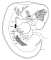
Category:Carnegie Embryo 109: Difference between revisions

From Embryology
mNo edit summary
mNo edit summary
 
(No difference)

Latest revision as of 09:46, 20 October 2020

Carnegie Collection Embryo No.109
Carnegie Collection Embryo No. 109

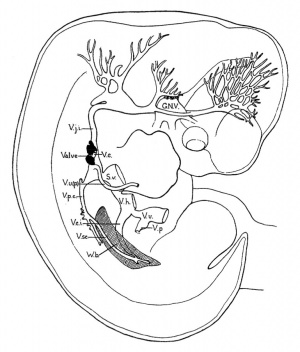
This Embryology category shows pages and images that relate to the Carnegie Collection Embryo No. 109. This embryo was classified as Carnegie stage 18 occurring during Week 7.

Links: Carnegie stage 18 | Keibel Mall Fig 488 | Keibel Mall Fig 489 | Keibel Mall Fig 491 | Carnegie Collection


Stage 18 Links: Week 7 | System Development | Lecture - Limb | Lecture - Head Development | Lecture - Sensory | Science Practical - Head | Science Practical - Sensory | Science Practical - Urogenital | Carnegie Embryos | Madrid Embryos | Category:Carnegie Stage 18 | Next Stage 19
  Historic Papers: 1911 larynx | 1948 stage 15-18


Serial No. Size (mm) Grade Fixative Embedding Medium Plane Thinness (µm) Stain Semi. ducts P.-M. duct (mm) Year Notes
109 E., 12.0* Ch.,30 Poor Alc. P Transverse 20 Al. coch. 1 0.4 1897 Tubal. Least—advanced third


Carnegie Collection Embryos - Stage 18 
Serial No. Size (mm) Grade Fixative Embedding Medium Plane Thinness (µm) Stain Semi. ducts P.-M. duct (mm) Year Notes
109 E., 12.0* Ch.,30 Poor Alc. P Transverse 20 Al. coch. 1 0.4 1897 Tubal Least—advanced third
144 E., 16.0* Ch, 40x30x30 Good Formalin P Sagittal 40 Al. eoch. 3 0.85 1899 Most—advanced third
175 E., 13.0 Ch, 30x25x25 Poor Alc. P Transverse 20 Al. coch. 2 0.6 1900 Tubal Partly macerated
296 E., 17.0 Poor Ale. P Coronal 20 Various 3 0.85 1905 Most—advanced third
317 E., 16.0 Good Formalin P Coronal 20 (Stain - Haematoxylin Eosin) or. G. 2 0.7 1905 Middle third
351 E.,14.0* Good Formalin P Coronal 250 Slightly carmine— 2 038 1904 Injected (Berlin blue)
406 E., 16.0 Ch., 40x40x40 Good Formalin P Sagittal 20 (Stain - Haematoxylin Eosin) 3 0.7 1907 Operative. Most—advanced third
423 E., 15.2 Good Formol—Zenker P Transverse 50 Carmine 3 0.85 1904
424 E., 172 Good Formalin P Transverse 50 Carmine 3 10 1904 Double infection. Advanced
492 E, 16.8 Ch, 40 x 40 Exc. Zenker P Coronal 40 Al. coch. v 3 0.7 1911 Injected (India ink)
511 E., 160* Ch., 3?* 32x32 Good Ale. P Sagittal 40 Al. coch. 3 1.1 1911 Head injured. Most advanced in group
670 E, 12.5 Poor Ale. P Sagittal 50 (Stain - Haematoxylin Eosin) 3 10 1913 Tubal Advanced
719 E, 15.0 Ch, 50x50x50 Good Formalin P Trans 40 Al. coch. 2 0.6 1913 Median in group
733 E., Ch., 4Sx40x2S ISO Poor Formalin P Sagittal 50 Al. coch. 2 0.6 1913 Median in group
841 E. 15.0 Ch., 18 x 16x9 Good Formalin P Coronal & Trans, 20 10 (Stain - Haematoxylin Eosin), carmine 2 0.32 1914 Operative. Head cut separately
899 E, 160* Ch. 50 x 18 x IS Good Bouin P Sagittal S0 Al. coch. 3 0,65 1914 Tubal Head injured
991 E. l?.0 Good Formalin P Sag so R, V, Gieson 3 0.9 1914 Advanced
1909 E., 14.6 Good Formalin P Coronal 20 Al. coch,or. G. 1 0.3 1917 Less advanced
2673 E.,15.5 Good Formalin P Transverse 40 Al. coch. 2 0.52 1919 Median in group
4430 E., 14.0 Ch, 51 x40x21 Exc. Corros. acetic P Transverse 15 Al. coch,or. G. 3 0.9 1923 Most—advanced third
5542B E., 16.0 Ch, 37x32x25 Good Formalin P Transverse 40 Al. coch. 2 0.7 1927 Other twin abnormal
5747 E, 15.2 Ch, 32x27x25 Poor Alc.—formol P Sagittal 25 Al. coch. 2 0.25 1928 Least—advanced or middle third
5935A E, 13.5 Ch, 40x30x30 Good Formalin P Coronal 40 Al. coch. 1 0.38 1929 Other twin stunted
6522 E, 13.2* Good Corros. acetic C—P Coronal 10 Al. coch. 3 0.8 7 Middle or most—advanced third
6524 E, 11.7* Exc. Corros. acetic C—P Transverse 10 Al. coch. 1 0.4 ? Least—advanced third
6525 E, 13.8* Exc. Corros. acetic C—P Sagittal 8 Al. coch. 2 0.42 ? Weak staining
6527 E, 14.4* Exc. Corros. acetic C—P Transverse 15 Al. coch. 2 0.67 ? Mechanical damage
6528 E, 13.4* Exc. Corros. acetic C—P Coronal 8 Al. coch. 1 0.33 ? Least—advanced third
6529 E, 15.6* Good Corros. acetic C—P Coronal 10 Al. coch. 2 0.4 .5 Middle third
6533 E, 12.5* Good Corros. acetic C—P Sagittal 6, 8, 10 Al. coch. 2 0.45 ? Middle third
6551 E, 18.0 Poor Formalin p Coronal 40 (Stain - Haematoxylin Eosin) 3 0.8 1932 Tubal
7707 E, 14.5 Ch ,37x32 Exc. Bouin C—P Transverse 10 (Stain - Haematoxylin Eosin), phlox. 2 0.54 1939 Operative. Middle third
8097 E, 15.5 Ch, 37x25x21 Good Formalin C—P Transverse 10 (Stain - Haematoxylin Eosin) 1 0.19 1942 Least advanced in group
8172 E, 16.5 Exc. Bouin C—P Transverse 20 (Stain - Haematoxylin Eosin) 3 0.58 1943 Operative. Very advanced
8235 E, 14.0* Good Bouin C—P Sagittal 10 (Stain - Haematoxylin Eosin) Mallory 2 0.25 1944 Tubal
8355 E, 15.0 Ch, 23 Exc. Formalin C—P Coronal 10 Azan 1946 Tubal. Duplicated spinal cord caudally
8812 E, 12_9 Exc Formalin C—P Transverse 10 (Stain - Haematoxylin Eosin) 1950 Rubella. Medical abortion. Midbrain punctured
8945 E, 13.9 Good Zenker p Transverse 8 Borax, carm. 1952 Univ. Chicago No. H 1254
9107 E, 17.0 Ch, 38x28x22 Good Bouin p Transverse 15 Borax, carm. 1918 Univ. Chicago No. H 516
9247 E, 15.0 Exc. Bouin C—P Sagittal 8 Azan 1954 Tubal
Abbreviations
  • Size - E. is the greatest length of the embryo and Ch. is the mean diameter of the chorion.
  • Grade - total grade of the specimen and includes both its original quality and the condition of the mounted sections.
  • Embedding medium - paraffin (P) or a combination of celloidin and paraffin (C-P).
  • Fixative - formalin (Formol), alcohol and formalin (Alc, formol), Bouin (Bouin solution)
  • Stain -
  • ? - unknown or not determined.

References

Bardeen CR. and Lewis WH. The development of the limbs, body-wall and back. (1901) Amer. J Anat. 1: 1-36.

Lewis WH. The development of the arm in man. (1902) Amer. J Anat. 1(2): 145-184.

Bardeen CR. Development of the thoracic vertebrae in man. (1905) Amer. J Anat. 4: 163-174.

Sabin FR. The lymphatic system in human embryos, with a consideration of the morphology of the system as a whole. (1909) Amer. J Anat. 9(1): 43–91.

Streeter GL. On the development of the membranous labyrinth and the acoustic and facial nerves in the human embryo. (1906) Amer. J Anat. 6:139-165.

Whitehead RH. and Waddell JA. The early development of the mammalian sternum (1911) Amer. J Anat. 12: 89-106.

Lisser H. Studies on the development of the human larynx. (1911) Amer. J Anat. 12: 27-66.

Mall FP. On the fate of the human embryo in tubal pregnancy. (1915) Contrib. Embryol., Carnegie Inst. Wash. Publ. 221, 1: 1-104.


Limb

Lewis WH. The development of the arm in man. (1902) Amer. J Anat. 1(2): 145-184.

Embryo CIX measures V. B. 10.5 mm. and X. B. 11 mm. in lengtli and is about five weeks old. There is a marked advance over the preceding stage. Cartilage has made its appearance both in the vertebrae and in portions of the arm skeleton. There is considerable difference in the character of the cartilage of the vertebra from that in the arm. The latter seems more advanced and lias more the appearance of true hyaline cartilage. It is possible that the cartilage appears first in the arm, though I have not been able to examine intervening stages to determine this with certainty. Other portions of the arm skeleton are in the precartilage and condensed tissue stages. Both cartilage and precartilage are surrounded in most places by a distinct perichondrium. This takes a very deep stain with the alum carmine. This perichondrium shades off into the condensed tissue of the carpus, which is like that composing the skeletal core in the preceding stage. This again shades into the even less differentiated tissue of the digits, which is at a.bont the same stage of development as the hand plate of the preceding stage, and it in turn shades off into the surrounding mesenchyma.

The muscles in the arm region show very different degrees of development. Those derived from the muscle plate system are in advance of most of the others. The trapezius, levator scapulae and serratus magnus are about as far advanced as those from the muscle plate system, they show distinct muscle fibers and are for the most part quite sharply limited from the surrounding loose mesenchyma. In position they correspond with their premuscle masses of the preceding stage. The pectoral muscle is next in advance and the latissimus dorsi next. These two muscles grow from the humeral region towards their future attachments on the body wall. It is this portion which lies farthest from the humerus which seems to show the most advance in fibrillation and the sharpest limitation from the surrounding mesenchyma. At the humeral end these muscles gradually shade into a condensed mesenchyma, which fuses with neighboring muscle and skeletal elements. Both muscles correspond in position to their premuscle masses of the preceding stage. As in the preceding stage, embryo CLXIII, the trapezius and serratus premuscle masses were in advance of the pectoral and latissimus; in embryo CIX we find the same relation still continues.

The remaining muscles of the arm apparently develop in situ from the premuscle sheath and undergo practically no migrations. They do not appear to be as far advanced as any of the above mentioned muscles. Of these muscles developing from, the arm premuscle sheath, the more proximal ones are more developed than the ones more distal. In the scapulo-humeral region most of the muscles show partial fibrillation, while those in the palm of the hand are in about the same condition as the proximal portion of the premuscle sheath in the preceding stage.

The fibrillation, position and nerve supply have made it possible to determine the presence of most of the muscles of the arm.

The Skeletal, System

The Vertebral Column

The intervertebral discs are composed of condensed mesenchyma, the cells having a concentric arrangement about the chorda. The vertebral bodies between the discs are each composed of two masses of cartilage, one on either side of the chorda. Tliey are surrounded by a perichondrium. Along the ventral surface of the vertebral coluuni is a layer of dense mesenchyma, which probably represents both perichondrium and the anterior common ligament. The neural processes, composed of condensed mesenchyma, are clearly defined. They are continuous with the discs and form a wide, shallow groove for the spinal cord. The transverse processes arise by two roots, one from the base of the neural process and the other from the disc. They are of condensed mesenchyma.

The Ribs. - The ribs are more sharply defined than in CLXIII. They are of condensed tissue except for a small area near the head, which is of precartilage. They extend farther into the body Avail than in the preceding stage.


Fig. 9. Skeleton of the arm region of embryo CIX, lateral view. X 12 diameters.

The Arm Skeleton

The Scapula is composed of precartilage and has greatly altered in shape. It lies in the region of the lower four cervical and first one or two thoracic vertebrae. From the anterior border, which corresponds to the spine, springs the large curved acromion process. On the median surface at the junction of the humerus with the scapula arises the large hooked coracoid process. Eunning across the median surface of the scapula to the vertebral border is a slight ridge which separates the supraspinatus from the subscapularis muscles and corresponds to the future anterior border. The condensed tissue is thickened on the medial surface into a perichondrium, while on the lateral surface the precartilage shades off into the surrounding mesenchyma.

The Clavicle

A rather poorly defined mass of condensed tissue continues from the tip of the acromion toward the tip of the first rib, extending for about one-third this distance. This mass represents the clavicle. From it a mass of ill-def]ned tissue extends to the coracoid process and represents the coraco-clavicular ligament.


The Humerus is directly continuous with the scapula and root of the coracoid process. No signs of joint surfaces or cavity are present. Both ends of the shaft are enlarged and Ihe distal end shows both external and internal condyles. The core of the shaft is of hyaline cartilage; this is surrounded by very thick perichondrium, which shades ofE into the condensed tissue of each end in which is enclosed an area of precartilage. The distal end seems more advanced than the proximal.

The Radius and Ulna are continuous with the distal end of the humerus, no indications of joint surfaces or cavities being present.


Fig. 10. Outline of the arm region of embryo CIX, lateral view from Plate IV. Bardeen and Lewis, Vol. I, No. 1, this Journal. X 12 Diameters.


There is more flexion at the elbow than in CLXIII. The forearm occupies a position about half way between pronation and supination. The core of each shaft is composed of hyaline cartilage. This is surrounded by a very thick perichondrium, which continues into the condensed tissue at either end of the bone, in which precartilage is enclosed. The hand-plate is continuous with the distal ends of the radius and ulna. It is composed of condensed mesenchyma. There are several centers of increased condensation which I believe must correspond to the carpal bones, namely, the scaphoid, lunar, pyramidal, trapezium, trapezoid, OS magnum and unciform. The scaphoid is in line with the radius and the lunar with the ulna, while the pyramidal is at the ulnar side of the carpus, and as the metacarpal V continues from it more than the uniform the whole hand has a peculiar bend toward the ulnar side. From the carpus five masses of condensed tissue project. They shade off into the surrounding mcsenchyma which fills the distal end of the arm. The condition of these finger masses corresponds to the condition of the hand-plate in CLXIII. There is not the sliglitest indication of segmentation into metacarpals and phalanges. The radial of the five projections probably consists of both trapezium and metacarpal I, which have not yet shown signs of separate centers of condensation.

The Muscular System

The muscle plate system has become differentiated into several muscles, namely, the deep dorsal muscles, the intercostals, the abdominal muscles and the deep ventral neck muscles.

The infrahyoid muscles correspond in position and nerve supply with the infrahyoid premuscle mass of the preceding stage. They extend nearly to the region where the median end of the clavicle will eventually extend.

The trapezius muscle has extended posteriorly to the level of the fifth cervical vertebra. Its posterior end lies near to the lateral surface of the body and is connected to the tips of the neural processes as far posteriorly as the second thoracic vertebra by a considerable interval of fascia. As the muscle passes anteriorly it lies deeper and deeper from the surface, being separated from it by the platysma and facial muscles. Its ventral border is free from attachment to the scapula and clavicle. At the level of the second cervical vertebra it is joined by the stemomastoid muscle, which has ascended from the more ventral neck region. The nerve supply is as in the adult.

The rhomboid mass lies in the region of the Y and A^I cervical vertebrae. It connects with the fascia passing to the dorsal tips of the neural processes but has no scapular attachment. A branch of the fifth cervical nerve supplies it.

The levator scapula; and serratus anterior muscles form a continuous fibrillated mass, extending from the first cervical vertebra to the ninth rib. It occupies much the same position that its premuscle mass did in embryo CLXIII except that the posterior end now extends to the ninth rib. Digitations go to all the cervical transverse processes and to each of the anterior nine ribs. The anterior and posterior digitations are very slender and contain but few fibers. The thickest part of the- muscle lies in the scapular region. There is no scapular attachment. The ventral edge of the muscle lies at about the same level as the dorsal edge of the scapula but in a more median plane. Branches from the second to the seventh cervical nerves supply the muscle. The first three penetrate directly into the muscle. The last three form a trunk which runs along the lateral surface of the muscle as far as the fourth rib.

The pedoralis major and minor "^ are united into a common muscle mass, which is well differentiated from the surrounding tissue. It forms a thick oval mass, which extends from the level of the second rib to the proximal portion of the humerus. The greater part of the muscle thus lies anterior to the first rib. As the mass bends towards the humerus it is attached also to the clavicle. So probably both sterno-costal and clavicular portions are present. The median side of the mass bulges towards the coracoid process and represents the minor. Most of the mass shows distinct fibrillation, but toward the humerus this passes into the condensed tissue which is not sharply outlined from the surrounding structures. The position of the pectoral muscle corresponds to the position of the pectoral premuscle mass in embryo CLXIII. Branches from the median side of the brachial plexus supply the pectoral. Two from the external cord contain fibers from the fifth, sixth and seventh cervical nerves. Two come from the inner cord. Within the muscle complicated anastomoses occur from which fibers spread out in all directions.

The muscles thus far considered were fairly definite, and, as we have seen, come from quite definite premuscle masses. The remaining muscles of the arm are in process of differentiation from the arm premuscle sheath. The exact limits of the individual muscles are almost impossible to determine.

The deltoid muscle extends from the acromion and clavicle and fascia over the infraspinatus to the humerus. It is very closely connected with the infraspinatus and only by the difference in the nerve supply can the two be separated. The position of the teres minor is also only indicated by its nerve and not by any line of separation between it and the infraspinatus or deltoid. The origin of part of the deltoid from the acromion and clavicle helps to distinguish some of its fibers, but a short distance from this origin no line of separation can be made between it and the infra- and supraspinatus muscles. Condensed tissue connects it with the triceps and pectoral muscles. The circumflex nerve supplies this muscle and also sends a branch to fibers which are closely associated with the infraspinatus and probably constitute the teres minor muscle.

That portion of the infraspinatus which lies on the lateral surface of the scapula is fairly distinct except where the deltoid and teres minor muscles join it. The portion of tlie supraspinatus on the anterior onefourth of the median surface of the scapula is distinct, but after it passes the acromion it is inseparably' connected with the infraspinatus and deltoid and pectoral muscles. These muscles shade off into the 23roximal end of the humerus. The main portion of each of these muscles contains muscle fibers. The suprascapular nerve supplies the supra- and infraspinatus muscles.

23 Lewis, Observations on the pectoralis major muscle in man, Johns Hopkins TTosp. Bui., Vol. XII, 1901.


The subscapularis muscle arises from the posterior one-half of the median surface of the scapula and passes beneath the coracoid process to the humerus. The circumflex nerve separates a portion of it from the teres major muscle, but the scapular portions of the two are closely imited, as is also the long head of the triceps. A branch from the circumflex and another from the posterior cord of the brachial plexus supply the subscapularis.

The teres major and latissimus dorsi muscles are closely associated at "their humeral end. The latissimus dorsi' lies in the lateral thoracic region, extending posteriorly as far as the fourth rib. It has no attachments to the ribs or vertebral column. The two muscles are inserted together into the proximal portion of the humerus. The teres major arises from the axillary border of the scapula near its posterior angle. The common portion of the latissimus and teres passes close to the posterior cord of the brachial plexus, from which a large branch is given off that runs into the latissimus and has a brush-like ending near the posterior limit of the muscle. A smaller branch of the posterior cord is given off to the teres major.

The triceps muscle extends along the posterior and lateral surfaces of the humerus, extending from the scapula to the ulna. Indications of the three heads are present. The portion of the muscle lying near the insertion of the latissimus dorsi and the infraspinatus muscles is not sharply defined from them. The musculo-spiral nerve passes through the muscle and gives branches to it.

The biceps and coracohracJnaUs muscles lie along the median side of the humerus, extending from the coracoid process to the radius. The two heads of the biceps are quite closely united nearly to their origins, Avhich are but a short distance apart. The portion of the coracoid process from which the long head arises must ultimately become a portion of the head of scapula. The attachment of the coracobrachialis to the humerus is by condensed tissue, as is the distal end of the biceps to the radius. The distal end of the biceps blends with the brachialis and the flexor mass. The musculo-cutaneous pierces this group and gives off branches to it.


The brachialis muscle is closely attached to the distal one-half of the hmnerns over the anterior and median surfaces. It is also closely attached to the overlying biceps muscle and it is impossible to determine just the line between the two or between it and the brachioradialis muscle. It is also impossible to determine the exact line between the muscle and the underlying perichondrium. It is closely associated with the triceps on one side and the deltoid on the other. The main portion of the muscle is fibrillated and is inserted into the ulna by condensed tissue, which is closely associated with the, flexor mass of the forearm. The musculo-cutaneous nerve gives off a large branch which has a brush-like endins; within the muscle.


Fig. 11. Outline of the arm region of embryo CIX, median view, from Plate V. Bardeen and Lewis, Vol. I, No. 1, this Journal. X 15 diameters.


The flexor muscle mass of the forearm forms a thick layer over the median surface of the ulna, radius, carpus and proximal end of the metacarpus. It is with considerable difficulty that I have separated this mass into two layers. The superficial layer is smaller in extent and lies in the proximal region of the forearm. It is connected with the radial portion of the forearm by a condensed tissue mass and distally fuses with the deep layer to become continuous with the condensed tissue of the digits. The median nerve passes through the proximal portion and then comes to lie between the two layers. From its position and relation to the median nerve I believe this to be the layer from which the flexor carpi radialis, flexor sublimis digitorum, pronator teres and palniaris long-us muscles differentiate. Branches from the median nerve supply this layer. Both layers arise partly from the inner condyle of the humerus, and are continuous more or less with the muscles of the upper arm. The deep layer is closely attached to the perichondrium of the forearm and hand. It is wider in extent than the superficial and shows indications of separations into muscles. Tlie portion for the flexor carpi nlnaris shows most advance. The extension into the hand probably constitutes the portion from which the interossei and lumbrical muscles and flexor tendons develop. It is continuous with the condensed tissue of the digits. The portion on the forearm forms the flexor profundus digitorum, flexor pollicis longus, flexor carpi ulnaris and pronator quadratus muscles. Both the ulnar and median nerves supply the deep layer.

The extensor mass of the forearm is farther advanced than the flexor. It can be differentiated into three groups of muscles which accord well with the adult groups. The first group, the largest and most superficial, extends from the lateral condyle to the proximal ends of the digits, where it blends with the condensed mesenchyma. It is a thin layer and spreads out over the ulnar two-thirds of the forearm and is quite closely applied to the perichondrium and cpndensed mesenchyma of the skeletal structures beneath. A portion of it overlaps the second and third groups. It is the still undifferentiated extensor communis digitorum, extensor carpi ulnaris, and extensor minimi digiti. It is supplied by branches of the posterior interosseus nerve.

The second group occupies the proximal portion of the radial side of the forearm. It arises in connection with the first group from the external condyle and adjoining portion of the humerus. The muscle mass passes distally along the radius and soon divides into two parts between which the radial nerve passes. The radial part fuses with the condensed tissue of the distal end of the radius. It is the brachioradialis muscle. The second part passes beneath the third group and fuses with the condensed mesenchyma at the proximal ends of the second and third digits. It is the extensor carpi radialis longior et hrevior muscle. Branches of the musculospiral nerve supply this second group.

The third group arises beneath the first from the ulna and radius. Its fibers pass toward the radial side of the forearm, passing from beneath the first group and over the second group, and finally end in the condensed tissue of the first and second digits. The portion to the second digit is closely fused with the portion of the first group which goes to this digit. This group is quite closely applied to the underlying skeletal condensed tissue. The third group represents the abductor pollicis longus, extensor pollicis brevis, extensor pollicis longus and extensor indicis proprius. Branches of the musculospiral nerve supply this group.

The supinator I believe must arise in connection with the third group, judging from its position and the direction of its fibers.

The muscle fibers of the extensor groups do not extend as far distally as do those of the flexor mass.

The Nerves

The enormous size of the lower cervical nerves attracts the attention at once. In the plates and figures they are given in their true proportion to the other structures. The main portion of the brachial plexus has but a very slight posterior inclination.

A branch from the V cervical supplies the rhomboid muscle mass.

The V, VI, VII and VIII cervical and I thoraeic nerves unite to form the brachial plexus. The IV cervical does not connect with the plexus. The main portion of the plexus forms a continuous sheet in which indications of the three cords can be seen. The V and VI unite before joining the others and from this union is given off the -suprascapular. It leaves the trunk at right angles and has the appearance of having its proximal end dragged distally to\\'ard the arm by the main portion of the plexus. The VIII and I thoracic unite before joining the plexus. The continuous sheet formed by these five nerves soon splits into a lateral (dorsal) and median (ventral) division. The lateral corresponds to the posterior cord and from it arise the circumflex, subscapular and musculospiral nerves. These nerves take the normal course found in adult and supply the same muscles as in adult. Cutaneous branches are also given ofl^. The median sheet of the plexus quickly divides into several bundles. The anterior one corresponds to the distal end of the external cord. From it are given off the musculo-cutaneous, two branches to the pectoral mass, and one head of the median nerve. The posterior division corresponds to the distal end of the inner cord. From it arise branches to the pectoral mass, the inner head of the median, the ulnar and internal cutaneous nerves. The distal end of the median splits into a peculiar fan-like arrangement of its branches. Both median and ulnar give branches to the deep flexor mass and anastomose within the mass.

I have attempted to trace the origin of the fibers in the main nerves of the arm. The results are given in the following table:

Cervical. Thoracic.

Suprascapular V, VI ?

Subscapularis V, VI, VII

Long thoracic VII, VIII I

Anterior thoracics V, VI, VII, VIII I

Musculo-cutaneous V, VI, VII ?

Median V, VI, VII, VIII I

Circumflex V, VI, VII

Musculospiral V, VI, VII, VIII I

Ulnar VI?, VII, VIII I


Larynx

Lisser H. Studies on the development of the human larynx. (1911) Amer. J Anat. 12: 27-66.

Carnegie Embryo 109 10.5 mm. Transverse sections, 20 micron

(Embryo 109 measures V. B. 10.5 mm and N. B. 11 mm in length and is about 5 weeks old.)

The Cartilages

The cartilages of the larynx, or better what become the cartilages of the larynx, are at this stage but very imperfectly formed, as is to be expected, since they reveal themselves for the first time in a 10.5 mm. human embryo. Consequently their appearance is introduced by condensations of mesenchyma, very well termed "pre cartilage." No true cartilage formation, whatsoever, has occurred at this time in any of the laryngeal cartilages, nor yet in the hyoid bone and styloid process. I have investigated the sections of 8, 8.5, 9, and 10 mm. embryos for earlier traces of these structures, but have been consistently unsuccessful, so that I place the first recognizable stage of the larynx skeleton (if one may be permitted to use such an expression) at 10.5 mm. in man. Of course, it is well known that all the structures of the larynx have their ultimate foundation in the gill arches, and I refer those interested in this very early stage of the subject to Frazaer's interesting studies ; but I would be skeptical of finding, wdth the present means of study at our disposal, actual structural status, as regards separate cartilage and muscle masses, earlier* than 10.5 mm. in man.


The cricoid-cartilage

{Figs. 1-2.) Contrary to expectations, the cricoid cartilage does not appear to develop from two lateral portions of condensed mesenchyma, separate and independent of each other, which later grow together ventrally and dorsally about the lumen of the larynx; nor as one might be led to anticipate, form a large area posterior or dorsal to the larnyx lumen (this portion ater becoming more prominent by far than the anterior arcus). But at this stage, there is a predominance of condensed mesenchyma about the ventral portion or arcus, which fades of laterally, and then becomes more emphasized again, by greater compactness and deeper stain, dorsal or posterior to the lumen, but not so extensively nor so well marked as ventrally. This is true provided one considers the deeper staining, more strikingly isolated portions of condensed mesenchyma, as the anlage. So that it seems reasonable to assume that the cricoid cartilage originates from an anlage primarily ventral in the position of what later becomes the anterior arcus, and also, though less prominently from a posterior portion, of itself perhaps, originating from two slightly separated posteriorly lateral portions. The lateral portions then, develop by a welding of the anterior and posterior portions lateralward. At this stage then, the ventral portion of the cricoid cartilage is appreciably advanced in development over the rest of this structure. It does seem difficult to reconcile this with the appearance, for instance, in the 20 mm. stage, when the ventral portion still persists as condensed mesenchyma, while the lateral parts have undergone considerable chondrification.

The arytenoid cartilages

It is doubtful whether these structures can be determined at this early period in development; probably not. There are two faint, indefinite masses which suggest beginning condensation, but as there is a possibility of these being a part of the superior portion of the cricoid, no positive assertions can be made as to their independence.


Thyreoid cartilage

This cartilage like the cricoid, makes its initial appearance at this stage. I have looked in vain for its rudiments in earlier human embryos. There has been quite a lengthy discussion, during most of the nineteenth century, in which many have participated, as to whether the thryeoid cartilage develops from two lateral anlages which grow around and fuse ventrally, or whether there is in addition to these lateral anlages, a third one a pars intermedia. Nicolas, in his excellent resume of the subject treats this very fully. My observations inclined to the former view, as in all the stages studied, the lateral portions depict a decidedly more advanced stage of development; yet, I must add that I have found no stage, where the lateral halves alone were present, and in which there was no indication whatsoever of a ventral condensation. Fig. 2 shows the thjTCoid cartilage as it exists in a 10.5 mm. embryo. It will be noticed that there is no interruption in the continuity between the two lateral portions via the ventral part ; but it will also be seen that the lateral parts exhibit a denser condensation. There is no suggestion of an inferior or superior cornu. The thryeoid cartilage merely looks like a horse shoe mass of condensed mesenchyma.


The hyoid hone and styloid process

These bodies are easily made out existing as pure condensed mesenchyma. .The styloid process is more advanced than the greater cornu of the hyoid at this time.

The epiglottis. This cartilage can be discerned, much flattened in comparison with the adult type. It shows as a beginning area of condensation.

The Musculature

The eight muscles are first visible at this period of development, not all as individual muscle masses however, although it is true that one or two of the muscles can be clearly differentiated from the rest. It is curious to note that the laryngeal musculature shows more advanced differentiation than the pharyngeal constrictors, as shown by clearer outline and more extensive fibrillation. The intrinsic oesophageal musculature however is farther developed. The sphincter formation, to which attention has been called by so many writers, can be recognized at this stage, and in a few sections there is some tendency to continuity between the fibres of the outer pharyngeal ring and the inner laryngeal one. Probably too much stress has been laid on this structure, perhaps by reason of the fact that an analogous muscle has been found in lower animals. At any rate, the muscles of the larynx differentiate themselves much earlier than has been previously believed. For instance, the crico artyaenoideus posterior (fig. 1) is unmistakably isolated at this stage, well defined of good size, and abundant fibrillation ; nor does it include any other laryngeal muscle. There is also a muscle mass with some fibrillation, not so large, but plainly evident, which apparently includes the crico aryteanoideus lateralis and thyreo arytaenoideus (fig. 2), principally the former. It is placed on the lateral surface of the cricoid cartilage. The inter-arytaenoideus; if it exists at all, does not appear as one muscle. In the position where one would expect to find it, there is continuity of the larynx and pharynx lumina. But just at the point where these join there are muscle fibres on either side, but which do not unite. These may later bridge across to form the m, interarytaenoideus, but of that point, I am by no means certain. No trace was found of the m. aryepiglotticus or the m. thyreoepiglotticus. The m. cricothyroideus » is fairly well developed, though not nearly so far advanced in form and size as the m. cricoary taenoideus posterior. It is the only one that shows any tendency to relation with the pharyngeal musculature, and Frazaer considers them to have the same origin. In general the musculature of the larynx is rather better defined than the cartilages at this period. Strazza in 1888, completed the only really valuable work done on the development of the human laryngeal musculature. Nothing of importance has been added since his paper. He thinks that the laryngeal, tongue and pharyngeal musculature develop out of one and the same muscle mass, which in the early embryo develops from an isolated 'muscle island,' which exists independently of the muscle plates. And that the premuscle tissue of the tongue and larynx is a continuous one, the latter merely lying inside the former. In the region of the epiglottis and larynx, he thinks, is also contained the premuscles masses, though he cannot differentiate them at all in his youngest embryo (12 to 13 mm). He associates the simultaneous development of the tongue and larynx musculature from the same source, with the fact of the union in speech between the muscles of the tongue and larynx. This is an attractive theory, but my observations cannot substantiate his statement. There is no indication that the larynx muscles develop from the myotomes, on the contrary, they apparently arise from the ventral visceral mesenchyme which continues up into the floor of the mouth. Bvt in this 10.5 mm. embryo in which even certain larynx muscles can he isolated, there is no association with the tongue musculature, and hut little with the pharyngeal set. In earlier stages the cells which are to form the premuscle masses cannot be distinguished by our present methods from other cells of the condensed mesenchyme of this region.

The Nerves

The n. laryngeus superior can be traced to the vicinity of the greater cornu of the hyoid, and the wing of the thyreoid, but I could not follow either the motor branch to its inervation of the crico thyreoideus muscle, or the sensory portion, within the larynx. Bits of tissue, were seen that might be nerve tissue, but I cannot be certain of this branch of the vagus any further than to its proximity to the hyoid and thyreoid. The nerve Recurrens, later the nerve laryngeus inferior (fig. 1) is better developed and can be followed clearly to its innervation of the crico artyaenoideus posterior ; but I cannot trace it to the other muscles, nor to any anastomosis with the n. laryngeus superior.


Normal Embryos Over 9 mm Long

Normal Embryos Over 9 mm Long

Mall FP. On the fate of the human embryo in tubal pregnancy. (1915) Contrib. Embryol., Carnegie Inst. Wash. Publ. 221, 1: 1-104.

A more advanced stage is seen in a similar specimen (No. 109) which contains an embryo 10.5 mm. long. This also shows a considerable amount of blood between the villi, but most of it seems to be fresh. A portion of the clot is stratified, but the rest does not show any marked signs of coagulation. Part of the stratified clot has within it a group of leucocytes. The chorion appears to be normal, and where it comes in contact with the tube wall its trophoblast is extremely active. It forms a wall which prevents further hemorrhage from the vessels of the tube into the intervillous spaces. Various stages of this process are shown in the figures of this specimen. In plate 1, figure 2, the trophoblast within the sinuses is shown. Most of the endothelial lining of the veins has been destroyed, but part still remains, as shown in the figure. An earlier stage of this process of destruction of the wall of the blood vessels is shown in plate 1, figure 1. Here are found all stages of trophoblastic development, from the tips of the villi to the free cells within the blood vessels. A somewhat more advanced stage is shown in plate 1, figure 3. The wall of the venous sinus has been completely destroyed and in its place are strands of trophoblast. Between the lumen and the villi is a pronounced vacuolated syncytium partly filled with blood. This specimen illustrates beautifully the dam thrown up by the trophoblast to prevent hemorrhage from the open veins. The blood between the villi is well encircled with trophoblastic cells; at some points the leucocytes have invaded it. A large clot, which is about 15 mm. long and 2 to 5 mm. thick, separates in part the chorion and the villi from the tube wall. On the chorionic side it is covered with an incomplete layer of trophoblastic cells, as shown in plate 4, figure 3. These seem to be invading the blood clot. Individual cells are often found far from their main strand, as shown in this figure. Undoubtedly we have here a clot of blood which the trophoblast is attempting to destroy. There is no indication of fresh hemorrhages in any of the sections. The trophoblast is extensive and vacuolated, forming a wall which protects nearly all of the intervillous spaces.


Carnegie Collection - Carnegie Embryos Sorted by Stage 
Carnegie Collection - Stage 3 
Serial No. Normality No. of Blastomeres Size of Fixed Specimen (µm) Fixative Embedding Medium Thinness (µm) Stain Year Notes
8663 normal 107 103x80 Alc. & Bouin C-P 6 (Stain - Haematoxylin Eosin) 1949 Hertig etal.(1954)[1]
8794 normal 58 108x86 Alc. & Bouin C-P 6 (Stain - Haematoxylin Eosin) 1950 Hertig etal.(1954)[1]
Abbreviations
  • Embedding medium - paraffin (P) or a combination of celloidin and paraffin (C-P).
  • Fixative - formalin (Formol), alcohol and formalin (Alc, formol), Bouin (Bouin solution)
  • ? - unknown or not determined.
Carnegie Collection - Stage 5 
Serial No. Stage Grade Fixative Embedding Medium Thinness (µm) Stain Year Notes
8020 5a Exc. Alc. & Bouin C-P 6 (Stain - Haematoxylin Eosin) 1942 Hertig and Rock (1945a)[2]
8155 5a Exc. Bouin C-P 6 (Stain - Haematoxylin Eosin) 1943 Hertig and Rock (1949)[3]
8225 5a Exc. Alc. & Bouin C-P 6 (Stain - Haematoxylin Eosin) 1944 Hertig and Rock (1945b)[4]
8004 5b Exc Alc. & Bouin C-P 6 (Stain - Haematoxylin Eosin) 1942 Hertig and Rock (1945a)[2]
8171 5b Exc Alc. C-P 6 (Stain - Haematoxylin Eosin) 1943 Hertig and Rock (1949)[3]
8215 5b Exc Alc. & Bouin C-P 6 (Stain - Haematoxylin Eosin) 1944 Hertig and Rock (1945c)[5]
9350 5b Exc Bouin ? ? (Stain - Haematoxylin Eosin) 1955 Heuser (1956)[6]
4900 5c Poor p P 10 p 1925 Incomplete. Streeter (1926)[7]
7699 5c Exc. Bouin C-P 6 (Stain - Haematoxylin Eosin) 1939 Hertig and Rock (1941)[8]
7700 5c Exc. Bouin C-P 6 (Stain - Haematoxylin Eosin) 1938 Hertig and Rock (1941)[8]
7771 5c Exc. Bouin C-P 10 (Stain - Haematoxylin Eosin) 1940 Abnormal
7950 5c Exc. Alc. & Bouin C-P 6 (Stain - Haematoxylin Eosin) 1941 Hertig and Rock (1944)[9]
8000 5c Poor Alc. & Bouin C-P 8 (Stain - Haematoxylin Eosin) 1942 Abnormal
8139 5c Exc. ? C-P 6 (Stain - Haematoxylin Eosin) 1943 Incomplete. Marchetti (1945)[10]
8299 5c Exc. Alc. & Bouin C-P 6 (Stain - Haematoxylin Eosin), phlox. 1945 Abnormal
8329 5c Exc. Alc. & Bouin C-P 6 (Stain - Haematoxylin Eosin), phlox. 1945 Abnormal
8330 5c Exc. Alc. & Bouin C-P 6 (Stain - Haematoxylin Eosin), phlox. 1945
8370 5c Poor Alc. & Bouin C-P 6 (Stain - Haematoxylin Eosin), phlox. 1946 Abnormal
8558 5c Exc. Alc. & Bouin C-P 6 (Stain - Haematoxylin Eosin) 1947
Stage 5 was originally subdivided into 3 sequential parts a, b, c.

Abbreviations

  • Grade - total grade of the specimen and includes both its original quality and the condition of the mounted sections.
  • Embedding medium - paraffin (P) or a combination of celloidin and paraffin (C-P).
  • Fixative - formalin (Formol), alcohol and formalin (Alc, formol), Bouin (Bouin solution)
  • ? - unknown or not determined.
References
  1. 1.0 1.1 Hertig AT. Rock J. Adams EC. and Mulligan W.J. On the preimplantation stages of the human ovum: a description of four normal and four abnormal specimens ranging from the second to the fifth day of development. (1954) Carnegie Instn. Wash. Publ. 603, Contrib. Embryol., 35: 199-220.
  2. 2.0 2.1 Hertig AT. and Rock J. Two human ova of the pre-villous stage, having a developmental age of about seven and nine days respectively. (1945) Contrib. Embryol., Carnegie Inst. Wash. Publ. 557, 31: 65-84.
  3. 3.0 3.1 Hertig, A. T., and Rock, J. 1949. Two human ova of the pre-villous stage, having a developmental age of about eight and nine days respectively. Carnegie Instn. Wash. Publ. 583, Contrib. Embryol., 33, 169-186
  4. Hertig AT. and Rock J. On a normal human ovum not over 7.5 days of age. (1945) Anat. Rec. 91: 281.
  5. Hertig, A. T., and Rock J. 1945c. On a normal ovum of approximately 9 to 10 days of age. Anat. Rec, 91,281.
  6. Heuser, C. H. 1956. A human ovum with an estimated ovulation age of about nine days. Anat. Rec, 124, 459.
  7. Streeter, G. L. 1926. The "Miller" ovum—the youngest normal human embryo thus far known. Carnegie Instn. Wash. Publ 363, Contrib. Embryol., 18, 31-48.
  8. 8.0 8.1 Hertig, A. T., and Rock, J. 1941. Two human ova of the pre-villous stage, having an ovulation age of about eleven and twelve days respectively. Carnegie Instn. Wash. Publ. 525, Contrib. Embryol., 29, 127-156.
  9. Hertig, A. T., and Rock, J. 1944. On the development of the early human ovum, with special reference to the trophoblast of the previllous stage: a description of a normal and 5 pathologic human ova. Amer. J. Obstet Gynecol, 47, 149-184.
  10. Marchetti AA. A pre-villous human ovum accidentally recovered from a curettage specimen. (1945) Contrib. Embryol., Carnegie Inst. Wash. Publ. 557, 31: 107-115.
Carnegie Collection - Stage 6 
Serial No. Grade Fixative Embedding Medium Thinness (µm) Stain Year Notes
6026 Poor ? ? 6? (Stain - Haematoxylin Eosin) 1929 Lockyer embryo. Abnormal. Ramsey (1937)
6734 Poor Zenker—acetic P 10 (Stain - Haematoxylin Eosin) 1934 Yale embryo. Ramsey (1938)
6900 Poor Formol P 15 (Stain - Haematoxylin Eosin) 1940 Linzenmeier (1914)
7634 Poor Formol P 10 (Stain - Haematoxylin Eosin) etc. 1940 Torpin embryo. Kraflca (1941)
7762 Good Zenker—formol P 8 ? 1940 Wilson (1945)
7800 Exc. ? C—P 8 (Stain - Haematoxylin Eosin) 1940 Abnormal
7801 Exc. Bouin C—P 8 (Stain - Haematoxylin Eosin) 1940 Heuser et al. (1945)
7850 Exc. Alc. & Bouin C—P 6 (Stain - Haematoxylin Eosin) 1940 Abnormal
8290 Exc. Bouin C—P 8 H. phlox. 1944
8360 Exc. Alc. & Bouin C—P 6 (Stain - Haematoxylin Eosin), phlox. 1944
8362 Poor p C—P 6 (Stain - Haematoxylin Eosin), phlox 1944
8672 Exc. Ale. & Bouin C—P 6 (Stain - Haematoxylin Eosin) 1949
8819 Exc. Formol—chrom. subl. C—P 8 (Stain - Haematoxylin Eosin) 1951 Edwards-Jones-Brewer (H1496). Brewer (1937, 38)[1][2]
8905 Poor Ale. p 6 (Stain - Haematoxylin Eosin), phlox. 1951 Abnormal
8910 Good Formol C—P 8 (Stain - Haematoxylin Eosin), phlox. 1951
9222 Good Bouin C—P 6 & 10 Azan 1954 Abnormal. Possibly stage 7
9250 Exc. Bouin p 8 (Stain - Haematoxylin Eosin) 1954
9595 Poor p p 8 (Stain - Haematoxylin Eosin) 1958
10003 Good Bouin p 5 Various 1963
Abbreviations
  • Grade - total grade of the specimen and includes both its original quality and the condition of the mounted sections.
  • Embedding medium - paraffin (P) or a combination of celloidin and paraffin (C-P).
  • Fixative - formalin (Formol), alcohol and formalin (Alc, formol), Bouin (Bouin solution)
  • ? - unknown or not determined.
References
Carnegie Collection - Stage 7 
Serial No. Grade Fixative Embedding Medium Thinness (µm) Stain Year Notes
7802 Exc. Alc. & Bouin C-P 6 (Stain - Haematoxylin Eosin) 1940 Heuser et al. (1945)
8206 Good p C-P 6 (Stain - Haematoxylin Eosin) 1943
8361 Good Bouin C-P 10 p 1946 Abnormal
8602 Exc. Alc. C-P 8 (Stain - Haematoxylin Eosin) 1948
8752 Exc. ? C-P 10 (Stain - Haematoxylin Eosin) 1950
8755 Exc. Formol C-P 10 (Stain - Haematoxylin Eosin) 1950
9217 Exc. p P 10 (Stain - Haematoxylin Eosin) 1954
Abbreviations
  • Grade - total grade of the specimen and includes both its original quality and the condition of the mounted sections.
  • Embedding medium - paraffin (P) or a combination of celloidin and paraffin (C-P).
  • Fixative - formalin (Formol), alcohol and formalin (Alc, formol), Bouin (Bouin solution)
  • ? - unknown or not determined.
Carnegie Collection - Stage 8 
Serial No. Grade Fixative Embedding Medium Plane Thinness (µm) Stain Year Notes
1399 Poor Formol P Transverse 10 (Stain - Haematoxylin Eosin) etc. 1916 "Mateer embryo" described by Streeter (1920a)[1]
3412 Poor Formol P Transverse 5-15 Al. coch. E. au., or. 1921
5960 Good Kaiserling P Transverse 5 Al. coch. & eosin 1929 Heuser (1932b)[2]
6630 Poor Formol P Oblique 6 (Stain - Haematoxylin Eosin) 1932
6815 Poor Formol P Oblique 10 Al. coch., or. G 1933
7170a and b 7545 Poor Alc. C-P Transverse 6 (Stain - Haematoxylin Eosin) 1935 Twins
7568 Poor Formoi C-P Transverse 6 (Stain - Haematoxylin Eosin) 1938
7640 Good Formol & Bouin P Transverse 10 (Stain - Haematoxylin Eosin) 1939 George (1942)[3]
7666 Exc. Formol-chrom. subl. C-P Transverse 6 (Stain - Haematoxylin Eosin) 1939 "H. 1515"
7701 Exc. ? C-P Transverse 8 (Stain - Haematoxylin Eosin) 1939
7822 Good Formoi C-P Transverse 10 (Stain - Haematoxylin Eosin) 1940
7949 Good Zenker p Sagittal 10 (Stain - Haematoxylin Eosin) etc. 1941
7972 Good Alc. & Bouin C-P Sagittal 6 (Stain - Haematoxylin Eosin) 1942
8255 Exc. Bouin C-P Sagittal 8 (Stain - Haematoxylin Eosin), phlox. 1944 Slides showing embryo returned to Dr. Patten in 1962
8320 Good Formol C-P Sagittal 8 (Stain - Haematoxylin Eosin), phlox. 1945
8352 Good Formol C-P Transverse 8 (Stain - Haematoxylin Eosin), phlox. 1946
8371 Poor Alc. & Bouin C-P Sagittal 8 (Stain - Haematoxylin Eosin), phlox. 1946
8671 Exc. Alc. & Bouin C-P Sagittal 6 (Stain - Haematoxylin Eosin), phlox. 1949
8725 Exc. Alc. & Bouin C-P Sagittal 6 (Stain - Haematoxylin Eosin), phlox. 1949 Preparation method described by Heard (1957)[4]
8727 Exc. Alc. & Bouin C-P Transverse 8 (Stain - Haematoxylin Eosin), phlox. 1949 Germ disc folded, possibly double (Hertig, 1968, fig. 180)[5]
8820 Good Zenker-formol ? Transverse 10 (Stain - Haematoxylin Eosin) 1951 "Jones-Brewer I" (H. 1459) described by Jones and Brewer (1941)[6]
9009a and b 9123 Good Formol C-P Sagittal 6 (Stain - Haematoxylin Eosin) 1952 Twins described briefly by Heuser (1954)[7]
8371 Good Formol C-P Sagittal 6 (Stain - Haematoxylin Eosin) 1953
9251 Good ? C-P Sagittal 10-12 Azan, H. & phlox. 1954
9286 Exc. Formol C-P Transverse 8 Azan 1955
10157 Exc. Formol C-P Transverse ? Cason 1967
10174 Exc. Bouin p Transverse 8 Cason 1967
Abbreviations
  • Grade - total grade of the specimen and includes both its original quality and the condition of the mounted sections.
  • Embedding medium - paraffin (P) or a combination of celloidin and paraffin (C-P).
  • Fixative - formalin (Formol), alcohol and formalin (Alc, formol), Bouin (Bouin solution)
  • ? - unknown or not determined.
References
  1. Streeter GL. A human embryo (Mateer) of the pre-somite period. (1920) Contrib. Embryol., Carnegie Inst. Wash. Publ. 272, 9: 389-424.
  2. Heuser, C. H. 1932b. A presomite human embryo with a definite chorda canal. Carnegie Instn. Wash. Publ. 433, Contrib. Embryol., 23, 251-267.
  3. George, W. C. 1942. A presomite human embryo with chorda canal and prochordal plate. Carnegie Instn. Wash. Publ. 541, Contrib. Embryol., 30, 1-7.
  4. Heard OO. Methods used by C. H. Heuser in preparing and sectioning early embryos. (1957.) Carnegie Instn. Wash. Publ. 611, Contrib. Embryol., 36, 1-18.
  5. Hertig, A. T. 1968. Human Tropboblast. Thomas, Springfield, Illinois.
  6. Jones HO. and Brewer JI. A human embryo in the primitive-streak stage (Jones-Brewer ovum I). (1941) Carnegie Instn. Wash. Publ. 525, Contrib. Embryol., 29: 157-165.
  7. Heuser, C. H. 1954. Monozygotic twin human embryos with an estimated ovulation age of 17 days. Anat. Rec, 118, 310.
Carnegie Collection - Stage 9 
Serial No. Pairs of somites Size (mm) Grade Fixative Embedding Medium Plane Thinness (µm) Stain Year Notes
1878 2-3 Embryo, 1.38
Ch., 12x10.5x7.5
Good Formalin P Coronal 10 (Stain - Haematoxylin Eosin) 1917 Described by Ingalls (1920).[1]
5080 1 Embryo, 1.5
Ch., 14.5
Poor Formalin P Transverse 10 Al. coch. 1926 Studied by Davis (1927).[2]
7650 2-3 Embryo, 2-3 Good Alc & Bouin C-P Transverse 6 (Stain - Haematoxylin Eosin) 1939 Said to be female[3]
Abbreviations
  • Grade - total grade of the specimen and includes both its original quality and the condition of the mounted sections.
  • Embedding medium - paraffin (P) or a combination of celloidin and paraffin (C-P).
  • Fixative - formalin (Formol), alcohol and formalin (Alc, formol), Bouin (Bouin solution)
  • ? - unknown or not determined.
References
  1. Ingalls, N.W. 1920. A human embryo at the beginning of segmentation, with special reference to the vascular system. Carnegie Instn. Wash. Publ. 274, Contrib. Embryol., 11, 61-90.
  2. Davis, C. L. 1927. Development of the human heart from its first appearance to the stage found in embryos of twenty paired somites. Carnegie Instn. Wash. Publ 380, Contrib. Embryol., 19, 245-284.
  3. PARK WW. (1957). The occurrence of sex chromatin in early human and macaque embryos. J. Anat. , 91, 369-73. PMID: 13448995
Carnegie Collection - Stage 10 
Serial No. Pairs of somites Size (mm) Grade Fixative Embedding Medium Thinness (µm) Stain Year Notes
391 8 E, 2 Ch., 14 Good Formalin P 10 Al. coch. 1907 Monograph by Dandy (1910)[1]
1201 7 E,2 Ch.. 144 Good Formalin P 8 H. & or. G. 1915 Univ. Chicago No. H 87
2795 4-5 E,2 Poor Alc. P 6 Al coch,or.G. 1919
3707 12 E, 1 5 Good Formalin P 12.5 I. H. 1921 Univ. Calif. No. H 197
3709 4 E. 1.4 Ch.. 14.8 Poor Formalin P 10 Erythrosin 1921 Univ. Chicago No H 279
3710 12 E., 3.6 Ch., 19.0 Good Formalin C-P 10 H. & or. G. 1921 Univ. Chicago No. H 392
4216 8 E, 2 Ch, 9.8 Good Formalin P 15 ? 1923 Monograph by Payne (1925)[2]
5074 10 E., 3.3 Ch., 10.8 Exc. Bouin P 10 Al. coch. 1925 Univ. Rochester No. H 10. Monograph by Corner (1929)[3]
6330 7 E, 2.83 Good P 5 Ehr. H. 1931 Univ. Chicago No. H 1404
6740 12 E., 2.2 Good p C-P 8 ? 1933 Litzenberg embryo. Studied by Boyden (1940)
7251 8 E., 1.27 Good Formalin C-P 10 (Stain - Haematoxylin Eosin) 1941 "Singapore embryo." Univ. Cambridge No. H 98.
Studied by Wilson (1914)[4]
8244 6 E., 1.55 Ch, 8,5 Good Alc. C-P 8 (Stain - Haematoxylin Eosin) phlox. 1944
9870 12 Ch, ca. 8 Good Zenker P 5 Various, chiefly carmine 1952 Univ. Chicago. No. H 637. Dicephaly
Abbreviations
  • Grade - total grade of the specimen and includes both its original quality and the condition of the mounted sections.
  • Embedding medium - paraffin (P) or a combination of celloidin and paraffin (C-P).
  • Fixative - formalin (Formol), alcohol and formalin (Alc, formol), Bouin (Bouin solution)
  • ? - unknown or not determined.
  • All Carnegie stage 10 embryos are cut in Transverse plane.
Carnegie Collection - Stage 11 
Serial No. Pairs of somites Size (mm) Grade Fixative Embedding Medium Plane Thinness (µm) Stain Year Notes
12 14 E, 2.1 Ch, 13 Poor P Transverse 10 Al. carm. 1893
164 18 E, 3.5 Ch, 14 Good Formalin P Transverse 20 Al. carm. 1913
318 13/14 E, 2.5 Ch, 16 Good P Transverse 25 Al. carm. 1905
470 17 E, 4.3 Ch, 16 Good Formalin P Transverse 10 Al. carm. . 1910
779 14 E, 2.75 Good C Transverse 15 Al. coch. 1913 Dysraphism. Noted by Dekaban (1964)[5]
1182b E, 3 Ch, 15x12x5 Good Formalin ? Transverse 20 Al. carm. 1915
2053 20 E, 3.1 Ch, 12 Exc. Formalin P Transverse 10 Al. coch. 1918 Most advanced in group. Ag added to slide 2
Monographs by Davis (1923)[6] and Congdon (1922)[7]
4315 17 E, 4.7 Ch, 23x10.4X11 Excellent ? C-P Transverse 10 I.H. & E. 1923 Univ. Chicago No. 951. Wen (1928)[8]
4529 14 E, 2.4 Ch, 21 Excellent Formalin P Transverse 10 Al. coch, or. G. 1924 Heuser (1930)[9]
4783 13 E, 2.3 Fair ? ? Transverse 5 I.H. 1924 Wallin (1913)[10]
4877 13 E, 2 Ch, 15 Good Formalin P Transverse 15 Al. coch. 1925
5072 17 E, 3 Good Formalin P Transverse 10 (Stain - Haematoxylin Eosin) 1925 Tubal Type specimen. Atwell (1930)[11]
6050 19/21 E.,3 Ch, 10 Good Formalin C-P Coronal 10 Al. coch. 1930 Advanced
6344 13 E, 2.5 Ch, 17 Excellent Formalin C-P Transverse 6 Al. coch. 1931 Least advanced in group
6784 17 E, 5 Ch, 16 Excellent Formalin C-P Transverse 6 I.H, or. G. 1933
7358 16 E, ? Ch, 15 Poor Alc, formol p Oblique 25 (Stain - Haematoxylin Eosin) 1936
7611 16 E., 2.4 Ch., 12 Excellent Bouin C-P Transverse 8 (Stain - Haematoxylin Eosin) 1938
7665 19 E., 4.36 Excellent ? C-P Transverse 6 1939 Univ. Chicago No. H 1516
7702 17 E, 3.7 Ch., 14 Good Formalin C-P Transverse 10 Al. coch. 1940 Returned to B M Patten
7851 13 E., 4.3 Ch, 18 Excellent Formalin C-P Transverse 8 (Stain - Haematoxylin Eosin) 1940 Slightly injured
8005 16/17 E, 3 Excellent Bouin C-P Transverse 8 (Stain - Haematoxylin Eosin) 1942 Tubal
8116 17 E, 14 Ch.. 17 Good Formalin p Sagittal 8 Azan 1953
8962 15 E, 1.55 Good ? * Sagittal ? ? 1952 Tubal Univ. Chicago No. H 810
Abbreviations
  • Size - E. is the greatest length of the embryo and Ch. is the mean diameter of the chorion.
  • Grade - total grade of the specimen and includes both its original quality and the condition of the mounted sections.
  • Embedding medium - paraffin (P) or a combination of celloidin and paraffin (C-P).
  • Fixative - formalin (Formol), alcohol and formalin (Alc, formol), Bouin (Bouin solution)
  • ? - unknown or not determined.
References
  1. Dandy WE. A human embryo with seven pairs of somites measuring about 2 mm in length. (1910) Amer. J Anat. 10: 85-109.
  2. Payne, F. 1925. General description of a 7-somite human embryo. Carnegie Instn. Wash. Publ. 361, Contrib. Embryol., 16,115-124.
  3. Corner GW. A well-preserved human embryo of 10 somites. (1929) Contrib. Embryol., Carnegie Inst. Wash. Publ. 394, 20:81-102.
  4. Wilson JT. Observations upon young human embryos. (1914) J Anat Physiol., 48(3): 315-51 PMID 17233002 PMC1288949
  5. Dekaban AS. and Bartelmez GW. Complete Dysraphism in 14 Somite Human Embryo. (1964) A Contribution To Normal And Abnormal Morphogenesis. Am. J. Anat. 115: 27-38. PMID 14199785
  6. Davis CL. Description of a human embryo having twenty paired somites. (1923) Carnegie Instn. Wash. Publ. 332, Contrib. Embryol., 15: 1-51.
  7. Congdon ED. Transformation of the aortic-arch system during the development of the human embryo. (1922) Contrib. Embryol., Carnegie Inst. Wash. Publ 277, 14:47-110.
  8. Wen IC. The anatomy of human embryos with seventeen to twenty-three pairs of somites (1928) J. Comp. Neural., 45: 301-376.
  9. Heuser CH. A human embryo with 14 pairs of somites. (1930) Carnegie Instn. Wash. Publ. 414, Contrib. Embryol., Carnegie Inst. Wash. 22:135-153.
  10. Wallin IE. A human embryo of thirteen somites. (1913) Amer. J Anat. 15(3): 319-331.
  11. Atwell WJ. A human embryo with seventeen pairs of somites. (1930) Contrib. Embryol., Carnegie Inst. Wash. Publ. 407, 21: 1-24.
Carnegie Collection Embryos - Stage 12 
Serial No. Pairs of somites Size (mm) Grade Fixative Embedding Medium Plane Thinness (µm) Stain Year Notes
209 ca_24 EH3 Ch.,15 Poor Alc P Coronal 50 Al. coch. 1902
250 19? E , 2 (11, 10x9x9 Poor p ? Sagittal 20 Al. coch. p
384 P E 2_5 Ch.,13 Poor Formalin P Transverse 10 H.&E. 1907 Macerated. Narrow yolk stalk
486 21 E.,4 Ch., 22 Good Corros. acetic P Transverse 10 Al. Coch. 1911
1062 29 E.,4.5 Ch., 20 Good Formalin P Transverse 20 Al. coch. 1915 Transitional to next stage
2197 ? E_,5_3 Ch., 19.5 Poor Formalin P Transverse 10 Al. coch, or. G. 1918
4245-7 ca.24 E.,3.5 Ch., 24 Good Alc, formol P Transverse 10 Al. coch. 1923 Caudal neuropore widely open
4479 P E.,5 .8 Ch , 17 Poor Formalin P Transverse 40 Al. coch. 1923 Macerated. Upper lirnb buds not visible
4736 26 E.,3.0 Ch.,20 Good Formalin P Coronal 10 Al. coch. 1924 No upper limb buds. Caudal neuropore closed
4759 ? E.,4.5 Ch.,15 Good Formalin P Transverse 15 (Stain - Haematoxylin Eosin) 1924 Neural tube folded
4784 23 E_,3 Good P P Transverse 10 p 1924
5035 25-28 E.,3.8 Ch.,18 Good Formalin C-P Transverse 10 Al. coch. 1925
5048 ca_25 E_,3_5 Good Formalin C-P Transverse 10 Al. coch. 1925 Tubal Injured
5056 25 E,,3 Ch.,12 Good Formalin P Transverse 10 Al. coch. 1925
5206 ? E.,4 Ch., 51x31x30 Poor ?? P Transverse 20 Al. coch. 1926 Tubal
5300 ? E., 4.5 Ch,16.5 Poor Formalin P Transverse 20 Al. coch. 1926 Autopsy. Partly macerated
5923 28 E.,4 Ch.,15 Exc. Formalin P Transverse 10 Al. coch. 1929
6097 25 E,3.4 Ch., 12.5 Exc. Formalin C-P Transverse 10 Al. coch, eosin 1930 Tubal Ag added to slides 1-3
6144 27 E. 3.3 Ch.,11 Good Lysol—Zenker C—P Transverse 10 Al. coch. 1930
6488 28 E, 32 Ch,22 Good Formalin C—P Transverse 10 Al. coch. 1932
6937 26 E.,3 Ch , 12 Poor Formalin C—P Coronal 10 I.H.,or.G. 1934 Tubal Caudal neuropore closed
7724 ca.29 E,3.5 Ch.,18 Good Formalin C—P Sagittal 8 (Stain - Haematoxylin Eosin) 1940 Caudal end broken
7852 25 E , 3.7 Ch,26 Exc. Formalin C—P Transverse 10 (Stain - Haematoxylin Eosin) 1940 Typical for stage 12
7999 ca.28 E,3.2 Ch , 15 Exc. Bouin C-P Transverse 10 (Stain - Haematoxylin Eosin) 1942 Caudal defect
8505a 24 Ch, 23.5 Exc. Formalin C-P Transverse P H. Phlox. 1947
8505b 23 Ch,24 Exc. Formalin C-P Sagittal p Azan 1947 Twins
8941 28 E,4.9 Ch, 35 Exc. Zenker C-P Transverse 6 I.H. 1927 Univ. Chicago No. H 1261
8942 25 E, 38 Ch, 35 Exc. Formal-Zenker C-P Coronal 5 11-1. 1930 Univ. Chicago No. H 1382
8943 22 E. 3.9 Ch, 20.4 Exc. Formal-Zenker C-P Transverse 8 H.&E. 1934 Univ. Chicago No. H 1481
8944 25 E,4 Ch,,25 Exc. Formal-Zenker C-P Sagittal 8 I.H. 1936 Univ. Chicago No. H 1514
8963 22 E, 3.8 Ch , 14.5 Fair Formalin C-P Transverse 10 I.H. 1928 Univ. Chicago. No. H 1093 Studied by Wen (1928)[1]
8964 23 E,2.8 Ch - 25 Poor Formalin p Transverse 8 I.H. 1928 Univ_ Chicago No. H984 Studied by Wen (1928)[1]
9154 24 E, 5 4 Exc Formalin C-P Transverse I.H. & phlox. 1953
Abbreviations
  • Size - E. is the greatest length of the embryo and Ch. is the mean diameter of the chorion.
  • Grade - total grade of the specimen and includes both its original quality and the condition of the mounted sections.
  • Embedding medium - paraffin (P) or a combination of celloidin and paraffin (C-P).
  • Fixative - formalin (Formol), alcohol and formalin (Alc, formol), Bouin (Bouin solution)
  • ? - unknown or not determined.
References
Carnegie Collection Embryos - Stage 13 
Serial No. Size (mm) Grade Fixative Embedding Medium Plane Thinness (µm) Stain Year Notes
1 E.,4.5 Ch., 30x30 Poor Salicylic acid P Transverse 10 Hemat. 1887 Obtained by Mall while student
19 E., 5.5 Ch., 18x14 Poor p ? Transverse 20 Al. coch. 1895
98 E., 4 Ch., 24x16x9 Poor p P Transverse 20 Al. coch. 1896
76 E., 4.5 Ch., 22x20 Poor Alc. P Transverse 20 Al. coch. 1897
112 E., 4 Poor p P Sagittal 10 Al. coch. p
116 E., 5 Poor p ? Sagittal 10 Al. coch. 1898
148 E.,4.3 Ch., 17x14x10 Poor Alc. P Coronal 10 (Stain - Haematoxylin Eosin) 1899 Abnormal. Nasal discs fused
186 E.,3.5 Ch., 25x20x15 Poor Alc. P Transverse 20 Al. coch. 1901
239 E., 3.0 Poor Formalin P Transverse 10 (Stain - Haematoxylin Eosin) 1903
248 E., 4.5 Ch., 30x23x15 Poor p ? Coronal 50 Al. coch. 1904
407 E.,4 Ch., 14x13X7 Poor Formalin ? Transverse 40 Al. coch. 1907
463 E., 3.9 Ch., 17x12x7 Good Formalin P Coronal 10 Al. coch. 1910
523 E., 5 Ch., 25x25x15 Fair Formalin P Transverse p Al. coch. 1911
588 E., 4.0 Ch., 19x15x8 Good Corros. acetic P Coronal 15 (Stain - Haematoxylin Eosin) 1912 Advanced
786 E., 4.5 Ch., 19x10x10 Poor Alc. P Sagittal 15 Al. coch. 1913
800 E., 6.0 Good Corros. acetic P Transverse 10 H 1913 Curettage. Anencephaly
808 E.,4.0 Poor Corros. acetic P Transverse 15 Al. coch. 1914 Tubal Incomplete
826 E., 5.0 Ch., 13x13x9 Good Formalin P Transverse 20 Al. coch. 1914 Shrunken
836 E.,4.0 Ch., 22x18x11 Exc. Corros. acetic P Transverse 15 Al. coch. 1914 Less advanced
963 E.,4.0 Ch., 23x18x16 Good Formalin P Coronal 20 Al. coch. 1914
1075 E.,6.0 Ch., 46x32x20 Exc. Corros. acetic P Coronal 20 (Stain - Haematoxylin Eosin) or. G. 1915 Most advanced in group
3956 E., 4.0 Poor Formalin P Transverse 20 Al. coch. 1922 Tubal Incomplete
4046 E.,5 Ch., 22x20x20 Poor Formalin P Transverse 50 Al. coch. 1922
5541 E., 6.0 Ch., 35x30x20 Good Formalin P Transverse 10 Al. coch., eosin 1927
5682 E., 5.3 Ch., 29x25x13 Poor Formalin P Coronal 20 Al. coch. 1928
5874 E., 4.8 Exc. Bouin P Transverse 10 (Stain - Haematoxylin Eosin) 1929 Hysterotomy. Bromides only
6032 E., 5.8 Ch., 30x24x13 Poor Formalin P P ? p 1929 Not good enough to cut
6469 E., 5.0 Ch., 25x18x18 Poor Formalin P P P P 1932 Fragmented on cutting. Not saved
6473 E., 5.0 Ch, 30x30x15 Exc. Formalin C-P Coronal 6 Al. coch. 1932 Less advanced. Ag added
7433 E., 5.2 Ch., 15x13x13 Exc. Formalin C-P Coronal 8 (Stain - Haematoxylin Eosin) 1937 Tubal
7618 E, 48 Ch, 18x15x15 Exc. Bouin C-P Coronal 10 (Stain - Haematoxylin Eosin) 1939 Hystereaomy. Advanced. Ag added
7669 E, 5.0 Ch., 23x16x14 Good Formalin C-P Coronal 6 (Stain - Haematoxylin Eosin) 1939 Hysterectomy. Least advanced in group, Ag added
7889 E, 4.2 Exc. Bouin C-P Coronal 6 (Stain - Haematoxylin Eosin) 1941 Hysterectomy
8066 E,53 Ch , 20x18xI8 Exc. Bouin C-P Transverse 8 (Stain - Haematoxylin Eosin) 1942 Hysterectomy. Ag added to slide 2
8119 E., 5.3 Ch., 32x28x6.5 Exc. Bouin C-P Transverse 8 (Stain - Haematoxylin Eosin) 1943 Hysterectomy
8147 E., 5.2 Ch., 27x21x19 Poor Formalin ? ? p p 1943 Tubal Not cut
8239 E., 4.3 Exc. Bouin C-P Sagittal 8 H. phlox. 1944
8372 E., 5.6 Exc. Alc.& Bouin P Transverse 10 Azan 1946
8581 E., 4.8 Good Kaiserling C-P Sagittal 8 Azan 1948 Most-advanced third
8967 E., 5.7 Exc. Acetic Zenker C-P Transverse 6 (Stain - Haematoxylin Eosin) 1931 Head injured. Univ. Chicago No. H1426
9296 E,4.5 Exc. C-P Coronal 8 Azan 1955
9297 E., 4.5 Exc. C-P Sagittal 8 Azan 1955
9697 E., 5.5 Bouin 1956 not cut
Abbreviations
  • Size - E. is the greatest length of the embryo and Ch. is the mean diameter of the chorion.
  • Grade - total grade of the specimen and includes both its original quality and the condition of the mounted sections.
  • Embedding medium - paraffin (P) or a combination of celloidin and paraffin (C-P).
  • Fixative - formalin (Formol), alcohol and formalin (Alc, formol), Bouin (Bouin solution)
  • Stain -
  • ? - unknown or not determined.
Carnegie Collection Embryos - Stage 14 
Serial No. Size (mm) Grade Fixative Embedding Medium Plane Thinness (µm) Stain Year Notes
4 E.,7 Poor p P Transverse 10 Al. coch. 1892
18 E.,7 Ch., 18x18 Poor p P Transverse 20 Al. coch. 1895
80 E., 5.0 Ch., 24x18x8 Good Alc. P Transverse 20 Al. coch. 1897
187 E.,7 Ch., 35x30x25 Poor ? P Sagittal 20 Al. coch. 1902
1208 E.,7 Ch., 22x11Xx1 Poor ? P Sagittal 20 Al. coch. 1902
245 E.,6 Ch., 13x12x10 Poor Formol, Zenker ? Transverse 5 (Stain - Haematoxylin Eosin) 1904
372 E.,7 Fair p P Transverse 10 H.-Congo red 1902
380 E,6 Ch., 20x20x14 Poor p P Sagittal 20 (Stain - Haematoxylin Eosin) 1906
387 E.,7 Ch., 45x40x50 Good Formalin P Transverse 20 (Stain - Haematoxylin Eosin) 1907
442 E.,6 Ch., 25x20 Poor Formalin P Coronal? 50 Al. coch. 1908
552 E.,6 Ch, 40x28Xx8 Poor p P Sagittal 40 Al. coch. 1911 Possible anencephaly
560 E., 7.0 Ch, 24x24 Poor Formalin P Coronal 40 Al. coch. 1912
676 E., 6.0 Ch, 35x20x17 Good Carnoy P Tr.-Coronal 20 (Stain - Haematoxylin Eosin) 1913 Possible spina bifida
873 E,6.0 Ch., 35x28x16 Poor Formalin P Sagittal 20 Al. coch. 1914
988 E,6.0 Ch., 38x30x23 Good Formol-corros. acetic P Transverse 20 Al. coch. 1914
1380 E , 5.7 Ch, 36x24x24 Exc. Formalin P Coronal 20 Al. coch. 1916
1620 E, 6.6 Ch, 35x30x8 Good Formalin P Sagittal 20 Al. coch. 1916
?? E, 6.68 Fair ? ? Transverse 6 Al. coch., or. G 1919
2841 E , sy Ch. 35x21 Good Alc. P Transverse 20 (Stain - Haematoxylin Eosin) or.G 1920
3360 E.6.0 Good Formalin C Transverse 20 (Stain - Haematoxylin Eosin) or.G 1920 In myomatous uterus. Advanced.
3805 E., 5.9 Exc. Bouin P Transverse 15 (Stain - Haematoxylin Eosin) 1921 Evans embryo No. 168. Serial bromides only
3960 E., 5.5 Good Formalin C-P Coronal 20 Al. coch. 1922 Blood vessels naturally injected
4154 E, 6.8 Ch., 33x31x20 Poor Alc. C-P Transverse 8 (Stain - Haematoxylin Eosin) 1923
4245-6 E., 7.0 Good Formalin P Transverse 15 Al. coch. 1923 Univ. Pennsylvania No. 40. Ag added
4692 E., 6.5 Ch., 32x23 Good Formalin C-P Sagittal 10 (Stain - Haematoxylin Eosin) 1924
4672 E,8.2 Ch., 40X34X25 Good Formalin P Transverse 20 Al. coch. 1924 Advanced
4805 E., 7.3 Ch., 15X8X9 Good Formalin C-P Transverse 10 (Stain - Haematoxylin Eosin) 1924 Tubal
5437 E., 7.0 Good Formalin C-P Transverse 8 (Stain - Haematoxylin Eosin) 1927 Advanced
5654 E., 5.0 Ch., 30x23x17 Good Formalin P Transverse 10 Al. coch., eosin 1928 Less advanced
5787 E., 6.8 Ch., 32x30x23 Good Formalin P Sagittal 10 Al. coch., eosin 1928
6428 E., 7.0 Ch., 30X28X25 Good Formalin C-P Coronal 6, 10 Al. coch. 1931 Advanced
6500 E, 4.9* Good Souza? C-P Sagittal 10 Al. coch. 1931 E. Leitz, Berlin
6502 E., 6.7* Exc. Souza? C-P Transverse 5, 10 (Stain - Haematoxylin Eosin) 1931 E. Leitz, Berlin. Ag added to slides 1-25
6503 E., 6.3* Exc. Souza? C-P Coronal 10 Al. coch. 1931 E. Leitz, Berlin
6739 E.,8 Poor Formalin C-P Sagittal 20 (Stain - Haematoxylin Eosin) 1933
6830 E,5.5 Ch., 47x23x15 Exc. Formalin C-P Coronal 8 (Stain - Haematoxylin Eosin) 1933
6848 E., 7.8 Good Formalin C-P Coronal 10 (Stain - Haematoxylin Eosin) 1934 Tubal
7324 E, 6.6 Ch., 17x13x10 Good Formalin C-P Transverse 8 (Stain - Haematoxylin Eosin) 1936 Low implantation
7333 E, 6.3 Good Formalin C-P Transverse 8 (Stain - Haematoxylin Eosin) 1936
7394 E, 7.2 Ch., 45x20x20 Exc. Formalin C-P Transverse 8 (Stain - Haematoxylin Eosin) 1937
7400 E, 6.3 Ch., 35x25x20 Good Formalin C-P Coronal 10 (Stain - Haematoxylin Eosin) 1937
7522 E., 7.7 Ch., 33x16x16 Good Formalin C-P Transverse 8 (Stain - Haematoxylin Eosin) 1938 Natural blood injection
7598 E., 7.0 Ch., 30x30x25 Poor Alc. C-P Transverse 10 (Stain - Haematoxylin Eosin) 1938 Macerated
7667 E., 5 Ch., 16x14x12 Fair Formalin P Transverse 8 (Stain - Haematoxylin Eosin), phlox. 1939
7829 E., 7.0 Exc. Bouin C-P Transverse 8 (Stain - Haematoxylin Eosin) 1940 Advanced
7870 E,7.2 Ch., 25x20x13 Exc. Bouin C-P Transverse 8 (Stain - Haematoxylin Eosin) 1941 On borderline of next stage. Ag added
8141 E,7.3 Ch.,33x28 Exc. C-P Coronal 8 (Stain - Haematoxylin Eosin) 1943 Shrinkage cracks in brain
8306 E.5.3 Ch., 27 Exc. Bouin C-P Transverse 10, 20 (Stain - Haematoxylin Eosin), phlox. 1945
8308 E, 5.85 Ch., 27x18x18 Exc. Formol & Bouin C-P Sagittal 8 Azan 1945
8314 E,8 Ch.,23x22 Exc. Formol C-P Transverse 8 Azan 1945
8357 E., 6.5 Good Formol C-P Sagittal 8 Azan 1946
8552 E,6.5 Exc. Alc. & Bouin C-P Transverse 8 Azan 1947
8999 E,6 Ch.,16x15 Exc. Alc. & Bouin C-P Sagittal 8 Azan 1952
9695 E,8.5 ? 1955 Not cut
Abbreviations
  • Size - E. is the greatest length of the embryo and Ch. is the mean diameter of the chorion.
  • Grade - total grade of the specimen and includes both its original quality and the condition of the mounted sections.
  • Embedding medium - paraffin (P) or a combination of celloidin and paraffin (C-P).
  • Fixative - formalin (Formol), alcohol and formalin (Alc, formol), Bouin (Bouin solution)
  • Stain -
  • ? - unknown or not determined.
Carnegie Collection Embryos - Stage 15 
Serial No. Size (mm) Grade Fixative Embedding Medium Plane Thinness (µm) Stain Year Notes
2 E., 7.0 Ch., 25x25x25 Good Alc. P Transverse 15 Al. carm. 1888 Least-advanced third
88 E.,8 Ch., 30x28x15 Poor Alc. P Coronal P Al. coch. 1897
113 E.,8 241 E,6.0 Poor P p Sagittal 10 Borax carmine ?
241 E,6.0 Good Formalin P Transverse 10 H. & Congo red 1904
371 E,6.6 Good Formalin P Sagittal 10 Al. coch. 1913 Shrunken and cracked
389 E., 9 Poor p p Sagittal 20 (Stain - Haematoxylin Eosin) 1907 Tubal
721 E., 9.0 Ch., 30x20x10 Exc. Zenker formol P Transverse 15 (Stain - Haematoxylin Eosin) 1913 Median in group
810 E., 7.0 Ch, 30x25x15 Good Alc. P Sagittal 20 Al. coch 1913
855 E.,7.5 Poor Formalin P Transverse 100 Al. coch. 1914 Pathological between limbs
1006 E,9.0 Ch., 37x26x22 Poor Formalin P Coronal 20 (Stain - Haematoxylin Eosin) or. G. 1914 Operative. Most-advanced third
1091 E,7.2 Ch., 28x26x20 Poor P P Coronal 20 Al. coch. 1915 Macerated
1354 E,7.8 Ch, 35x30x25 Good Formalin P Sagittal 20 Al. coch. 1916 Least-advanced third
1767 E , 11.0 Ch, 41x23x5 Good Formalin P Sagittal 40 (Stain - Haematoxylin Eosin) or. G. 1917 Most-advanced third
2743 E., 7.2 Ch., 19xl8x14 Poor Formalin P Transverse 20 Al. coch. 1919 Macerated. Least-advanced third
3216 E, 6.5 Ch, 30x30x5 Good Formalin P Transverse 20 Al. coch. 1920 Hysterectomy. Least-advanced third
3385 E,83 Ch., 25x20x16 Exc. Corros. acetic P Trans. 20 (Stain - Haematoxylin Eosin) or. G. 1921 Some sections lost. Most-advanced third. Ag added
3441 E,8.0 Ch., 25x24x20 Good Formalin P Sag. 10 Al. coch. 1921
3512 E,8,5 Ch., 33x28x25 Good Formalin P Trans. 10 Al. coch. 1921
3952 E,6,7 Ch., 30x25x15 Good Formalin P Cor. 15 Al. coch. 1922 Median in group
4602 E,9.3 Ch,, 33x30x26 Good Formalin P Sag. 15 Al. coch. 1924 Medical abortion
4782 E,9.0 Ch., I4xl3x11 Poor Formalin P Cor. 20 Al. coch. 1924
5772 E, 8 Poor ? P Cor. 15 Al. coch. eosin 1928
Template:CE5?92 E, 3 Good Corros. acetic C-P Cor. 10 Al. coch. phlox 1929 Transitional to next stage
6223 E ? Poor Alc. C-P Sag. 8 Or. G. 1930 Fragmented sections. Not saved
6504
6506
6508
6595
????
Abbreviations
  • Size - E. is the greatest length of the embryo and Ch. is the mean diameter of the chorion.
  • Grade - total grade of the specimen and includes both its original quality and the condition of the mounted sections.
  • Embedding medium - paraffin (P) or a combination of celloidin and paraffin (C-P).
  • Fixative - formalin (Formol), alcohol and formalin (Alc, formol), Bouin (Bouin solution)
  • ? - unknown or not determined.
Carnegie Collection Embryos - Stage 16 
Serial No. Size (mm) Grade Fixative Embedding Medium Plane Thinness (µm) Stain Year Notes
163 E., 9.0 Ch., 35x35x20 Good Formol P Transverse 20 Al. coch. 1899 Used by Bardeen and Lewis
221 E., 7.5 Ch., 40x33x33 Poor Formalin P Sagittal 20 Al. coch. 1903 Macerated
383 E., 7.0 Ch., 15x15x15 Poor Formalin P Transverse 10 Al. carm., H. & Congo red 1904
397 E., 8.0 Ch., 15x15x15 Poor Formalin P Transverse 10 (Stain - Haematoxylin Eosin) 1907
422 E., 9.0 Ch., 30x30x30 Poor Alc. P Transverse 40 Al. coch. 1910 Tubal. Partly macerated
559 E., 8.6 Ch., 20x15x12 Good Formalin P Transverse 20 H. & Congo red 1911 Cyclopia. Formerly listed as stage 17
589 E., 11 Ch., 30x13x13 Poor p P Sagittal 50 Al. coch. 1912
617 E., 7.0 Ch., 18x14x12 Good Formalin P Transverse 15 Al. coch. 1912 Median in group
636 E., 10 Ch.,28x28x22 Poor Formalin P Transverse 50 Al. coch. 1913 Macerated
651f E., 7 Ch., 25x20x15 Poor p p p p p 1913 Spina bifida
675 E., 10 Ch., 50x30x25 Poor Formalin P Sagittal 100 Carmine 1915 Abnormal head and limbs
792 E., 8.0 Ch., 40x30x30 Good Formalin P Transverse 20 Al. coch. 1913 Advanced
887 E, 9.0 Ch., 31x28x17 Good Formalin P Transverse 40 Al. coch. 1914 Near next stage
1121 E., 11.8 Good Corros. acetic P Coronal 40 Al. coch. 1915 Operative. Median in group
1197 E., 10.0 Ch., 23x19x15 Good Formalin C Sagittal 20 (Stain - Haematoxylin Eosin), or. G. 1915 Advanced
1544 E., 7.2 Good Zenker P Sagittal 20 Al. coch. 1916 Tubal. Mechanical injury
1836 E., 11.0 Good Formalin P Transverse 20 (Stain - Haematoxylin Eosin) 1917 Most-advanced third
4677 E., 9.5 Ch., 48x36x30 Good Formalin P Transverse 10 Al. coch. 1924 Median in group
5515 E., 12.0 Ch., 47x37x25 Good Formalin C-P Transverse 10 (Stain - Haematoxylin Eosin) 1927 Near next stage
6054 E., 7,0 Ch., 21x17x12 Good Formalin C-P Transverse 8 (Stain - Haematoxylin Eosin) 1930 Least-advanced third
6507 E., 9.0* Excellent Corros. acetic C-P Coronal 10 & 8 Al. coch. p Middle or most-advanced third
6509 E. 8.1* Excellent Corros. acetic C-P Coronal 10 Al. coch. p Least-adianced or middle third
6510 E., 10.1* Excellent Corros. acetic C-P Coronal 10 Al. coch. p Close to No. 6507. Ag added
6511 E, 8.1* Good Corros. acetic C-P Sagittal 10 Al. coch., iron H. p Surface injured by fixative. Most-advanced third
6512 E., 7.0* Excellent Corros. acetic C-P Transverse 10 Al. coch. p Least-advanced third. Borderline
6513 E., 7.2* Good Corros. acetic C-P Coronal 10 Al. coch. p Surface injured by fixative. Least advanced in group
6514 E, 9 0* Poor Corros. acetic C-P Sagittal 10 Al. coch. p Most-advanced third
6516 E., 10 5* Good Corros acetic C-P Sagittal 8 Al. coch. p Most-advanced third. Double left kidney and ureter
6517 E., 10.5* Excellent Corros. acetic C-P Transverse 8 Al. coch. ? Close to No. 6516
6686 E., 11.0 Ch., l7x17xP Poor Formalin C-P Coronal 20 Al. coch. 1933 Tubal. Partly macerated
6750 E., 10.0 Good Formalin C-P Transverse 10 H. & phlox. 1933 Tubal. Advanced
6909 E., 11.0 Good Bouin C-P Coronal 10 (Stain - Haematoxylin Eosin) 1934 Tubal. Advanced
6931 E., 8.8 Ch., 3""x 33x16 Good Formalin C-P Coronal 10 Al coch., phlox. 1934 Least-advanced third. Type specimen
6950 E.. 9 0 Ch., 3lx20x18 Good Formalin C-P Transverse 10 (Stain - Haematoxylin Eosin) 1934 Tubal. Partly fragmented
7?15 E., 9.7 Exc Bouin C-P Coronal 10 H. & phlox 1935 Operative. Less advanced
7629 E., 11.5 Ch., 31x31 Good Formalin C-P Coronal 10 Al. coch., phlox 1939 Hysterectomy. Most advanced in group
7804 E., 9.5 Ch., 26x21x16 Good Formalin C-P Transverse 10 (Stain - Haematoxylin Eosin) 1940 Least-advanced third
7897 E., 12.2 Ch., 31x24x23 Good Formalin C-P Transverse 10 (Stain - Haematoxylin Eosin) 1941 Tubal. Advanced
8098 E., 10.0 Ch., 30 Good Formalin C-P Coronal 6 (Stain - Haematoxylin Eosin) 1942 Tubal. Median in group
8112 E., 10.9 Excellent Bouin C-P Coronal 8 (Stain - Haematoxylin Eosin) 1943 Most-advanced third
8179 E., 11.9 Ch., 23x18x17 Good Formalin C-P Coronal 10 (Stain - Haematoxylin Eosin) 1943 Tubal
8436 E., 10.9 Ch., 13x15x1? Good Formalin P Coronal 10 Azan 1946 Advanced
8692 E., 10 Good Bouin P Transverse 10 (Stain - Haematoxylin Eosin) 1949 Rubella. Medical abortion. Mechanically damaged
8697 E., 11.3 Poor Formalin C-P Transverse 10 (Stain - Haematoxylin Eosin) 1949 Perhaps stage 17
8773 E, 11 Excellent Bouin P Coronal 10 Azan 1950
8971 E., 10 Ch., 20.5x14.5x13.7 Poor Formalin Transverse 15 (Stain - Haematoxylin Eosin) 1932 Synophthalmia. Univ. Chicago No. H 1439
Template:CE9055 E., ca. 10 Excellent Bouin P Transverse 20 Azan & Ag 1953 Damaged
9229 E, 9.5 Excellent Formalin P Transverse 6 Ag & (Stain - Haematoxylin Eosin) 1954 Stage 15, 16, or 17? Mislaid
Abbreviations
  • Size - E. is the greatest length of the embryo and Ch. is the mean diameter of the chorion.
  • Grade - total grade of the specimen and includes both its original quality and the condition of the mounted sections.
  • Embedding medium - paraffin (P) or a combination of celloidin and paraffin (C-P).
  • Fixative - formalin (Formol), alcohol and formalin (Alc, formol), Bouin Bouin solution)
  • Stain -
  • ? - unknown or not determined.
Carnegie Collection Embryos - Stage 17 
Serial No. Size (mm) Grade Fixative Embedding Medium Plane Thinness (µm) Stain Year Notes
353 E, 11.0 Ch., 40x35x20 Good Formalin P Coronal 10 (Stain - Haematoxylin Eosin) 1906 Very advanced
485 E, 13.0 Ch., 33x25 Exc. Formalin P Coronal 40 Al. coch. 1911 Injected (India ink)
544 E., 11.5 Ch., 30 Good Zenker-Formol P Sagittal 40 Al. coch. 1911 Operative Injected (India ink)
562 E, 13.0 Ch., 28x17x17 Poor Formalin P Sagittal 100 Al. coch. 1912 Advanced
623 E 10.1 Good Alc. P Transverse 20 H. & Congo red 1912 Operative. Median in group
695 E, 13.5 Ch., 40x40x17 Poor Formalin P Transverse 10 H. & Congo red 1913 Macerated
916 E, 11.0 Ch, 30x30x16 Good Bouin C Transverse 40 H.&E, or. G. 1915 Most-advanced third
940 E, 14.0 Ch, 28x23x21 Good Formalin C Transverse 40 H.&E, or. G. 1914 Advanced
1232 E, 14.5 Ch., 35x35x30 Poor Formalin P Coronal 40 Al. coch. 1915 Close to No.1267A
1267A E-a 145 Ch., 35x30x26 40 Good Formalin C Sagittal 20 (Stain - Haematoxylin Eosin). or. G 1915 Excellent CN.S.
1267B ?? E, 125 Good Formalin p Sagittal 20 (Stain - Haematoxylin Eosin) 1917 Tubal
5642 E, 11.5 Ch.. 33x30x17 Good Formalin p Transverse 15 Al. coch. 1928 Right upper limb injured
5893 E.. 13.2 Good Formalin C-P Transverse 20 Al. coch. 1929 Most advanced in group
6258 E. 14.0 Ch 48x35x25 Good Formalin C-P Transverse 10 (Stain - Haematoxylin Eosin) 1930 Median in group
6519 E.. 10.8* Exc Corrov acetic C-P Sagittal 8 Al. coch. ? Least-advanced or middle third
6520 E., 14.2* Exc. Corros. acetic C-P Transverse 10 Al. coch. P Median in group. Ag added to slides 1-25
6521 E., 13.2* Exc. Corros. acetic C-P Transverse 8-18 Al. coch. ? Sections vary in thinness
6631 E., 13.0 Good Formalin C-P Coronal 10 (Stain - Haematoxylin Eosin) 1932 Tubal. Advanced
6742 E, 11.0 Ch.,50x40x15 Good Formalin C-P Transverse 12 H. & phlox. 1933 Good primary germ cells
6758 E., 12.8 Good Formalin C-P Transverse 10 H. & phlox. 1933 Least—advanced third
7317 E, 10.0* Good P Coronal 10 (Stain - Haematoxylin Eosin) 1936 His embryo “Ru." Every third section
7436 E., 13.0 Good Formalin C-P Coronal 30 Al. coch. 1937 Most-advanced third
8101 E., 13.0 Exc. Bouin C-P Transverse 10 (Stain - Haematoxylin Eosin) 1943 Operative
8118 E., 12.6 Exc. Bouin C-P Coronal 10 (Stain - Haematoxylin Eosin) 1943 Middle third
8253 E., 11.2 Ch.,30x20x10 Good Bouin C-P Coronal 10 Al. coch., phlox. 1944 Operative. Least advanced in group
8789 E., 11.7 Exc. Bouin C-P Sagittal 10 Azan 1950
8969 E., 11.2 Exc. ? p Transverse 15 Azan 1919 Univ. Chicago No. H566
8998 E., 11.0 Exc. ? C-P Coronal 10 Azan 1952
9100 E., 12.0 Ch., 12x13x10 Exc. Formol-chrom. subl. C-P Sagittal 10 Azan 1933 Univ. Chicago No. H1475
9282 E, 12.0 Ch., 16 Good Ale. p Transverse 15 Ag 1955 Mislaid
Abbreviations
  • Size - E. is the greatest length of the embryo and Ch. is the mean diameter of the chorion.
  • Grade - total grade of the specimen and includes both its original quality and the condition of the mounted sections.
  • Embedding medium - paraffin (P) or a combination of celloidin and paraffin (C-P).
  • Fixative - formalin (Formol), alcohol and formalin (Alc, formol), Bouin (Bouin solution)
  • Stain -
  • ? - unknown or not determined.
Carnegie Collection Embryos - Stage 18 
Serial No. Size (mm) Grade Fixative Embedding Medium Plane Thinness (µm) Stain Semi. ducts P.-M. duct (mm) Year Notes
109 E., 12.0* Ch.,30 Poor Alc. P Transverse 20 Al. coch. 1 0.4 1897 Tubal Least—advanced third
144 E., 16.0* Ch, 40x30x30 Good Formalin P Sagittal 40 Al. eoch. 3 0.85 1899 Most—advanced third
175 E., 13.0 Ch, 30x25x25 Poor Alc. P Transverse 20 Al. coch. 2 0.6 1900 Tubal Partly macerated
296 E., 17.0 Poor Ale. P Coronal 20 Various 3 0.85 1905 Most—advanced third
317 E., 16.0 Good Formalin P Coronal 20 (Stain - Haematoxylin Eosin) or. G. 2 0.7 1905 Middle third
351 E.,14.0* Good Formalin P Coronal 250 Slightly carmine— 2 038 1904 Injected (Berlin blue)
406 E., 16.0 Ch., 40x40x40 Good Formalin P Sagittal 20 (Stain - Haematoxylin Eosin) 3 0.7 1907 Operative. Most—advanced third
423 E., 15.2 Good Formol—Zenker P Transverse 50 Carmine 3 0.85 1904
424 E., 172 Good Formalin P Transverse 50 Carmine 3 10 1904 Double infection. Advanced
492 E, 16.8 Ch, 40 x 40 Exc. Zenker P Coronal 40 Al. coch. v 3 0.7 1911 Injected (India ink)
511 E., 160* Ch., 3?* 32x32 Good Ale. P Sagittal 40 Al. coch. 3 1.1 1911 Head injured. Most advanced in group
670 E, 12.5 Poor Ale. P Sagittal 50 (Stain - Haematoxylin Eosin) 3 10 1913 Tubal Advanced
719 E, 15.0 Ch, 50x50x50 Good Formalin P Trans 40 Al. coch. 2 0.6 1913 Median in group
733 E., Ch., 4Sx40x2S ISO Poor Formalin P Sagittal 50 Al. coch. 2 0.6 1913 Median in group
841 E. 15.0 Ch., 18 x 16x9 Good Formalin P Coronal & Trans, 20 10 (Stain - Haematoxylin Eosin), carmine 2 0.32 1914 Operative. Head cut separately
899 E, 160* Ch. 50 x 18 x IS Good Bouin P Sagittal S0 Al. coch. 3 0,65 1914 Tubal Head injured
991 E. l?.0 Good Formalin P Sag so R, V, Gieson 3 0.9 1914 Advanced
1909 E., 14.6 Good Formalin P Coronal 20 Al. coch,or. G. 1 0.3 1917 Less advanced
2673 E.,15.5 Good Formalin P Transverse 40 Al. coch. 2 0.52 1919 Median in group
4430 E., 14.0 Ch, 51 x40x21 Exc. Corros. acetic P Transverse 15 Al. coch,or. G. 3 0.9 1923 Most—advanced third
5542B E., 16.0 Ch, 37x32x25 Good Formalin P Transverse 40 Al. coch. 2 0.7 1927 Other twin abnormal
5747 E, 15.2 Ch, 32x27x25 Poor Alc.—formol P Sagittal 25 Al. coch. 2 0.25 1928 Least—advanced or middle third
5935A E, 13.5 Ch, 40x30x30 Good Formalin P Coronal 40 Al. coch. 1 0.38 1929 Other twin stunted
6522 E, 13.2* Good Corros. acetic C—P Coronal 10 Al. coch. 3 0.8 7 Middle or most—advanced third
6524 E, 11.7* Exc. Corros. acetic C—P Transverse 10 Al. coch. 1 0.4 ? Least—advanced third
6525 E, 13.8* Exc. Corros. acetic C—P Sagittal 8 Al. coch. 2 0.42 ? Weak staining
6527 E, 14.4* Exc. Corros. acetic C—P Transverse 15 Al. coch. 2 0.67 ? Mechanical damage
6528 E, 13.4* Exc. Corros. acetic C—P Coronal 8 Al. coch. 1 0.33 ? Least—advanced third
6529 E, 15.6* Good Corros. acetic C—P Coronal 10 Al. coch. 2 0.4 .5 Middle third
6533 E, 12.5* Good Corros. acetic C—P Sagittal 6, 8, 10 Al. coch. 2 0.45 ? Middle third
6551 E, 18.0 Poor Formalin p Coronal 40 (Stain - Haematoxylin Eosin) 3 0.8 1932 Tubal
7707 E, 14.5 Ch ,37x32 Exc. Bouin C—P Transverse 10 (Stain - Haematoxylin Eosin), phlox. 2 0.54 1939 Operative. Middle third
8097 E, 15.5 Ch, 37x25x21 Good Formalin C—P Transverse 10 (Stain - Haematoxylin Eosin) 1 0.19 1942 Least advanced in group
8172 E, 16.5 Exc. Bouin C—P Transverse 20 (Stain - Haematoxylin Eosin) 3 0.58 1943 Operative. Very advanced
8235 E, 14.0* Good Bouin C—P Sagittal 10 (Stain - Haematoxylin Eosin) Mallory 2 0.25 1944 Tubal
8355 E, 15.0 Ch, 23 Exc. Formalin C—P Coronal 10 Azan 1946 Tubal. Duplicated spinal cord caudally
8812 E, 12_9 Exc Formalin C—P Transverse 10 (Stain - Haematoxylin Eosin) 1950 Rubella. Medical abortion. Midbrain punctured
8945 E, 13.9 Good Zenker p Transverse 8 Borax, carm. 1952 Univ. Chicago No. H 1254
9107 E, 17.0 Ch, 38x28x22 Good Bouin p Transverse 15 Borax, carm. 1918 Univ. Chicago No. H 516
9247 E, 15.0 Exc. Bouin C—P Sagittal 8 Azan 1954 Tubal
Abbreviations
  • Size - E. is the greatest length of the embryo and Ch. is the mean diameter of the chorion.
  • Grade - total grade of the specimen and includes both its original quality and the condition of the mounted sections.
  • Embedding medium - paraffin (P) or a combination of celloidin and paraffin (C-P).
  • Fixative - formalin (Formol), alcohol and formalin (Alc, formol), Bouin (Bouin solution)
  • Stain -
  • ? - unknown or not determined.
Carnegie Collection - Stage 19 
Serial No. Size (mm) Grade Fixative Embedding Medium Plane Thinness (µm) Stain Score Sex Year Notes
17 E, 18 Ch, 40x30x20 Poor Alc. P 50, 100 Al. carm. 16.5 Male 1894
43 E, 16 Good Alc. P 50 Al. coch. 10 Male 1894
293 E, 19 Poor Ale. P Sagittal 50 Coch. 16.5 S 1905
390 E, 19 Good Formol? P Sagittal 20, (Stain - Haematoxylin Eosin) 11.5 Male 1906 Tubal Injected

50

409 E.18 Ch, 50x40x40 Good Formalin P Transverse 20 Copper, iron H. & erythrosin 14.5 Male 1907
432 E..18.5 Ch , 45x35x20 Good Formalin P Sagittal 20 H. & Congo red 13.5 Male 1910 Tubal
576 E. 17 Ch, 60x40 Good Formalin P Sagittal 15, 20 (Stain - Haematoxylin Eosin) 14.5 d 1912 Tubal
626 E., 21.5 Ch., 40x30x21 Good Formalin P Transverse 100 Al. coch. 14_5 6 1913
6??8 E, 20 Ch, ca. 30 Poor Formalin P Sagittal 50 Al. coch. 12 9 1913 Head damaged
709 E, 19 Ch. 40x35x25 Poor Alc. P Coronal 40 Al. coch, Lyons blue 15 49 1913
837 E. 21 Ch. 65x45x Good Formalin P Sagittal 40 Al. coch. 14.5 P 1914
1324 E., 18 50x30x18 Good Formalin C Coronal 40 (Stain - Haematoxylin Eosin), aur, or. G 125 79 1915
1332 E., 19 Ch., 40x43x22 Poor Formalin C Coronal 40 (Stain - Haematoxylin Eosin) aur, or. G. 15 Male 1915
1390 E., 18 Ch, 40x38x15 Good Formalin P Sagittal 20 Al. coch. 10_5 Male 1915 Tubal
1534 E., 13 Ch.,35x31x25 Poor Formalin P 53% 50 Al. coch. 13.5 F 1916 Protruding midbrain
2114 E., 19.3 Ch., 49x42x33 Good Formol P Transverse 40 A1. coch. 12 M 1918
4405 E., 15.5 Good Formalin P Transverse 10 Coch, Mallory 13.5 <3 1923 Midbrain injured
4501 E, 18 Exc. Bouin P Transverse 15 Coch, or. G. 14.6 1924 Cystic left kidney
5609 E., 18 Exc. Formalin P Coronal 25 A1. coch. 13.5 Male
6150 E., 17 Ch., 40x39x30 Good Bouin C-P Transverse 15 (Stain - Haematoxylin Eosin) 16.5 Male 1930 Tubal
6824 E., 18.5 Ch., 45x40x25 Good Formalin C-P Sagittal 12 (Stain - Haematoxylin Eosin) 14.5 Female 1933
7900 E., 16.5 Good Bouin C-P Sagittal 20 (Stain - Haematoxylin Eosin), phlox. 11.5 . . 1941 Tubal
8092 E., 16.3 Ch., 52 x 47 Exc. Bouin C-P Transverse 20 (Stain - Haematoxylin Eosin), phlox. 13 Male 1942
8913 E.,? Ch, 34 Poor Formalin p Transverse 10 Alan . 7 1951 rubella. Medical abortion. Isolated head damaged
8965 E, 19.1 Ch, 42x32x19 Good Formol—Zenker C-P Transverse 10 Borax, carm, or. G. 1952 Univ. Chicago No. H 173
9097 E, 21 Exc. Formol—glucose C-P Coronal 10 Azan ? . ? 1930 Univ. Chicago No H 1380
9113 E, 185 Ch, 24 Exc. Formalin C-P Transverse 10 Alan > 6 1953 Rubella. Medical abortion
9325 E, 17.0 Ch, 32x28x20 Good Formalin —acetic p Transverse 15& 8-10 Azan & Ag ? - 1955 Tubal
Abbreviations
  • Size - E. is the greatest length of the embryo and Ch. is the mean diameter of the chorion.
  • Grade - total grade of the specimen and includes both its original quality and the condition of the mounted sections.
  • Embedding medium - paraffin (P) or a combination of celloidin and paraffin (C-P).
  • Fixative - formalin (Formol), alcohol and formalin (Alc, formol), Bouin (Bouin solution)
  • Stain -
  • ? - unknown or not determined.
Carnegie Collection - Stage 20 
Serial No. Size (mm) Grade Fixative Embedding Medium Plane Thinness (µm) Stain Point Score Sex Year Notes
240 E, 20 Ch, 50x40x30 Poor Formalin P Coronal 20 Iron H. 27 d 19??
256 E, 21 poor Alc p Sagittal 25 Coch. 23 M 1904 Tubal. Partial anencephaly
353 E, 20 Poor Alc P Sagittal 20 H. & Congo red 22,5 M 1906
431 E, 19 Ch, 30x25x25 Good Formalin P Sagittal 20 H. & Congo red 25,5 M 1908 Tubal
437 E, 23 Ch., 80x60x50 Poor Formalin P Sagittal 50 Coch 24 M 193?
453 E, 23 Ch, 60x40x30 Poor Formalin P Sagittal 20 H. & Congo red 23-5 ? 1910 Injected
460 E, 21 Exc. Bichlor. acetic P Transverse 40 (Stain - Haematoxylin Eosin), coch, 24.5 M 1910 injected
462 E, 20 Ch, 50x40x30 Exc. Formalin P Transverse 40 Al, coch 23.5 F 1910
635B E, 22 Poor Alc P Transverse 50 Al, coch 26.5 M 1913
657 E, 25 Ch, 35x20x15 Poor Formalin C Sagittal 40 Al, coch 26.5 M 1913 Tubal
966 E, 23 Ch, 51x38x13 Exc Bichlor. acetic P Coronal 40 (Stain - Haematoxylin Eosin), aur, or. G 25 M 1911 Tubal
1134B E, 23 Poor Formalin p Sagittal 100 Al. coch., 22 - 1915
1266 E, 23.1 Poor Formalin C-P Sagittal 25 Al. coch., (Stain - Haematoxylin Eosin) aur, or G 20.5 F 191?
2393 Ch, 61.5x50x35 Poor
3527 E., 22 Ch. 32x30x10 Good Formalin P Sagittal 25 Al. coch., 28 ? 1921
4059 E, 21.6 Good Formalin P Coronal 15 Al. coch., Mallory 29.5 $ 1922
4148 E, 21 Ch. 45x34x30 Good Formalin p Coronal 15 A1. coch., Mallory 19 ? 1922
4361 E, 22 Ch., 52x42x23 poor Formalin 9 Transverse 20 Coch. 24 8 1923
6202 E, 21 Ch., 35x35x22 Exc Bouin P Sagittal 20 (Stain - Haematoxylin Eosin) 20.5 8 1930 Tubal
6426 E 21.5 Good Formalin C—P Transverse 20 (Stain - Haematoxylin Eosin) 21 3 1931
7274 E, 18.5 Ch., 48x44x35 Exc Bouin C—P Transverse 20 (Stain - Haematoxylin Eosin), phlox. 20 M 1936
7906 E19.5 Exc Bouin C—P Coronal 20 (Stain - Haematoxylin Eosin) 22 8 1941 Left renal agenesis
8517 E., 20.8 Exc. Bouin C—P Coronal 20 (Stain - Haematoxylin Eosin) 24 8 1943
8226 E, 18.0 Exc. Bouin C—P Sagittal 10 Alan ? 3 1944
Abbreviations
  • Size - E. is the greatest length of the embryo and Ch. is the mean diameter of the chorion.
  • Grade - total grade of the specimen and includes both its original quality and the condition of the mounted sections.
  • Embedding medium - paraffin (P) or a combination of celloidin and paraffin (C-P).
  • Fixative - formalin (Formol), alcohol and formalin (Alc, formol), Bouin (Bouin solution)
  • Stain -
  • ? - unknown or not determined.
Carnegie Collection - Stage 21 
Serial No. Size (mm) Grade Fixative Embedding Medium Plane Thinness (µm) Stain Point Score Sex Year Notes
22 E, 20 Ch, 35x30x30 Good Alc. P Transverse 50 Al. coch. 34.5 Female 1895
57 E, 23 Ch., ca. 30 Poor Alc. P Sagittal 50 Al. coch. 36 Male 1896
128 E, 20 Ch., 50x43 Good Formalin P Coronal 50 Al. coch. 33 Female 1898
229 E, 19 Poor Alc. P Sagittal 50 Al. coch. 33 Female 1903
349 E, 24 Good Zenker C Coronal 250 Unstained 36 ? 1905 Double vascular injection
455 E, 24 Ch., 42x34x20 Good Alc. P Transverse 30 (Stain - Haematoxylin Eosin) 36.5 Male 1910
632 E, 24 Ch., 60x50x30 Good Bichlor. acetic P Sagittal 40, 100, 250 Al. coch. 33 Female 1913 Injected
903C E, 23.5 Good Formalin P Transverse 40 Al. coch. 38.5 Female 1914
1008 E, 26,4 Good Formalin P Sagittal 40 Al. coch. 39 ?? 1914
1358F E, 23 Good Formalin P Sagittal 40 Al. coch. 37.5 Female 1916
2937 E,, 24.2 Good Bouin P Transverse 50 (Stain - Haematoxylin Eosin) aur., or. G. 39 Female 1920
3167 E., 24.5 Ch., 60x50x40 Poor Bichlor, acetic, formol P Transverse 20 Al. coch. 32 Male 1920
4090 E, 22.2 Ch.. 66x46x30 Good Formalin P Transverse 40 Al. coch. 30 Female 1922
4160 E,25 Poor Formalin P Sagittal 25 (Stain - Haematoxylin Eosin) 39 Male 1923 Tubal
4960 E.22 Ch,, 47x42x28 Good Formalin P Transverse 15 Al. coch., Mallory 31.5 Female 1925
5??6 E. 215 Good Formalin P Sagittal 20 (Stain - Haematoxylin Eosin) 34 Female 1927
6531 E,22 Poor Glacial acetic, C-P Transverse 10 (Stain - Haematoxylin Eosin) 31.5 Female 1931 Leitz Collection
7254 E,225 Exc Bouin C-P Transverse 20 (Stain - Haematoxylin Eosin) 33.5 Male 1936
7592 E,22-> Exc. Bouin C-P Transverse 20 (Stain - Haematoxylin Eosin) 36 Female 1937
7864 E., 24 Exc, Formalin C-P Frontal 20 (Stain - Haematoxylin Eosin) 32.5 Male 1941
8553 E., 22 Exc Bouin C-P Transverse 12 (Stain - Haematoxylin Eosin) 38 Female 1947
9614 E,,22 5 Exc Bouin P Coronal 10 &15 Azan ? ? 1958 Rubella. Hysterectomy
Abbreviations
  • Size - E. is the greatest length of the embryo and Ch. is the mean diameter of the chorion.
  • Grade - total grade of the specimen and includes both its original quality and the condition of the mounted sections.
  • Embedding medium - paraffin (P) or a combination of celloidin and paraffin (C-P).
  • Fixative - formalin (Formol), alcohol and formalin (Alc, formol), Bouin (Bouin solution)
  • Stain -
  • ? - unknown or not determined.
Carnegie Collection - Stage 22 
Serial No. Size (mm) Grade Fixative Embedding Medium Plane Thinness (µm) Stain Point Score Sex Year Notes
392 E., 23 Ch., 45x45x25 Poor ... P Sagittal 50 Al. coch. 42 Female 1907 Brödel Collection. Injected
405 E., 26 Good Formalin C Sagittal 40 Carmine 42.5 Male 1907
464 E.,26 Ch., 45x40x30 Good Formol? alc? P Sag. 100 Al. coch. 44.5 Male 1910
584A E.,25 Ch., 50x42x40 Poor Formalin P Sagittal 50 Al. coch. 41 ? 1913
630 E., 25 Poor Formalin P Transverse 100 Al. coch. 46 Male 1913
840 E, 24.8 Good Formalin P Transverse 50 Al. coch. 44.5 Female 1914
875 E, 27 Ch., 40x28x22 Good Formalin P Sagittal 40 Al. coch. 45 Male 1914
895 E., 26 Ch., 67x62x54 Good Formalin P Transverse 25 Al. coch. 46.5 Female 1914
1315 E.,25 Good Formalin P Sagittal 50 Al. coch. 40.5 Female 1915 Spina bifida and anencephaly
1458 E., 27.5 Ch, 45x45x30 Exc. Formalin C Sagittal 50 (Stain - Haematoxylin Eosin) aur, or. G. 45.5 Male 1916
1894 E, 24.6 Good Formalin c Sagittal 40,80 (Stain - Haematoxylin Eosin) aur, or. G. 41 Female 1917
2206 E, 27 Ch, 50x30x18 Poor Formalin p Transverse 40 (Stain - Haematoxylin Eosin) 44.5 Male 1918
3681 E, 26.3 Ch, 36x36x34 Good Formalin p Transverse 25 Al. coch. 44.5 Male 1921
4304 E,25 Ch, 66x45x45 Good Bouin p Transverse 20 (Stain - Haematoxylin Eosin) 44.5 Female 1923 Injected
4339 E, 24.5 Good Formalin p Transverse 15 Al. coch, Mallory 46,5 Female 1923
4476 E., 26.2 Good Bouin p Transverse 40 (Stain - Haematoxylin Eosin) 46 Female 1923 Tubal
4638 E, 23.4 Exc. Bouin p Transverse 15,20 Al. coch, or. G. 41.5 Male 1924
6701 E, 24 Poor Formalin p Coronal 20 (Stain - Haematoxylin Eosin) 41 Female 1933
6832 E, 25.8 Exc. Bouin C-P Coronal 20 (Stain - Haematoxylin Eosin) 42 Female 1934
8394 E, 25.3 Ch, 48x50x34 Exc. Bouin C-P Transverse 20 (Stain - Haematoxylin Eosin), Masson 44.5 Female 1946
8948 E, 26.7 Ch, 61x51x50 Poor Formol-Zenker p Transverse 15 Ag ? ? 1952
Abbreviations
  • Size - E. is the greatest length of the embryo and Ch. is the mean diameter of the chorion.
  • Grade - total grade of the specimen and includes both its original quality and the condition of the mounted sections.
  • Embedding medium - paraffin (P) or a combination of celloidin and paraffin (C-P).
  • Fixative - formalin (Formol), alcohol and formalin (Alc, formol), Bouin (Bouin solution)
  • Stain -
  • ? - unknown or not determined.
Carnegie Collection - Stage 23 
Serial No. Size (mm) Grade Fixative Embedding Medium Plane Thinness (µm) Stain Point Score Sex Year Notes
45 E,28 Ch, 40x35x20 Poor ? P Coronal/Transverse 50 Al. coch. 51.5 Female 1895
75 E,30 Good Alc. P Sagittal 50 Coch. 57 Male 1897
86 E,30 Good ? ? Coronal 50 Coch. Male 1897 May be an early fetus
100 E,27 Poor ? P Sagittal 50 Al. coch. 57.5 ? 1897
108 E, 28 (est.) Poor Piurosulph. acid P Sagittal 45 Borax carm. 52.5 Male 1897
227 E30 Ch, 60x40x20 Poor Formalin P Sagittal 50, 100 Al. coch. 54 Female 1903
417 E,32 Ch., 0x60x40 Good Formalin P Transverse 100 Al. coch. 58.5 Female 1907
756A E, 27 Ch. 60x45x35 Good Formalin P Coronal 50 Al. coch. 56 Male 1913
882 E , 28 Ch, 80x80x40 Good Formalin P Transverse 40 Multiple 53 8 1913
950 E, 29 Good Formol P Transverse 50 Al. coch. 54 Male 1914
1199 E.,26 Ch,, 60x40x30 Good Formalin C Coronal 40 (Stain - Haematoxylin Eosin) aur , or. G. 54.5 Male 1915
1535 E , 28 Ch. 50x45 x15 Poor Formalin P Transverse 40 (Stain - Haematoxylin Eosin) 495 Female 1916
1945 E., 27.3 Ch., 83x53x22.5 Good Formalin C-P Transverse 50 (Stain - Haematoxylin Eosin) aur., or. G. 48 Male 1917
2561 E., 27 .5 Good Formalin C-P Transverse 25 (Stain - Haematoxylin Eosin) aur., or. G. 48 .5 Male 1919
4205 E., 29.5 Good Bouin P Transverse 50 A1. coch. 55.5 Female 1923
4289 E., 32.2 Ch., 52x35x25 Good Formalin P Transverse 15, 20 A1. coch., Mallory 59 Female 1923
4525 E., 30 Good Formalin P Sagittal 20 (Stain - Haematoxylin Eosin) 57 Male 1924
4570 E, 30.7 Ch., 52X50X28 Exc. Bouin P Transverse. 15 (Stain - Haematoxylin Eosin) , phlox. 55 Male 1924
5154 E.,32 Good Bouin P Transverse 20 (Stain - Haematoxylin Eosin) 59.5 Male 1926
5422 E., 27 Good Formalin P Sagittal 40 (Stain - Haematoxylin Eosin) 52.5 Female 1927
5621A E., 27.5 Good Formalin P Transverse 20 (Stain - Haematoxylin Eosin) 52.5 Male 1927 Other twin has spina bifida and fused kidneys
5725 E, 23 Good Formalin P Coronal 25 (Stain - Haematoxylin Eosin) aur., or. G. 50.5 Female 1928
6573 E,31.5 Good Bouin C Transverse 20 (Stain - Haematoxylin Eosin) 58.5 Female 1932
7425 E, 27 Exc. Bouin C-P Coronal 20 (Stain - Haematoxylin Eosin) 47 Female 1937 Ag added
9226 E, 31 Exc. Formalin C—P Transverse 12 Azan ? Female 1954
D.122 E, 27 Exc. ? ? Transverse 19 Ag ? ? 1976 Yntema and Truex
Abbreviations
  • Size - E. is the greatest length of the embryo and Ch. is the mean diameter of the chorion.
  • Grade - total grade of the specimen and includes both its original quality and the condition of the mounted sections.
  • Embedding medium - paraffin (P) or a combination of celloidin and paraffin (C-P).
  • Fixative - formalin (Formol), alcohol and formalin (Alc, formol), Bouin (Bouin solution)
  • Stain -
  • ? - unknown or not determined.
Carnegie Collection - Fetal  
Serial No. Size CRL (mm) Grade Fixative Embedding Medium Plane Thinness (µm) Stain Point Score Sex Year Notes
95 40 catalogued as CRL 40 but development suggests 50 stage. Spinal cord - Kunitomo (1920)[1] Colon - Lineback (1920)[2]
96 50 Brain venous sinuses - Streeter (1915)[3] Spinal cord - Kunitomo (1920)[1] Brain vascular - Streeter (1921)[4] Brain weight - Jenkins (1921)[5]
142 125 Spinal cord - Kunitomo (1920)[1]
145 33 Spinal cord - Kunitomo (1920)[1]
184 50 34 vertebrae, 31 spinal ganglia, Spinal cord - Kunitomo (1920)[1]
211 33 34 vertebra, 31 spinal ganglia, Spinal cord - Kunitomo (1920)[1]
217 45 Male Genital - Spaulding (1921)[6]
300 73 85 days, Bone ossification - Mall (1906)[7]
362 30 Spinal cord - Kunitomo (1920)[1]
448 52 Colon - Lineback (1920)[2]
449 36 Spinal cord - Kunitomo (1920)[1]
538
590 21 to 23 Male Genital - Spaulding (1921)[6]
607 37 Male Genital - Spaulding (1921)[6]
625 220 Temporomandibular joint - Moffatt (1957)[8]
662 80 Spinal cord - Kunitomo (1920)[1]
693 45 Male Genital - Spaulding (1921)[6]
847 58.8 Male Genital - Spaulding (1921)[6]
858 57.25 Temporomandibular joint - Moffatt (1957)[8]
922 37
928 120 Spinal cord - Kunitomo (1920)[1]
948 45 Male Genital - Spaulding (1921)[6]
972 37 34 vertebrae, 30 spinal ganglia, Spinal cord - Kunitomo (1920)[1]
1318 37 Temporomandibular joint - Moffatt (1957)[8]
1388 51 Female Genital - Spaulding (1921)[6]
1455 78.5 Temporomandibular joint - Moffatt (1957)[8]
1591 36 subcutaneous vascular plexus - Finley (1923)[9]
1656 67 34 vertebrae, Spinal cord - Kunitomo (1920)[1]
1686 40 Male Genital - Spaulding (1921)[6]
3990 49 Temporomandibular joint - Moffatt (1957)[8]
4473 43 20 Spinal cord meninges - Sensenig (1951)[10]
4475 48 20 Spinal cord meninges - Sensenig (1951)[10]
5652 49 Temporomandibular joint - Moffatt (1957)[8]
6581 75 Temporomandibular joint - Moffatt (1957)[8]
7218 80 20 um Spinal cord meninges - Sensenig (1951)[10]
1597b 47 Female Genital - Spaulding (1921)[6]
2250a 40 Female Genital - Spaulding (1921)[6]
2250b 36 Female Genital - Spaulding (1921)[6]
This table currently contains only has embryo number information.

Abbreviations

  • Size - E. is the greatest length of the embryo and Ch. is the mean diameter of the chorion.
  • Grade - total grade of the specimen and includes both its original quality and the condition of the mounted sections.
  • Embedding medium - paraffin (P) or a combination of celloidin and paraffin (C-P).
  • Fixative - formalin (Formol), alcohol and formalin (Alc, formol), Bouin (Bouin solution)
  • Stain -
  • ? - unknown or not determined.
References
  1. 1.00 1.01 1.02 1.03 1.04 1.05 1.06 1.07 1.08 1.09 1.10 1.11 Kunitomo K. The development and reduction of the tail and of the caudal end of the spinal cord (1920) Contrib. Embryol., Carnegie Inst. Wash. Publ. 272, 9: 163-198.
  2. 2.0 2.1 Lineback PE. Studies on the longitudinal muscle of the human colon, with special reference to the development of the taeniae. (1920) Contrib. Embryol., Carnegie Inst. Wash. Publ. 50
  3. Streeter GL. The development of the venous sinuses of the dura mater in the human embryo. (1915) Amer. J Anat.18: 145-178.
  4. Streeter GL. The developmental alterations in the vascular system of the brain of the human embryo. (1921) Contrib. Embryol., Carnegie Inst. Wash. 8:7-38.
  5. Jenkins GB. Relative weight and volume of the component parts of the brain of the human embryo at different stages of development. (1921) Contrib. Embryol., Carnegie Inst. Wash., 59: 5-54.
  6. 6.00 6.01 6.02 6.03 6.04 6.05 6.06 6.07 6.08 6.09 6.10 Spaulding MH. The development of the external genitalia in the human embryo. (1921) Contrib. Embryol., Carnegie Inst. Wash. Publ. 81, 13: 69 – 88.
  7. Mall FP. On ossification centers in human embryos less than one hundred days old. (1906) Amer. J Anat. 5:433-458.
  8. 8.0 8.1 8.2 8.3 8.4 8.5 8.6 Moffatt BC. The prenatal development of the human temporomandibular joint. (1957) Carnegie Instn. Wash. Publ. 611, Contrib. Embryol., 36: .
  9. Finley EB. The development of the subcutaneous vascular plexus in the head of the human embryo. (1923) Contributions to Embryology Carnegie Institution No. 71: 155-161.
  10. 10.0 10.1 10.2 Sensenig EC. The early development of the meninges of the spinal cord in human embryos. (1951) Contrib. Embryol., Carnegie Inst. Wash. Publ. 611.
Fertilization and Gestational Age - Crown-Rump Length (ultrasound
Fertilization Age
(days)
Gestational Age
GA (week.day)
Crown-Rump
Length (mm)
37 5.2 1
38 5.3 2
39 5.4 3
40 55 3
41 5.6 4
42    Week 4 6 4
43 6.1 5
44 6.2 6
45 6.3 7
46 6.4 8
47 6.5 9
48 6.6 10
49    Week 5 7 11
50 7.1 11
51 7.2 12
52 7.3 12
53 7.4 13
54 7.5 14
55 7.6 15
56    Week 6 8 17
57 8.1 18
58 8.2 19
59 8.3 20
60 8.4 21
61 8.5 22
62 8.6 22
63    Week 7 9 23
64 9.1 24
65 9.2 26
66 9.3 27
67 9.4 28
68 9.5 29
69 9.6 31
70    Week 8 10 34
71 10.1 36
72 10.2 37
73 10.3 38
74 10.4 39
75 10.5 39
76 10.6 40
77    Week 9 11 44
78 11.1 45
79 11.2 47
80 11.3 48
81 11.4 52
82 11.5 55
83 11.6 56
84    Week 10 12 57
85 12.1 58
86 12.2 60
87 12.3 61
88 12.4 63
89 12.5 64
90 12.6 65
91    Week 11 13 68
92 13.1 70
93 13.2 72
94 13.3 74
95 113.4 76
96 135 77
97 13.6 80
98    Week 12 14 81
99 14.1 84
100 14.2 85
101 14.3 86
102 14.4 87
Reference: Table data measured by ultrasound, adapted from Westerway (2015) PDF and[1]
Links: ultrasound | Fetal Development
Week: 1 2 3 4 5 6 7 8
Carnegie stage: 1 2 3 4 5 6 7 8 9 10 11 12 13 14 15 16 17 18 19 20 21 22 23

Cite this page: Hill, M.A. (2024, April 19) Embryology Carnegie Embryo 109. Retrieved from https://embryology.med.unsw.edu.au/embryology/index.php/Category:Carnegie_Embryo_109

What Links Here?
© Dr Mark Hill 2024, UNSW Embryology ISBN: 978 0 7334 2609 4 - UNSW CRICOS Provider Code No. 00098G
  1. Westerway SC, Davison A & Cowell S. (2000). Ultrasonic fetal measurements: new Australian standards for the new millennium. Aust N Z J Obstet Gynaecol , 40, 297-302. PMID: 11065037