Category:Cardiovascular
From Embryology
This Embryology category shows pages, images and media related to cardiovascular system development.
See also the narrower categories Category:Heart and Category:Blood.
Subcategories
This category has the following 13 subcategories, out of 13 total.
Pages in category 'Cardiovascular'
The following 200 pages are in this category, out of 519 total.
(previous page) (next page)2
A
- Template:Abbott Figures
- Template:Abbott1915
- Template:Abbott1915images
- Abnormal Development - Hypertension
- Advanced - Abnormalities
- Advanced - Cardiac Conduction
- Advanced - Cardiac Looping
- Advanced - Cardiac Looping 2
- Advanced - Cardiac Septation
- Advanced - Cardiac Septation 2
- Advanced - Heart Fields
- Advanced - Heart Tubes
- Advanced - Molecular Development
- Advanced - Outflow Tract
- Advanced - Valve Development
- Advanced Cardiac Embryology
- Template:Advanced Cardiac menu
- ANAT2241 Cardiovascular System
- ANAT2341 Lab 4 - Early Cardiovascular Development
- ANAT3241 Lab 11
- Template:Aorta
- Template:Aortic arch
- Template:Aortic stenosis
- Template:Arachnoid mater
- Template:Artery
- Atlas of the Development of Man 2 - Cardiovascular
- Template:Atrial septal defects
- Template:Atrial septum movie links
B
- Template:Bartelmez GW.
- BGDA Lecture - Development of the Embryo/Fetus 2
- Template:BGDA Practical 14 - Maternal Decidua Interactive
- Template:BGDA Practical 14 - Placental Cord Interactive
- Template:BGDA Practical 14 - Villi Interactive
- BGDA Practical 7 - Week 5
- BGDB Lecture - Heart Development
- Template:Blood
- Template:Blood cell images
- Template:Blood vessel
- Template:Blood vessel histology
- Template:Bone marrow
- Book - A Laboratory Manual and Text-book of Embryology 9
- Book - An experimental analysis of the origin of blood and vascular endothelium (1915)
- Book - Congenital Cardiac Disease (1915)
- Book - Congenital Cardiac Disease - Figures
- Book - Congenital Cardiac Disease 1
- Book - Congenital Cardiac Disease 10
- Book - Congenital Cardiac Disease 11
- Book - Congenital Cardiac Disease 12
- Book - Congenital Cardiac Disease 13
- Book - Congenital Cardiac Disease 14
- Book - Congenital Cardiac Disease 15
- Book - Congenital Cardiac Disease 2
- Book - Congenital Cardiac Disease 3
- Book - Congenital Cardiac Disease 4
- Book - Congenital Cardiac Disease 5
- Book - Congenital Cardiac Disease 6
- Book - Congenital Cardiac Disease 7
- Book - Congenital Cardiac Disease 8
- Book - Congenital Cardiac Disease 9
- Book - Contributions to Embryology Carnegie Institution No.32
- Book - Contributions to Embryology Carnegie Institution No.36
- Book - Contributions to Embryology Carnegie Institution No.65
- Book - Contributions to Embryology Carnegie Institution No.67
- Book - Early stages of vasculogenesis in the cat with especial reference to the mesenchymal origin of endothelium (1914)
- Book - Human Embryology and Morphology 16
- Book - Quain's Embryology 9
- Book - Text-Book of the Embryology of Man and Mammals 17-1
- Template:Brain Vascular System gallery
- Template:Brain Vascular System table1
- Template:Bremer1914 figures
C
- Template:Capillary
- Template:CapillaryEM links
- Template:Cardiac
- Cardiac Embryology
- Cardiac Muscle Histology
- Template:Cardiovascular
- Cardiovascular - Arterial Development
- Cardiovascular - Detailed Cardiac Development
- Cardiovascular - Venous Development
- Cardiovascular 3D stage 13 Movie
- Cardiovascular 3D stage 22 Movie
- Template:Cardiovascular abnormalities
- Cardiovascular System - Abnormalities
- Cardiovascular System - Atrial Septal Defects
- Cardiovascular System - Blood Development
- Cardiovascular System - Blood Vessel Development
- Cardiovascular System - Bone Marrow
- Cardiovascular System - Carnegie Stage 22
- Cardiovascular System - Circulation Development
- Cardiovascular System - Coarctation of the Aorta
- Cardiovascular System - Coronary Circulation Development
- Cardiovascular System - Developmental Shunts
- Cardiovascular System - Double Outlet Right Ventricle
- Cardiovascular System - Ductus Arteriosus
- Cardiovascular System - Ductus Venosus
- Cardiovascular System - Fetal Shunts
- Cardiovascular System - Foramen Ovale
- Cardiovascular System - Heart Development
- Cardiovascular System - Heart Histology
- Cardiovascular System - Heart Rate Development
- Cardiovascular System - Heart Valve Development
- Cardiovascular System - Hypoplastic Left Heart
- Cardiovascular System - Lymphatic Development
- Cardiovascular System - Movies
- Cardiovascular System - Patent Ductus Arteriosus
- Cardiovascular System - Spleen Development
- Cardiovascular System - Tetralogy of Fallot
- Cardiovascular System - Transposition of the Great Vessels
- Cardiovascular System - Tricuspid Atresia
- Cardiovascular System - Truncus Arteriosus
- Cardiovascular System - Ventricular Septal Defects
- Cardiovascular System Development
- Template:Carotid body
- Template:Cerebral Arterial Timeline table
- Template:CHARGE syndrome
- Chicken Aortic Arches Movie
- Template:Choroid plexus
- Template:Coarctation of the aorta
- Template:Common truncus
- Computed Tomography
- Template:Congdon1922 collapse table1
- Template:Congdon1922 table1
- Template:Coronary circulation
- Template:CVS cartoons
D
- Detailed Cardiac - Arterial Roots
- Detailed Cardiac - Atrioventricular Canal
- Detailed Cardiac - Atrioventricular Conduction Axis
- Detailed Cardiac - Atrioventricular Cushions
- Detailed Cardiac - Extrapericardial Arterial Channels
- Detailed Cardiac - Interventricular Communication
- Detailed Cardiac - Intrapericardial Arterial Trunks
- Detailed Cardiac - Pulmonary Vein
- Detailed Cardiac - Sinus Node
- Detailed Cardiac - Subpulmonary Infundibulum
- Detailed Cardiac - Superior Interatrial Fold
- Detailed Cardiac - Systemic Venous Sinus
- Development Animation - Heart Atrial Septation
- Development Animation - Heart Realign
- Developmental Signals - Slit2/Robo1
- Template:Doppler ultrasound
- Template:Double outlet right ventricle
- Template:Ductus arteriosus
- Template:Ductus venosus
E
- Electrocardiogram
- Electron Microscopy Virtual Slides
- Talk:Embryo Serial Sections
- Embryology History
- Special:Badtitle/NS501:Embryology History
- History:Embryology History
- Embryology History - Douglas Reid
- Embryology History - George Bartelmez
- Embryology History - Herbert Evans
- Template:Evans HM.
- Template:Evans1909 figures
F
H
- Template:Haematopoiesis
- Template:Heart
- Template:Heart abnormal cartoon gallery
- Template talk:Heart abnormal cartoon gallery
- Heart Atrial Septation Movie
- Template:Heart histology
- Heart Historic Movie 1
- Heart Historic Movie 1951
- Heart Historic Movie 2
- Heart Historic Movie 3
- Heart Historic Movie 4
- Heart Historic Movie 5
- Heart Historic Movie 6
- Heart Historic Movie 7
- Heart Historic Movie 8
- Template:Heart Links
- Heart Looping Movie
- Heart Outflow Septation Movie
- Template:Heart rate
- Heart Realign Movie
- Template:Heart terms
- Template:Heart valve
- Historic Animation - Heart 02
- Historic Animation - Heart 04
- Template:Historic Heart
- Template talk:Historic Heart
Media in category 'Cardiovascular'
The following 200 files are in this category, out of 695 total.
(previous page) (next page) Keith1902 fig204.jpg 918 × 700; 0 bytes
 Keith1902 fig205.jpg 1,079 × 800; 164 KB
 Keith1902 fig206.jpg 896 × 700; 105 KB
 Keith1902 fig207.jpg 703 × 550; 44 KB
 Keith1902 fig208.jpg 556 × 550; 45 KB
 Keith1902 fig209.jpg 926 × 650; 67 KB
 Keith1902 fig210.jpg 808 × 750; 65 KB
 Keith1921 fig026.jpg 995 × 346; 71 KB
 Keith1921 fig129.jpg 827 × 683; 96 KB
 Kollmann512.jpg 737 × 842; 100 KB
 Kollmann528.jpg 744 × 587; 91 KB
 Kollmann529.jpg 737 × 593; 94 KB
 Kollmann530.jpg 733 × 524; 83 KB
 Kollmann531.jpg 688 × 570; 88 KB
 Kollmann532.jpg 702 × 409; 59 KB
 Kollmann533.jpg 717 × 594; 76 KB
 Kollmann534.jpg 684 × 599; 74 KB
 Kollmann535.jpg 646 × 577; 81 KB
 Kollmann536.jpg 655 × 577; 64 KB
 Kollmann537.jpg 481 × 489; 45 KB
 Kollmann538.jpg 519 × 472; 42 KB
 Kollmann539.jpg 736 × 696; 132 KB
 Kollmann540.jpg 746 × 376; 62 KB
 Kollmann541.jpg 757 × 535; 74 KB
 Kollmann542.jpg 732 × 520; 74 KB
 Kollmann543.jpg 739 × 445; 63 KB
 Kollmann544.jpg 731 × 379; 74 KB
 Kollmann545.jpg 731 × 568; 107 KB
 Kollmann546.jpg 718 × 491; 99 KB
 Kollmann547.jpg 519 × 475; 57 KB
 Kollmann548.jpg 738 × 525; 72 KB
 Kollmann549.jpg 689 × 573; 75 KB
 Kollmann550.jpg 852 × 667; 156 KB
 Kollmann551.jpg 776 × 688; 126 KB
 Kollmann552.jpg 634 × 469; 47 KB
 Kollmann553.jpg 724 × 734; 83 KB
 Kollmann554.jpg 625 × 568; 54 KB
 Kollmann555.jpg 605 × 585; 61 KB
 Kollmann556.jpg 659 × 588; 86 KB
 Kollmann557.jpg 568 × 570; 82 KB
 Kollmann558.jpg 743 × 852; 170 KB
 Kollmann559.jpg 720 × 505; 76 KB
 Kollmann560.jpg 705 × 457; 62 KB
 Kollmann561.jpg 739 × 591; 82 KB
 Kollmann562.jpg 670 × 581; 68 KB
 Kollmann563.jpg 754 × 848; 143 KB
 Lewis1906 fig425.jpg 1,000 × 1,191; 200 KB
 Lewis1909 fig01.jpg 600 × 710; 60 KB
 Lewis1909 fig02.jpg 500 × 733; 67 KB
 Lewis1909 fig03.jpg 1,329 × 1,132; 189 KB
 Lewis1909 fig04.jpg 1,000 × 1,027; 180 KB
 Lewis1909 fig05.jpg 1,280 × 1,013; 374 KB
 Lewis1909 fig06.jpg 1,000 × 991; 110 KB
 Lymphatic vessel formation model.jpg 600 × 692; 179 KB
 Mall1912-fig01.jpg 969 × 1,100; 365 KB
 Mall1912-fig02.jpg 797 × 730; 180 KB
 Mall1912-fig03.jpg 627 × 600; 95 KB
 Mall1912-fig04.jpg 832 × 650; 151 KB
 Mall1912-fig05.jpg 900 × 836; 177 KB
 Mall1912-fig06.jpg 1,000 × 991; 184 KB
 Mall1912-fig07.jpg 652 × 600; 93 KB
 Mall1912-fig08.jpg 800 × 471; 82 KB
 Mall1912-fig09.jpg 700 × 800; 158 KB
 Mall1912-fig10.jpg 800 × 550; 136 KB
 Mall1912-fig11.jpg 1,000 × 725; 124 KB
 Mall1912-fig12.jpg 816 × 800; 212 KB
 Mall1912-fig13.jpg 800 × 631; 173 KB
 Mall1912-fig14.jpg 538 × 600; 66 KB
 Mall1912-fig15.jpg 600 × 385; 33 KB
 Mall1912-fig16.jpg 735 × 650; 59 KB
 Mall1912-fig17.jpg 574 × 500; 39 KB
 Mall1912-fig18.jpg 600 × 425; 38 KB
 Mall1912-fig19.jpg 900 × 629; 77 KB
 Mall1912-fig20.jpg 400 × 494; 56 KB
 Mall1912-fig21.jpg 760 × 582; 141 KB
 Mall1912-fig22.jpg 800 × 600; 116 KB
 Mall1912-fig23.jpg 800 × 594; 159 KB
 Mall1912-fig24-25.jpg 850 × 1,000; 241 KB
 Mall1912-fig26.jpg 800 × 769; 150 KB
 Mall1912-fig27.jpg 542 × 600; 41 KB
 Mall1912-fig28.jpg 731 × 1,000; 396 KB
 Mall1912-fig29.jpg 472 × 580; 47 KB
 Mall1912-fig30.jpg 438 × 500; 58 KB
 Mall1912-fig31-33.jpg 963 × 1,000; 94 KB
 Mall1912-fig34-37.jpg 1,000 × 774; 96 KB
 McClure1925 fig16.jpg 1,000 × 919; 212 KB
 Model coupling hematopoiesis with osteopoiesis.jpg 486 × 600; 44 KB
 Model for granulocytic nuclear lobulation.jpg 600 × 930; 85 KB
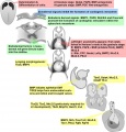 Molecular & Genetic Cardiac Development Factors.jpg 1,475 × 1,541; 269 KB
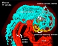
 Mouse E9 cervical intersomitic vessels A.jpg 1,714 × 1,142; 209 KB
 Mouse E9 cervical intersomitic vessels B.jpg 1,714 × 1,142; 175 KB
 Mouse E9 cervical intersomitic vessels C.jpg 1,714 × 1,142; 245 KB
 Mouse E9 cervical intersomitic vessels D.jpg 1,714 × 1,142; 147 KB
 Mouse E9 cervical intersomitic vessels.jpg 2,989 × 2,300; 777 KB
 Mouse embryo vascular.png 600 × 576; 518 KB
 Mouse heart E9.5.jpg 600 × 558; 31 KB
 Mouse hematopoietic stem cell.gif 600 × 595; 40 KB
 Mouse osteoblast 01.jpg 599 × 449; 118 KB
 Mouse osteoclast 01.jpg 600 × 453; 153 KB
 Mouse placenta blood vessel EM01.jpg 1,000 × 739; 132 KB
 Mouse-Cephalic-plexus-11somite 01.jpg 758 × 598; 106 KB
 Mouse-coronary vessel formation.jpg 800 × 282; 44 KB
 Mouse-heart E17.5.jpg 353 × 1,000; 145 KB
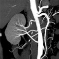 Multiple renal arteries 01.jpg 496 × 496; 40 KB
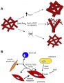 Notch and yolk sac blood vessels model.jpg 600 × 775; 97 KB
 NOTCH-endothelial-cartoon.jpg 1,280 × 888; 92 KB
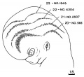 ORahilly1987 fig20-2.jpg 600 × 549; 46 KB
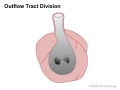 Outflow tract 001 icon.jpg 720 × 540; 33 KB
 Partial Anomalous Pulmonary Venous Drainage.jpg 290 × 350; 16 KB
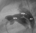 Patent ductus arteriosus angiogram.jpg 1,000 × 897; 91 KB
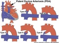 Patent ductus arteriosus classification.jpg 1,200 × 880; 138 KB
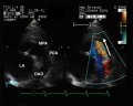 Patent ductus arteriosus echocardiogram.jpg 1,200 × 960; 133 KB
 Patent Ductus Arteriosus.jpg 294 × 350; 16 KB
 Patten024.jpg 1,047 × 451; 75 KB
 Patten047.jpg 785 × 765; 165 KB
 Patten048.jpg 797 × 857; 174 KB
 Patten049.jpg 807 × 864; 119 KB
 Patten050.jpg 800 × 890; 109 KB
 Patten054.jpg 764 × 1,060; 179 KB
 Persistent fifth aortic arch and patent ductus arteriosus CT01.jpg 589 × 800; 69 KB
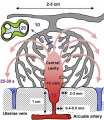 Placenta lobule blood flow cartoon.jpg 692 × 800; 134 KB
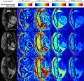 Placenta MRI01.jpg 1,280 × 1,227; 317 KB
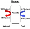 Placenta oxygen exchange levels.jpg 404 × 401; 18 KB
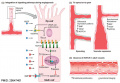
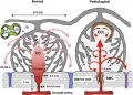 Placenta spiral artery conversion.jpg 592 × 423; 65 KB
 Placental cord vessels 01.jpg 650 × 488; 51 KB
 Placental cord vessels 02.jpg 800 × 477; 65 KB
 Platypus right auricle 01.jpg 800 × 640; 124 KB
 Platypus ventricular septum 01.jpg 683 × 800; 94 KB
 Postnatal persistant ductus venosus ultrasound 01.jpg 1,370 × 600; 96 KB
 Postnatal persistant ductus venosus ultrasound 02.jpg 671 × 600; 44 KB
 Postnatal persistant ductus venosus ultrasound 03.jpg 694 × 600; 54 KB
 Prentiss 286.jpg 799 × 933; 121 KB
 Pulmonary Atresia.jpg 301 × 350; 17 KB
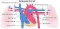 Pulmonary circulation cartoon.jpg 800 × 385; 65 KB
 Pulmonary Stenosis.jpg 289 × 350; 16 KB
 Quain594.jpg 592 × 1,000; 87 KB
 Quain595.jpg 1,200 × 803; 123 KB
 Quain596.jpg 869 × 819; 97 KB
 Quain597.jpg 811 × 1,200; 204 KB
 Robert Anderson.jpg 307 × 400; 16 KB
 Rugh 156.jpg 700 × 655; 86 KB
 Rugh 157.jpg 993 × 1,000; 183 KB
 Rugh 158.jpg 1,000 × 623; 158 KB
 Rugh 159.jpg 768 × 800; 87 KB
 Rugh 160.jpg 1,000 × 568; 97 KB
 Rugh 161.jpg 1,000 × 568; 104 KB
 Rugh 162.jpg 759 × 800; 75 KB
 Rugh 163.jpg 651 × 800; 75 KB
 Rugh 164.jpg 941 × 800; 108 KB
 Rugh 165.jpg 598 × 800; 132 KB
 Rugh 166.jpg 638 × 1,000; 175 KB
 Sabin1909 fig01-02.jpg 636 × 262; 19 KB
 Sabin1909 fig13.jpg 640 × 547; 114 KB
 Sabin1909 fig16.jpg 512 × 438; 120 KB
 Sabin1909 fig17.jpg 645 × 545; 80 KB
 Sabin1915 plate01.jpg 2,561 × 3,250; 922 KB
 Sabin1915 plate02.jpg 2,111 × 2,788; 609 KB
 Sabin1915 plate03.jpg 2,236 × 3,033; 1.08 MB
 Sabin1915 plate04.jpg 2,023 × 2,907; 797 KB
 Sabin1915 plate05.jpg 2,861 × 2,231; 1,013 KB
 Sabin1915 plate06.jpg 2,741 × 2,269; 926 KB
 Sabin1915 plate07.jpg 2,275 × 2,920; 1.16 MB
 Sabin1915.pdf ; 6.25 MB
 Schematic ECG normal and inverted T-wave.jpg 1,001 × 384; 32 KB
 Semilunar Valves.jpg 1,569 × 713; 93 KB
 Sinus venosus atrial septal defect 01.jpg 1,000 × 754; 90 KB
 Sinus venosus atrial septal defect 02.jpg 600 × 449; 29 KB
 Sinus venosus atrial septal defect 03.jpg 600 × 449; 31 KB
 Sinus venosus atrial septal defect 04.jpg 600 × 449; 31 KB
 Sinus venosus atrial septal defect 05.jpg 600 × 449; 25 KB
 Stage 13 image 022.jpg 1,000 × 473; 101 KB
 Stage 13 image 023.jpg 1,000 × 544; 110 KB
 Stage 13 image 060.jpg 1,000 × 486; 96 KB
 Stage 13 image 061.jpg 1,000 × 600; 101 KB
 Stage 13 image 066.jpg 1,000 × 579; 93 KB
 Stage 13 image 068.jpg 1,000 × 557; 97 KB
 Stage 13 image 069.jpg 1,000 × 581; 100 KB
 Stage 13 image 070.jpg 1,000 × 554; 101 KB
 Stage 13 image 071.jpg 1,000 × 526; 100 KB
 Stage 13 image 072.jpg 1,000 × 614; 116 KB
 Stage 13 image 073.jpg 1,000 × 619; 118 KB
 Stage 13 image 074.jpg 1,000 × 549; 113 KB
 Stage 13 image 075.jpg 1,000 × 567; 116 KB
 Stage 13 image 076.jpg 1,000 × 667; 120 KB
 Stage 13 image 077.jpg 1,000 × 612; 127 KB
 Stage 13 image 078.jpg 1,000 × 486; 81 KB
 Stage 13 image 079.jpg 1,000 × 409; 80 KB
 Stage 13 image 080.jpg 1,000 × 533; 91 KB
 Stage 13 image 081.jpg 1,000 × 449; 90 KB
 Stage 13 image 082.jpg 1,000 × 451; 90 KB
 Stage 13 image 083.jpg 1,000 × 440; 98 KB
 Stage 13 image 084.jpg 1,000 × 420; 101 KB
 Stage 13 image 085.jpg 1,000 × 434; 99 KB
 Stage 13 image 086.jpg 1,000 × 499; 97 KB
 Stage 13 image 087.jpg 1,000 × 493; 95 KB
 Stage 13 image 088.jpg 1,000 × 484; 97 KB
 Stage 13 image 089.jpg 1,000 × 498; 104 KB
 Stage 13 image 090.jpg 1,000 × 512; 103 KB
 Stage 13 image 091.jpg 1,000 × 470; 93 KB
 Stage 13 image 092.jpg 1,000 × 481; 86 KB


