Cat Development
| Embryology - 18 Apr 2024 |
|---|
| Google Translate - select your language from the list shown below (this will open a new external page) |
|
العربية | català | 中文 | 中國傳統的 | français | Deutsche | עִברִית | हिंदी | bahasa Indonesia | italiano | 日本語 | 한국어 | မြန်မာ | Pilipino | Polskie | português | ਪੰਜਾਬੀ ਦੇ | Română | русский | Español | Swahili | Svensk | ไทย | Türkçe | اردو | ייִדיש | Tiếng Việt These external translations are automated and may not be accurate. (More? About Translations) |
Introduction

Cats (Felis catus) are seasonally polyestrous animals that have multiple estrous cycles only during certain periods of the year.
The cat genome was initially sequenced in 2007[1] and has been recently annotated in August 2014.[2]
| Animal Development: axolotl | bat | cat | chicken | cow | dog | dolphin | echidna | fly | frog | goat | grasshopper | guinea pig | hamster | horse | kangaroo | koala | lizard | medaka | mouse | opossum | pig | platypus | rabbit | rat | salamander | sea squirt | sea urchin | sheep | worm | zebrafish | life cycles | development timetable | development models | K12 |
Some Recent Findings
|
| More recent papers |
|---|
 This table allows an automated computer search of the external PubMed database using the listed "Search term" text link.
More? References | Discussion Page | Journal Searches | 2019 References | 2020 References Search term: Cat Embryology <pubmed limit=5>Cat Embryology</pubmed> |
Developmental Timeline

Twenty-two stages have been described for the prenatal development of the domestic cat.[5]
The following data on early development is based upon the time after copulation[6]
oviduct embryo development
- 64 hours - 1 to 4 cells (17 of 20; 85.0%)
- 76 hours - 5 to 8 cells (18 of 28; 64.3% )
- 100 hours - 9 to 16 cells (14 of 24; 58.3%)
- 124 hours - morulae (15 of 21; 71.4% )
uterine embryo development
- 148 hours - compact morulae or early blastocysts
- days 12-14 - implantation occurs
Historic Development
- 1924 Cat Development: 1. Ovum of the Cat | 2. Process of Cleavage | 3. Formation of the Blastocyst | 4. Discussion | Plates | cat
Cat Ovary

Oocyte and Spermatozoa
The following scanning electron micrographs are from a recent paper on fresh and frozen cat oocytes.[7] Scale bar is 10 microns.


Genetics
Lineage: Eukaryota; Opisthokonta; Metazoa; Eumetazoa; Bilateria; Coelomata; Deuterostomia; Chordata; Craniata; Vertebrata; Gnathostomata; Teleostomi; Euteleostomi; Sarcopterygii; Tetrapoda; Amniota; Mammalia; Theria; Eutheria; Laurasiatheria; Carnivora; Feliformia; Felidae; Felinae; Felis; Felis catus
The cat genome was initially sequenced in 2007[1] and has been recently annotated in August 2014.[2]
- Mitochondria - entire mitochondrial genome 17,009 bp has been sequenced.
- Links: Genome Mitochondrial Genome
Early Development
Hill JP. and Tribe M. The early development of the cat (Felis domestica). (1924) Quart. J. Microsc. Sci., 68: 513-602.
Hill and Tribe in 1924[8] wrote a detailed description of oocyte to blastocyst development in the cat.
Placenta
- zonary placenta without cotyledons
- relatively small marginal hematoma
- materno-fetal barrier is endothelial-chorial
- superficially invasive into the endometrium but not into the myometrium
- placental labryrinth has characteristic giant cells
Placental cord
- two pairs of vessels in the cord
- two arteries and two veins
- allantoic duct
- cord average length 2 to 3 cm and 0.3 to 0.5 cm in diameter
- inserts at the margin of the zonary organ
- no spirals, no vitelline duct, and no additional vessels or structures
Additional Images
Historic Images
| Historic Disclaimer - information about historic embryology pages |
|---|
|
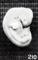
Fig. 210. Normal well-preserved cat fetus
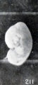
Fig. 211. Normal poorly preserved cat fetus of approximately the same length
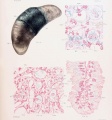
Plate 4. Cat Fetus and Placenta
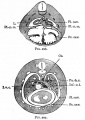
Fig. 297. Transverse section through the thoracic region of a cat embryo of 25 mm
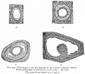
Fig. 331. Sections of a cat's ovary.
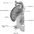
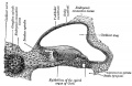
Fig. 903. Cat cochlear duct and ganglia


References
- ↑ 1.0 1.1 <pubmed>17975172</pubmed> Cite error: Invalid
<ref>tag; name 'PMID17975172' defined multiple times with different content - ↑ 2.0 2.1 Tamazian, G. etal., Annotated features of domestic cat - Felis cats genome. GigaScience 2014, 3:13
- ↑ Malandain E, Rault D, Froment E, Baudon S, Desquilbet L, Begon D & Chastant-Maillard S. (2011). Follicular growth monitoring in the female cat during estrus. Theriogenology , 76, 1337-46. PMID: 21798582 DOI.
- ↑ Inomata T, Ariga M, Sakita K, Kashiwazaki N, Ito J, Yokoh K, Ichikawa M, Ninomiya H & Inoue S. (2009). Development of external genitalia in fetal and neonatal domestic cats. J. Vet. Med. Sci. , 71, 139-45. PMID: 19262023
- ↑ Knospe C. (2002). Periods and stages of the prenatal development of the domestic cat. Anat Histol Embryol , 31, 37-51. PMID: 11841356
- ↑ Swanson WF, Roth TL & Wildt DE. (1994). In vivo embryogenesis, embryo migration, and embryonic mortality in the domestic cat. Biol. Reprod. , 51, 452-64. PMID: 7803616
- ↑ Hermansson U, Axnér E & Holst BS. (2007). Application of a zona pellucida binding assay (ZBA) in the domestic cat benefits from the use of in vitro matured oocytes. Acta Vet. Scand. , 49, 28. PMID: 17908298 DOI.
- ↑ Hill, J. P., and Tribe, M. 1924. The early development of the cat (Felis domestica). Quart. J. Microsc. Sci, 68, 513-602.
Reviews
Articles
Inomata T, Ninomiya H, Sakita K, Kashiwazaki N, Ito J, Ariga M & Inoue S. (2009). Developmental changes of Müllerian and Wolffian ducts in domestic cat fetuses. Exp. Anim. , 58, 41-5. PMID: 19151510
Inomata T, Ariga M, Sakita K, Kashiwazaki N, Ito J, Yokoh K, Ichikawa M, Ninomiya H & Inoue S. (2009). Development of external genitalia in fetal and neonatal domestic cats. J. Vet. Med. Sci. , 71, 139-45. PMID: 19262023
Ciani F, Cocchia N, Rizzo M, Ponzio P, Tortora G, Avallone L & Lorizio R. (2008). Sex determining of cat embryo and some feline species. Zygote , 16, 169-77. PMID: 18405438 DOI.
França LR & Godinho CL. (2003). Testis morphometry, seminiferous epithelium cycle length, and daily sperm production in domestic cats (Felis catus). Biol. Reprod. , 68, 1554-61. PMID: 12606460 DOI.
Knospe C. (2002). Periods and stages of the prenatal development of the domestic cat. Anat Histol Embryol , 31, 37-51. PMID: 11841356
Hill JP. and Tribe M. The early development of the cat (Felis domestica). (1924) Quart. J. Microsc. Sci., 68: 513-602.
Hill, J. P., and Tribe, M. 1924. The early development of the cat (Felis domestica). Quart. J. Microsc. Sci, 68, 513-602.
Search Pubmed
cat development | feline development
| Animal Development: axolotl | bat | cat | chicken | cow | dog | dolphin | echidna | fly | frog | goat | grasshopper | guinea pig | hamster | horse | kangaroo | koala | lizard | medaka | mouse | opossum | pig | platypus | rabbit | rat | salamander | sea squirt | sea urchin | sheep | worm | zebrafish | life cycles | development timetable | development models | K12 |
Glossary Links
- Glossary: A | B | C | D | E | F | G | H | I | J | K | L | M | N | O | P | Q | R | S | T | U | V | W | X | Y | Z | Numbers | Symbols | Term Link
Cite this page: Hill, M.A. (2024, April 18) Embryology Cat Development. Retrieved from https://embryology.med.unsw.edu.au/embryology/index.php/Cat_Development
- © Dr Mark Hill 2024, UNSW Embryology ISBN: 978 0 7334 2609 4 - UNSW CRICOS Provider Code No. 00098G