Cartilage Histology: Difference between revisions
No edit summary |
mNo edit summary |
||
| (8 intermediate revisions by 2 users not shown) | |||
| Line 1: | Line 1: | ||
{{Header}} | |||
== Introduction == | == Introduction == | ||
[[File:Articular_cartilage.jpg|thumb|Articular cartilage]] | [[File:Articular_cartilage.jpg|thumb|Articular cartilage]] | ||
| Line 7: | Line 8: | ||
[[Lecture - Musculoskeletal Development]] and notes on [[Musculoskeletal System - Bone Development|Bone Development]]. | [[Lecture - Musculoskeletal Development]] and notes on [[Musculoskeletal System - Bone Development|Bone Development]]. | ||
{{ | |||
{{Musculoskeletal Links}} | |||
==Chondroblasts and Chondrocytes== | ==Chondroblasts and Chondrocytes== | ||
| Line 43: | Line 45: | ||
</gallery> | </gallery> | ||
{{Bone Histology}} | {{Bone Histology}} | ||
===Human Fetal Head (12 week)=== | ===Human Fetal Head (12 week)=== | ||
| Line 92: | Line 81: | ||
:'''Links:''' [[Histology Stains]] | :'''Links:''' [[Histology Stains]] | ||
== | ==Historic Images== | ||
<gallery> | |||
File:Bailey116.jpg|Tarsal bones of a pig embryo | |||
File:Bailey117.jpg | |||
File:Bailey118.jpg|Bone deposited around trabeculae of cartilage | |||
File:Bailey119.jpg|Diagram representing growth in diameter of a long bone | |||
File:Bailey120.jpg|Longitudinal section from head of femur of young dog | |||
File:Streeter001.jpg|Section through lateral semicircular canal human fetus 33 mm | |||
</gallery> | |||
== Other Textbooks == | == Other Textbooks == | ||
| Line 113: | Line 106: | ||
* '''Pubmed''' [http://www.ncbi.nlm.nih.gov/sites/gquery?itool=toolbar&cmd=search&term=ossification ossification] | * '''Pubmed''' [http://www.ncbi.nlm.nih.gov/sites/gquery?itool=toolbar&cmd=search&term=ossification ossification] | ||
== External Links == | |||
{{External Links}} | |||
* Virtual Slidebox of Histology (USA) [http://www.path.uiowa.edu/cgi-bin-pub/vs/fpx_browse.cgi?cat=o_skeletal&div=nlm Skeletal system] | |||
* [http://www.e-radiography.net/articles/ossification/ossification.htm e-radiography Ossification] | |||
* UWA Blue Histology [http://www.lab.anhb.uwa.edu.au/mb140/CorePages/Cartilage/Cartil.htm Histology - Cartilage] | |||
== Terms == | == Terms == | ||
* '''haematopoiesis''' (Greek, ''haima'' = "blood"; ''poiesis'' = "to make") the process of blood cell formation. | * '''haematopoiesis''' (Greek, ''haima'' = "blood"; ''poiesis'' = "to make") the process of blood cell formation. | ||
* '''Howship's lacuna''' - (resorptive bay) the historic name for the shallow bay or cavity lying directly under an osteoclast. This is the site of bone matrix resorption. | * '''Howship's lacuna''' - (resorptive bay) the historic name for the shallow bay or cavity lying directly under an osteoclast. This is the site of bone matrix resorption. | ||
* '''lacuna''' - (Latin, ''lacuna'' = “ditch, gap” diminutive form of ''lacus'' = “lake”) lacunae is the plural, cavity in bone or cartilage for cell. | * '''lacuna''' - (Latin, ''lacuna'' = “ditch, gap” diminutive form of ''lacus'' = “lake”) lacunae is the plural, cavity in bone or cartilage for cell. | ||
Latest revision as of 18:03, 13 September 2016
| Embryology - 16 Apr 2024 |
|---|
| Google Translate - select your language from the list shown below (this will open a new external page) |
|
العربية | català | 中文 | 中國傳統的 | français | Deutsche | עִברִית | हिंदी | bahasa Indonesia | italiano | 日本語 | 한국어 | မြန်မာ | Pilipino | Polskie | português | ਪੰਜਾਬੀ ਦੇ | Română | русский | Español | Swahili | Svensk | ไทย | Türkçe | اردو | ייִדיש | Tiếng Việt These external translations are automated and may not be accurate. (More? About Translations) |
Introduction
Our adult skeleton forms from a larger number of developmental elements that are replaced and fuse. In development there are 2 separate signaling pathways for pattern formation and the formation of bone itself. Furthermore bone formation can be divided into 2 specific forms that occur in anatomically different regions. This practical class will describe the development and structure of bone and finish with a study of abnormalities associated with bone.
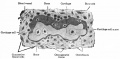
The image shown to the left shows a histological section through the developing lower limb at the level of a developing joint (knee), surrounding the developing bone is cartilage, skeletal muscles and connective tissue of the limb.
Lecture - Musculoskeletal Development and notes on Bone Development.
Chondroblasts and Chondrocytes
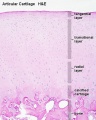
Articular cartilage
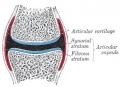
Synovial joint showing cartilage

Immature and mature cartilage forming cells located at articular cartilage regions.
Interstitial growth
- Occurs mainly in immature cartilage.
- Chondroblasts in existing cartilage divide and form small groups of cells (isogenous groups) which produce matrix to become separated from each other by a thin partition of matrix.
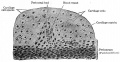
Appositional growth
- Occurs also in mature cartilage.
- Mesenchymal cells surrounding the cartilage in the deep part of the perichondrium (or the chondrogenic layer) differentiate into chondroblasts.
Cartilage Development

Endochondral ossification slides: Developing bone | Bone, Developing (LS, Femur) Cat H&E
Blue Histology - endochondral | Dev Biology - endochondral ossification | endochondral ossification animation


- Bone Histology: Cartilage Histology | Histology Stains | Histology | cartilage | bone | bone timeline
- Trabecular bone trabecular | lamellar | trabecular - overview HE | trabecular - low HE | trabecular - med HE
- Endochondral ossification primary ossification | endochondral ossification
- Intramembranous ossification intramembranous - VG low | intramembranous - VG high | intramembranous - HE low | intramembranous - HE high
Human Fetal Head (12 week)


Histology Stains
Alizarin Red
- an anthraquinone derivative used to identify calcium in tissue sections
- calcium forms an Alizarin Red S-calcium complex in a chelation process and the end product is also birefringent.
- reaction can also identify magnesium, manganese, barium, strontium, and iron may interfere
- these elements usually in too low concentration to interfere with the staining
H&E
- acronym for hematoxylin and eosin stain
- hematoxylin - basic dye which colors basophilic structures with blue-purple hue (nucleus, DNA, RNA)
- eosin Y - acidic alcohol-based which colors eosinophilic structures bright pink (cytoplasm, extracellular matrix, protein)
H&Van Gieson
- Van Gieson's Stain is a mixture of picric acid and acid fuchsin used for differential staining of collagen and other connective tissue.
- Nuclei - stains brownish black to black
- Collagen (fibrous connective tissue) - stains pink or deep red
- Muscle, Cytoplasm, RBC and Fibrin - stains yellow
- Links: Histology Stains
Historic Images
Tarsal bones of a pig embryo
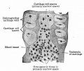
Bone deposited around trabeculae of cartilage
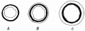
Diagram representing growth in diameter of a long bone
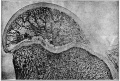
Longitudinal section from head of femur of young dog
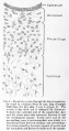
Section through lateral semicircular canal human fetus 33 mm


Other Textbooks
- Anatomy of the Human Body (H. Gray, 1918.) historical anatomy text Osteology
- Molecular Biology of the Cell Bone Is Continually Remodeled by the Cells Within It | Image: Figure 22-52. Deposition of bone matrix by osteoblasts | Image: Figure 22-56. The development of a long bone
- Molecular Cell Biology Mutations in Collagen Reveal Aspects of Its Structure and Biosynthesis
- The Cell- A Molecular Approach Steroid Hormones and the Steroid Receptor Superfamily
- Clinical Methods: The History, Physical, and Laboratory Examinations 100. Alkaline Phosphatase and Gamma Glutamyltransferase
- Endocrinology: An Integrated Approach by Nussey, S.S. and Whitehead, S.A. Endocrinology: Definition and causes of osteoporosis
- Developmental Biology 6th ed. by Gilbert, Scott F. Figure 14.13. Schematic diagram of endochondral ossification | Aging: The Biology of Senescence
Search
- Pubmed ossification
External Links
External Links Notice - The dynamic nature of the internet may mean that some of these listed links may no longer function. If the link no longer works search the web with the link text or name. Links to any external commercial sites are provided for information purposes only and should never be considered an endorsement. UNSW Embryology is provided as an educational resource with no clinical information or commercial affiliation.
- Virtual Slidebox of Histology (USA) Skeletal system
- e-radiography Ossification
- UWA Blue Histology Histology - Cartilage
Terms
- haematopoiesis (Greek, haima = "blood"; poiesis = "to make") the process of blood cell formation.
- Howship's lacuna - (resorptive bay) the historic name for the shallow bay or cavity lying directly under an osteoclast. This is the site of bone matrix resorption.
- lacuna - (Latin, lacuna = “ditch, gap” diminutive form of lacus = “lake”) lacunae is the plural, cavity in bone or cartilage for cell.
- lamellar bone - the highly organized strong bone matrix deposited in concentric sheets with a low proportion of osteocytes. Many collagen fibers parallel to each other in the same layer.
- osteon - (Haversian system) the functional unit of compact bone. Consists of a central canal (Haversian canal) surrounded by lamellar bone matrix within which osteocytes reside.
- resorptive bay - (Howship's lacuna) the shallow bay or cavity lying directly under an osteoclast. This is the site of bone matrix resorption.
- suture - in the skull a form of articulation where the contiguous margins of the bones are united by a thin layer of fibrous tissue.
- woven bone - the first deposited weaker bone matrix with many osteocytes and a matrix disorganized structure. Replaced by lamellar bone. Seen in developing, healing and bone disease.
Glossary Links
- Glossary: A | B | C | D | E | F | G | H | I | J | K | L | M | N | O | P | Q | R | S | T | U | V | W | X | Y | Z | Numbers | Symbols | Term Link
Cite this page: Hill, M.A. (2024, April 16) Embryology Cartilage Histology. Retrieved from https://embryology.med.unsw.edu.au/embryology/index.php/Cartilage_Histology
- © Dr Mark Hill 2024, UNSW Embryology ISBN: 978 0 7334 2609 4 - UNSW CRICOS Provider Code No. 00098G