Carnegie stage 7: Difference between revisions
mNo edit summary |
mNo edit summary |
||
| Line 2: | Line 2: | ||
== Introduction == | == Introduction == | ||
{| | {| | ||
| [[File:Stage7-bf1.jpg|300px]] | | valign=top|[[File:Stage7-bf1.jpg|300px]] | ||
| | | | ||
===Facts=== | ===Facts=== | ||
| Line 11: | Line 11: | ||
The initial images are displayed unlabeled to allow you to explore the embryo for yourself, linked labeled versions are also available for some images. | The initial images are displayed unlabeled to allow you to explore the embryo for yourself, linked labeled versions are also available for some images. | ||
=== | ===Summary=== | ||
Gastrulation is continuing as cells migrate from the epiblast, continuing to form mesoderm. | Gastrulation is continuing as cells migrate from the epiblast, continuing to form mesoderm. | ||
| Line 21: | Line 21: | ||
The notochord is a key to embryonic folding and regulation of ectoderm and mesoderm differentiation. It lies in the rostrocordal axis and the embryonic disc will fold either side ventrally, pinching off a portion of the yolk sac to form the lining of the gastrointestinal tract. | The notochord is a key to embryonic folding and regulation of ectoderm and mesoderm differentiation. It lies in the rostrocordal axis and the embryonic disc will fold either side ventrally, pinching off a portion of the yolk sac to form the lining of the gastrointestinal tract. | ||
See also [[#Events|Events]] | |||
:'''Links:''' [[Week 3]] | [[Gastrulation]] | [[Lecture - Week 3 Development|Lecture]] | [[ANAT2341_Lab_2|Practical]] | [[Carnegie_stage_8|Stage 8]] | :'''Links:''' [[Week 3]] | [[Gastrulation]] | [[Lecture - Week 3 Development|Lecture]] | [[ANAT2341_Lab_2|Practical]] | [[Carnegie_stage_8|Stage 8]] | ||
| Line 116: | Line 117: | ||
'''Scanning Electron Images:''' [[:File:Stage7-sem1.jpg|Embryonic Disc 1]] | [[:File:Stage7-sem2.jpg|Embryonic Disc 2]] | [[:File:Stage7-sem3.jpg|Embryonic Disc 3]] | [[:File:Stage7-sem4.jpg|Primitive Streak]] | '''Scanning Electron Images:''' [[:File:Stage7-sem1.jpg|Embryonic Disc 1]] | [[:File:Stage7-sem2.jpg|Embryonic Disc 2]] | [[:File:Stage7-sem3.jpg|Embryonic Disc 3]] | [[:File:Stage7-sem4.jpg|Primitive Streak]] | ||
==Events== | |||
* <ref name=“PMID”><pubmed></pubmed></ref> | |||
===References=== | |||
<references/> | |||
==Additional Images== | |||
===Historic Images=== | |||
{{Carnegie_stages}} | {{Carnegie_stages}} | ||
Revision as of 10:16, 4 April 2016
| Embryology - 24 Apr 2024 |
|---|
| Google Translate - select your language from the list shown below (this will open a new external page) |
|
العربية | català | 中文 | 中國傳統的 | français | Deutsche | עִברִית | हिंदी | bahasa Indonesia | italiano | 日本語 | 한국어 | မြန်မာ | Pilipino | Polskie | português | ਪੰਜਾਬੀ ਦੇ | Română | русский | Español | Swahili | Svensk | ไทย | Türkçe | اردو | ייִדיש | Tiếng Việt These external translations are automated and may not be accurate. (More? About Translations) |
Introduction

|
FactsHuman embryonic stage 7 occurs during week 3 between 15 to 17 days. The embryo is now 0.4 mm diameter in size. The initial images are displayed unlabeled to allow you to explore the embryo for yourself, linked labeled versions are also available for some images. SummaryGastrulation is continuing as cells migrate from the epiblast, continuing to form mesoderm. Mesoderm lies between the ectoderm and endoderm as a continuous sheet except at the buccopharyngeal and cloacal membranes. These membranes have ectoderm and endoderm only and will lie at the rostral (head) and caudal (tail) of the gastrointestinal tract. From the primitive node a tube extends under the ectoderm in the opposite direction to the primitive streak. This tube forms first the axial process then notochordal process, then finally the notochord. The notochord is a key to embryonic folding and regulation of ectoderm and mesoderm differentiation. It lies in the rostrocordal axis and the embryonic disc will fold either side ventrally, pinching off a portion of the yolk sac to form the lining of the gastrointestinal tract. See also Events
|
| Week: | 1 | 2 | 3 | 4 | 5 | 6 | 7 | 8 |
| Carnegie stage: | 1 2 3 4 | 5 6 | 7 8 9 | 10 11 12 13 | 14 15 | 16 17 | 18 19 | 20 21 22 23 |
- Carnegie Stages: 1 | 2 | 3 | 4 | 5 | 6 | 7 | 8 | 9 | 10 | 11 | 12 | 13 | 14 | 15 | 16 | 17 | 18 | 19 | 20 | 21 | 22 | 23 | About Stages | Timeline
Bright Field Lateral


Scanning EM

|

|
Human embryonic disc
|
Human embryonic disc
|

|

Primitive streak detail (left) |
Human embryonic disc
|
Kyoto Collection

|

|
View: embryonic disc, showing the epiblast viewed from the amniotic (dorsal) side. Head to tail orientation is Cranial (image top) and Caudal (image bottom).
Features: embryonic disc, primitive node, primative streak, primative groove, connecting stalk
Alternate View: embryonic disc, probably from from the ventral side. Showing the connecting stalk to the left.
Image source: The Kyoto Collection images are reproduced with the permission of Prof. Kohei Shiota and Prof. Shigehito Yamada, Anatomy and Developmental Biology, Kyoto University Graduate School of Medicine, Kyoto, Japan for educational purposes only and cannot be reproduced electronically or in writing without permission.
Carnegie Collection

Surface view of implantation site |

Surface lateral view of implantation site |


| Carnegie Collection - Stage 7 | ||||||||||
|---|---|---|---|---|---|---|---|---|---|---|
| Serial No. | Grade | Fixative | Embedding Medium | Thinness (µm) | Stain | Year | Notes | |||
| 7802 | Exc. | Alc. & Bouin | C-P | 6 | (Stain - Haematoxylin Eosin) | 1940 | Heuser et al. (1945) | |||
| 8206 | Good | p | C-P | 6 | (Stain - Haematoxylin Eosin) | 1943 | ||||
| 8361 | Good | Bouin | C-P | 10 | p | 1946 | Abnormal | |||
| 8602 | Exc. | Alc. | C-P | 8 | (Stain - Haematoxylin Eosin) | 1948 | ||||
| 8752 | Exc. | ? | C-P | 10 | (Stain - Haematoxylin Eosin) | 1950 | ||||
| 8755 | Exc. | Formol | C-P | 10 | (Stain - Haematoxylin Eosin) | 1950 | ||||
| 9217 | Exc. | p | P | 10 | (Stain - Haematoxylin Eosin) | 1954 | ||||
Abbreviations
| ||||||||||
| iBook - Carnegie Embryos | |
|---|---|

|
|
Photograph

|

|

|

|
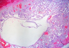
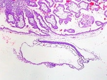
Image: Dr Ed Uthman (Houston, Texas) - other pathology images section of ectopic (tubal) pregnancy.
- Image Links: Original full image | Trilaminar embryo excerpt | Villi excerpt 1 | Villi excerpt 2
- Full Image versions: ExtraLarge 1712x1206px | Large 1024x721px | Medium 500x352px
Embryo Virtual Slide
|
|
Stage 7 Images
Bright Field Images: Stage7-bf1.jpg | Stage7-bf2.jpg | Stage7-bf3.jpg | Stage7-bf4.jpg
Scanning Electron Images: Embryonic Disc 1 | Embryonic Disc 2 | Embryonic Disc 3 | Primitive Streak
Events
References
- ↑ <pubmed></pubmed>
Additional Images
Historic Images
- Carnegie Stages: 1 | 2 | 3 | 4 | 5 | 6 | 7 | 8 | 9 | 10 | 11 | 12 | 13 | 14 | 15 | 16 | 17 | 18 | 19 | 20 | 21 | 22 | 23 | About Stages | Timeline
Glossary Links
- Glossary: A | B | C | D | E | F | G | H | I | J | K | L | M | N | O | P | Q | R | S | T | U | V | W | X | Y | Z | Numbers | Symbols | Term Link
Cite this page: Hill, M.A. (2024, April 24) Embryology Carnegie stage 7. Retrieved from https://embryology.med.unsw.edu.au/embryology/index.php/Carnegie_stage_7
- © Dr Mark Hill 2024, UNSW Embryology ISBN: 978 0 7334 2609 4 - UNSW CRICOS Provider Code No. 00098G