Carnegie stage 5: Difference between revisions
From Embryology
No edit summary |
|||
| Line 51: | Line 51: | ||
File:Stage5 bf22.jpg|Embryo No.7700 | File:Stage5 bf22.jpg|Embryo No.7700 | ||
File:Stage5 bf22b.jpg|Embryo No.7700 | File:Stage5 bf22b.jpg|Embryo No.7700 | ||
<gallery> | </gallery> | ||
{{Template:Carnegie_stages}} | {{Template:Carnegie_stages}} | ||
Revision as of 18:02, 23 September 2011

|
Facts: Week 1-2, size 0.1 - 0.2 mm
Features: implantation completed, inner cell mass, bilaminar embryo, trophoblast development
|
| File:Chorion 001 icon.jpg</wikiflv> |
Animation (left) shows the events following implantation and focuses on changes in the the spaces surrounding the embryonic disc, the extraembryonic coelom. The blastoceol cavity is converted into two separate spaces: the yolk sac and the chorionic cavity. The third space lies above the epiblast, the amniotic cavity.
|
| Quicktime movie |
Stage 5b
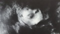
Embryo No.8004
Embryo No.8004
Embryo No.8004


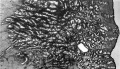
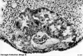
Stage 5c
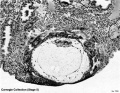
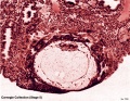
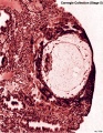
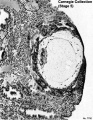
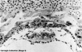
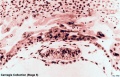
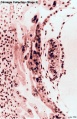
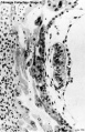
Embryo No.7700
Embryo No.7700
Embryo No.7700
Embryo No.7700
Embryo No.7700
Embryo No.7700
Embryo No.7700
Embryo No.7700


- Carnegie Stages: 1 | 2 | 3 | 4 | 5 | 6 | 7 | 8 | 9 | 10 | 11 | 12 | 13 | 14 | 15 | 16 | 17 | 18 | 19 | 20 | 21 | 22 | 23 | About Stages | Timeline
Cite this page: Hill, M.A. (2024, April 24) Embryology Carnegie stage 5. Retrieved from https://embryology.med.unsw.edu.au/embryology/index.php/Carnegie_stage_5
- © Dr Mark Hill 2024, UNSW Embryology ISBN: 978 0 7334 2609 4 - UNSW CRICOS Provider Code No. 00098G