Carnegie stage 3: Difference between revisions
From Embryology
No edit summary |
|||
| Line 1: | Line 1: | ||
==Introduction== | ==Introduction== | ||
The free-floating blastocyst has reached the uterine body still enclosed in the zone pellucida and "hatches" from this surrounding extracellular matrix. It is only after hatching that the blastocyst can attach to and then implant into the uterine wall. | |||
{| | {| | ||
| [[Image:CSt3.jpg]] | | [[Image:CSt3.jpg]] | ||
| Line 23: | Line 25: | ||
:'''Links:''' [[Lecture - Week 1 and 2 Development|Lecture]] | [[2011_Lab_2|Practical]] | [[Implantation]] | [[Week 1]] | :'''Links:''' [[Lecture - Week 1 and 2 Development|Lecture]] | [[2011_Lab_2|Practical]] | [[Implantation]] | [[Week 1]] | ||
{| | |||
| width= 200px | [[File:Human-blastocyst-day-3-6-icon.jpg|120px|link=Movie_-_Blastocyst_Development]] | |||
| width= 200px | [[File:Human_blastocyst_day_5-6.jpg|120px|link=Movie_-_Blastocyst_Contractions]] | |||
| width= 200px | [[File:Human blastocyst hatching movie icon.jpg|120px|link=Movie - Blastocyst Hatching]] | |||
|-bgcolor="FAF5FF" | |||
| [[Movie_-_Blastocyst_Development|Blastocyst Development]] | |||
Early development day 3 to 6 | |||
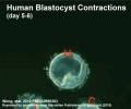
| [[Movie_-_Blastocyst_Contractions|Blastocyst Contractions]] | |||
(day 5 to 6) | |||
| [[Movie_-_Blastocyst_Hatching|Blastocyst Hatching]] | |||
(day 5 to 6) | |||
|} | |||
==Carnegie Collection== | ==Carnegie Collection== | ||
Revision as of 06:05, 25 October 2011
Introduction
The free-floating blastocyst has reached the uterine body still enclosed in the zone pellucida and "hatches" from this surrounding extracellular matrix. It is only after hatching that the blastocyst can attach to and then implant into the uterine wall.

|
Human Blastocyst "hatching" from zona pellucida (classified as Carnegie stage 3).
Blastocyst is too the right of image and zona pellucida is shown to the left of the image.
|


- Human blastocyst (day 5) still within zone pellucida.
Facts: Week 1, 4 - 5 days, size 0.1-0.2 mm
Features: zona pellucida, trophoblast shell, inner cell mass, blastoceol
- Links: Lecture | Practical | Implantation | Week 1
|
||
| Blastocyst Development
Early development day 3 to 6 |
Blastocyst Contractions
(day 5 to 6) |
Blastocyst Hatching
(day 5 to 6) |
Carnegie Collection

|

|
| 58 cell blastocyst (day 4) | 107 cell blastocyst (day 4.5) |
- Carnegie Stages: 1 | 2 | 3 | 4 | 5 | 6 | 7 | 8 | 9 | 10 | 11 | 12 | 13 | 14 | 15 | 16 | 17 | 18 | 19 | 20 | 21 | 22 | 23 | About Stages | Timeline
Cite this page: Hill, M.A. (2024, April 18) Embryology Carnegie stage 3. Retrieved from https://embryology.med.unsw.edu.au/embryology/index.php/Carnegie_stage_3
- © Dr Mark Hill 2024, UNSW Embryology ISBN: 978 0 7334 2609 4 - UNSW CRICOS Provider Code No. 00098G