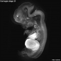
Carnegie stage 21
| Embryology - 24 Apr 2024 |
|---|
| Google Translate - select your language from the list shown below (this will open a new external page) |
|
العربية | català | 中文 | 中國傳統的 | français | Deutsche | עִברִית | हिंदी | bahasa Indonesia | italiano | 日本語 | 한국어 | မြန်မာ | Pilipino | Polskie | português | ਪੰਜਾਬੀ ਦੇ | Română | русский | Español | Swahili | Svensk | ไทย | Türkçe | اردو | ייִדיש | Tiếng Việt These external translations are automated and may not be accurate. (More? About Translations) |
Introduction

Facts
Week 8, 53 - 54 days, 22 - 24 mm
Gestational age GA week 10
Summary
- Ectoderm: sensory placodes, nasal pits moved ventrally, fourth ventricle of brain
- Mesoderm: heart prominence, ossification continues
- Head: nose, eye, external acoustic meatus
- Body: straightening of trunk, heart, liver, umbilical cord
- Limb: upper limbs longer and bent at elbow, foot plate with digital rays begin to separate, wrist, hand plate with webbed digits
See also Carnegie stage 21 Events
Features
- scalp vascular plexus, eylid, eye, nose, auricle of external ear, arm, elbow, wrist, knee, notch between digital rays, umbilical cord
- Identify: straightening of trunk, pigmented eye, eyelid, nose, external acoustic meatus, scalp vascular plexus, webbed digits, liver prominance, thigh, ankle, foot plate, umbilical cord
- Links: Week 8 | System Development | Lecture - Limb | Lecture - Head Development | Lecture - Sensory | Science Practical - Head | Science Practical - Sensory | Science Practical - Urogenital | Category:Carnegie Stage 21 | Stage 22
| Week: | 1 | 2 | 3 | 4 | 5 | 6 | 7 | 8 |
| Carnegie stage: | 1 2 3 4 | 5 6 | 7 8 9 | 10 11 12 13 | 14 15 | 16 17 | 18 19 | 20 21 22 23 |
- Carnegie Stages: 1 | 2 | 3 | 4 | 5 | 6 | 7 | 8 | 9 | 10 | 11 | 12 | 13 | 14 | 15 | 16 | 17 | 18 | 19 | 20 | 21 | 22 | 23 | About Stages | Timeline
Bright Field

|
Virtual Embryo Slides
|

Kyoto Collection

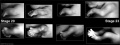
View: This is a dorsolateral view of embryo. Amniotic membrane removed.
| Central Nervous System | |
|---|---|

|

|
| left lateral view | medial view |
| scale bar = 1mm |

|

|

|
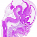
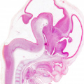
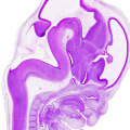
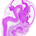
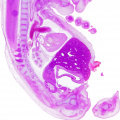
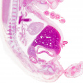
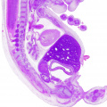
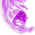
- Kyoto embryo 940 Histology
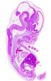
sagittal 1
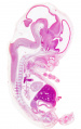
sagittal 2
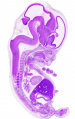
sagittal 3
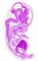
sagittal 4
upper
upper
upper
upper
lower
lower
lower
lower


Image source: The Kyoto Collection images are reproduced with the permission of Prof. Kohei Shiota and Prof. Shigehito Yamada, Anatomy and Developmental Biology, Kyoto University Graduate School of Medicine, Kyoto, Japan for educational purposes only and cannot be reproduced electronically or in writing without permission.
Carnegie Collection


- Carnegie stage 21: 7592 Right | 7592 Anterior | 7592 Left | 8553 Right | 8553 Anterior | 8553 Left | 4090 Right | 4090 Anterior | 4090 Left

| Carnegie Collection - Stage 21 | |||||||||||
|---|---|---|---|---|---|---|---|---|---|---|---|
| Serial No. | Size (mm) | Grade | Fixative | Embedding Medium | Plane | Thinness (µm) | Stain | Point Score | Sex | Year | Notes |
| 22 | E, 20 Ch, 35x30x30 | Good | Alc. | P | Transverse | 50 | Al. coch. | 34.5 | Female | 1895 | |
| 57 | E, 23 Ch., ca. 30 | Poor | Alc. | P | Sagittal | 50 | Al. coch. | 36 | Male | 1896 | |
| 128 | E, 20 Ch., 50x43 | Good | Formalin | P | Coronal | 50 | Al. coch. | 33 | Female | 1898 | |
| 229 | E, 19 | Poor | Alc. | P | Sagittal | 50 | Al. coch. | 33 | Female | 1903 | |
| 349 | E, 24 | Good | Zenker | C | Coronal | 250 | Unstained | 36 | ? | 1905 | Double vascular injection |
| 455 | E, 24 Ch., 42x34x20 | Good | Alc. | P | Transverse | 30 | (Stain - Haematoxylin Eosin) | 36.5 | Male | 1910 | |
| 632 | E, 24 Ch., 60x50x30 | Good | Bichlor. acetic | P | Sagittal | 40, 100, 250 | Al. coch. | 33 | Female | 1913 | Injected |
| 903C | E, 23.5 | Good | Formalin | P | Transverse | 40 | Al. coch. | 38.5 | Female | 1914 | |
| 1008 | E, 26,4 | Good | Formalin | P | Sagittal | 40 | Al. coch. | 39 | ?? | 1914 | |
| 1358F | E, 23 | Good | Formalin | P | Sagittal | 40 | Al. coch. | 37.5 | Female | 1916 | |
| 2937 | E,, 24.2 | Good | Bouin | P | Transverse | 50 | (Stain - Haematoxylin Eosin) aur., or. G. | 39 | Female | 1920 | |
| 3167 | E., 24.5 Ch., 60x50x40 | Poor | Bichlor, acetic, formol | P | Transverse | 20 | Al. coch. | 32 | Male | 1920 | |
| 4090 | E, 22.2 Ch.. 66x46x30 | Good | Formalin | P | Transverse | 40 | Al. coch. | 30 | Female | 1922 | |
| 4160 | E,25 | Poor | Formalin | P | Sagittal | 25 | (Stain - Haematoxylin Eosin) | 39 | Male | 1923 | Tubal |
| 4960 | E.22 Ch,, 47x42x28 | Good | Formalin | P | Transverse | 15 | Al. coch., Mallory | 31.5 | Female | 1925 | |
| 5??6 | E. 215 | Good | Formalin | P | Sagittal | 20 | (Stain - Haematoxylin Eosin) | 34 | Female | 1927 | |
| 6531 | E,22 | Poor | Glacial acetic, | C-P | Transverse | 10 | (Stain - Haematoxylin Eosin) | 31.5 | Female | 1931 | Leitz Collection |
| 7254 | E,225 | Exc | Bouin | C-P | Transverse | 20 | (Stain - Haematoxylin Eosin) | 33.5 | Male | 1936 | |
| 7592 | E,22-> | Exc. | Bouin | C-P | Transverse | 20 | (Stain - Haematoxylin Eosin) | 36 | Female | 1937 | |
| 7864 | E., 24 | Exc, | Formalin | C-P | Frontal | 20 | (Stain - Haematoxylin Eosin) | 32.5 | Male | 1941 | |
| 8553 | E., 22 | Exc | Bouin | C-P | Transverse | 12 | (Stain - Haematoxylin Eosin) | 38 | Female | 1947 | |
| 9614 | E,,22 5 | Exc | Bouin | P | Coronal | 10 &15 | Azan | ? | ? | 1958 | Rubella. Hysterectomy |
Abbreviations
| |||||||||||
| iBook - Carnegie Embryos | |
|---|---|

|
|
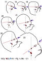
Hinrichsen Collection
| frontal section | respiratory excerpt |
|---|---|

|

|

|
- Links: frontal section | respiratory excerpt | labeled respiratory excerpt | Carnegie stage 21 | Week 8 | Hinrichsen Collection
Image source: The Hinrichsen Collection images are reproduced with the permission of Prof. Beate Brand-Saberi, Head, Department of Anatomy and Molecular Embryology, Ruhr-Universität Bochum. Images are for educational purposes only and cannot be reproduced electronically or in writing without permission.
Events
- Hearing - tip of the cochlea is recurved.
- Vision - (stage 19 -22) the eyelid folds develop into the eyelids and cover more of the eye as the palpebral fissure takes shape. The upper and the lower eyelids meet at the outer canthus in Stage 19. [2]
- Musculoskeletal
- Mandible bone-formation has taken place and formed a plate on the lateral side of Meckel's cartilage; this plate is in the substance of, and is surrounded by, that mesodermal condensation which outlines the mandible and precedes the formation of bone. The plate of bone is confined to the region of what approximately corresponds to the future body of the mandible, but the mesodermal condensation can be traced further backwards, always, of course, on the lateral side of Meckel's cartilage and the associated branches of the mandibular nerve. The condensation admittedly becomes gradually much less sharply defined but is still distinguishable from the surrounding tissue. Finally, some distance beyond the limit of bone-formation, the lateral pterygoid muscle can be seen running into and out- lining the terminal part of the mesodermal condensation. This is the first indication of the condylar process in my series (P1. 1, fig. 1). Meckel's cartilage with the inferior dental and lingual nerves on its upper surface lies medial to this rudimentary condylar area and inferior to the lateral pterygoid muscle.[3]
- Joint - knee posterior cruciate ligament present.[4]
- Cardiovascular
- Cerebral artery the formation of the anterior communicating artery.[5]
References
- ↑ Streeter GL. Developmental Horizons In Human Embryos Description Or Age Groups XIX, XX, XXI, XXII, And XXIII, Being The Fifth Issue Of A Survey Of The Carnegie Collection. (1957) Carnegie Instn. Wash. Publ. 611, Contrib. Embryol., 36: 167-196.
- ↑ <pubmed>7364662</pubmed>
- ↑ <pubmed>12980883</pubmed>| PMC1273756
- ↑ <pubmed>9185992</pubmed>
- ↑ <pubmed>26060802</pubmed>| J Stroke.
Additional Images

External ear Stages 14-23 and adult
Limbs Stage 20-23
Stage 21 Optical Projection Tomography
Head Stage 17-23


Historic Images
| Historic Disclaimer - information about historic embryology pages |
|---|
 |
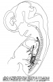
1909 Liver
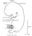
1921 Embryo
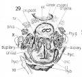
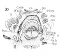
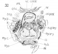
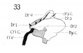
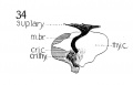
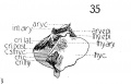
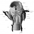
1911 Frontal section of human thyreoid cartilage
1911 Frontal section of human laryngeal muscles and cartilages.
1911 Frontal section of human cricoid cartilage
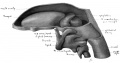
1911 Graphic reconstruction of cricoid cartilage posterior view
1911 Graphic reconstruction of cricoid cartilage, lateral view
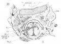
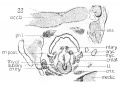
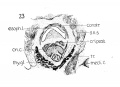
1911 Embryo no. 22 choudrification
1911 Graphic reconstruction thyreoid cartilage
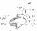
1911 Graphic reconstruction of laryngeal cartilages
F1911 Cross section of human superior laryngeal nerve and nerve recurrens.
1911 Cross section of human M. interarytaenoideus and aryepiglotticus
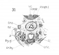
1911 Cross section low in laryngeal region
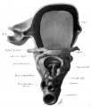
1911 Cross section of human thyreoid cartilage, cricoid cartilage, and hyoid bone
1911 Graphic reconstruction of nerve recurrens and its branches
1911 Graphic reconstruction of motor branch of superior laryngeal nerve
1911 Graphic reconstructions of laryngeal muscles, and cartilages, and their relations
1911 Wax model of laryngeal muscles and nerves
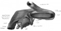
1911 Wax model of laryngeal region from below
1911 Wax model of laryngeal region from the left side
1911 Wax model of laryngeal region from below


- Carnegie Stages: 1 | 2 | 3 | 4 | 5 | 6 | 7 | 8 | 9 | 10 | 11 | 12 | 13 | 14 | 15 | 16 | 17 | 18 | 19 | 20 | 21 | 22 | 23 | About Stages | Timeline
Cite this page: Hill, M.A. (2024, April 24) Embryology Carnegie stage 21. Retrieved from https://embryology.med.unsw.edu.au/embryology/index.php/Carnegie_stage_21
- © Dr Mark Hill 2024, UNSW Embryology ISBN: 978 0 7334 2609 4 - UNSW CRICOS Provider Code No. 00098G