Carnegie stage 20: Difference between revisions
mNo edit summary |
mNo edit summary |
||
| Line 15: | Line 15: | ||
* [[Sensory - Hearing and Balance Development|hearing]] - otic capsule connected with the basal plate and with the future exoccipitals. Tip of the cochlea is elongated and curled. Tensor tympani and stapedius present. | * [[Sensory - Hearing and Balance Development|hearing]] - otic capsule connected with the basal plate and with the future exoccipitals. Tip of the cochlea is elongated and curled. Tensor tympani and stapedius present. | ||
See also [[#Events|Events]] | |||
See also [[Carnegie stage 20#Events|'''Carnegie stage 20 Events''']] | |||
===Features=== | ===Features=== | ||
* scalp vascular plexus, eylid, eye, nose, external acoustic meatus, auricle of external ear, arm, elbow, wrist, liver prominence, digital rays | * scalp vascular plexus, eylid, eye, nose, external acoustic meatus, auricle of external ear, arm, elbow, wrist, liver prominence, digital rays | ||
| Line 110: | Line 111: | ||
==Events== | ==Events== | ||
* [[Sensory_-_Vision_Development|Vision]] | * [[Sensory_-_Vision_Development|'''Vision''']] | ||
** (stage 19 -22) the eyelid folds develop into the eyelids and cover more of the eye as the palpebral fissure takes shape. The upper and the lower eyelids meet at the outer canthus in Stage 19. <ref name=“PMID7364662”><pubmed>7364662</pubmed></ref> | ** (stage 19 -22) the eyelid folds develop into the eyelids and cover more of the eye as the palpebral fissure takes shape. The upper and the lower eyelids meet at the outer canthus in Stage 19. <ref name=“PMID7364662”><pubmed>7364662</pubmed></ref> | ||
** The lens cavity is lost and a lens suture begins to form. The inner canthus is established. <ref name=“PMID7364662”><pubmed>7364662</pubmed></ref> | ** The lens cavity is lost and a lens suture begins to form. The inner canthus is established. <ref name=“PMID7364662”><pubmed>7364662</pubmed></ref> | ||
* [[Cardiovascular System Development|'''Cardiovascular''']] | |||
** [[Neural - Vascular Development|Cerebral artery]] the stem of the anterior cerebral artery gives rise to the olfactory artery.<ref name="PMID26060802"><pubmed>26060802</pubmed>| [http://synapse.koreamed.org/DOIx.php?id=10.5853/jos.2015.17.2.144 J Stroke.]</ref> | |||
===References=== | ===References=== | ||
Revision as of 12:21, 25 August 2016
| Embryology - 25 Apr 2024 |
|---|
| Google Translate - select your language from the list shown below (this will open a new external page) |
|
العربية | català | 中文 | 中國傳統的 | français | Deutsche | עִברִית | हिंदी | bahasa Indonesia | italiano | 日本語 | 한국어 | မြန်မာ | Pilipino | Polskie | português | ਪੰਜਾਬੀ ਦੇ | Română | русский | Español | Swahili | Svensk | ไทย | Türkçe | اردو | ייִדיש | Tiếng Việt These external translations are automated and may not be accurate. (More? About Translations) |
Introduction

Facts
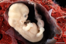
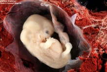
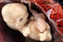
Week 8, 51 - 53 days, 18 - 22 mm
Gestational age GA week 10
Summary
- Ectoderm: sensory placodes, lens pit, otocyst, nasal pits moved ventrally, fourth ventricle of brain
- Mesoderm: heart prominence, ossification continues
- Head: forebrain, eye, external acoustic meatus
- hearing - otic capsule connected with the basal plate and with the future exoccipitals. Tip of the cochlea is elongated and curled. Tensor tympani and stapedius present.
See also Carnegie stage 20 Events
Features
- scalp vascular plexus, eylid, eye, nose, external acoustic meatus, auricle of external ear, arm, elbow, wrist, liver prominence, digital rays
- Identify: straightening of trunk, pigmented eye, eyelid, nose, external acoustic meatus, scalp vascular plexus, digital rays, liver prominance, thigh, ankle, foot plate, umbilical cord
- Links: Week 8 | System Development | Lecture - Limb | Lecture - Head Development | Lecture - Sensory | Science Practical - Head | Science Practical - Sensory | Science Practical - Urogenital | Historic - Skull Development | Category:Carnegie Stage 20 | Stage 21
| Week: | 1 | 2 | 3 | 4 | 5 | 6 | 7 | 8 |
| Carnegie stage: | 1 2 3 4 | 5 6 | 7 8 9 | 10 11 12 13 | 14 15 | 16 17 | 18 19 | 20 21 22 23 |
- Carnegie Stages: 1 | 2 | 3 | 4 | 5 | 6 | 7 | 8 | 9 | 10 | 11 | 12 | 13 | 14 | 15 | 16 | 17 | 18 | 19 | 20 | 21 | 22 | 23 | About Stages | Timeline
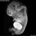
Bright Field


Virtual Embryo Slides
|
|
|
Kyoto Collection

View: This is a dorsolateral view of embryo. Amniotic membrane removed.
- Sagittal Histology
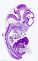
image 1
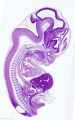
image 2
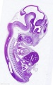
image 3
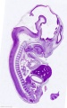
image 4
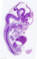
image 5
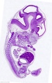
image 6


Image source: The Kyoto Collection images are reproduced with the permission of Prof. Kohei Shiota and Prof. Shigehito Yamada, Anatomy and Developmental Biology, Kyoto University Graduate School of Medicine, Kyoto, Japan for educational purposes only and cannot be reproduced electronically or in writing without permission.
Carnegie Collection


- Carnegie stage 20: 8517 Right | 8517 Anterior | 8517 Left | 7906 Right | 7906 Anterior | 7906 Left | 7274 Right | 7274 Anterior | 7274 Left
| Carnegie Collection - Stage 20 | |||||||||||
|---|---|---|---|---|---|---|---|---|---|---|---|
| Serial No. | Size (mm) | Grade | Fixative | Embedding Medium | Plane | Thinness (µm) | Stain | Point Score | Sex | Year | Notes |
| 240 | E, 20 Ch, 50x40x30 | Poor | Formalin | P | Coronal | 20 | Iron H. | 27 | d | 19?? | |
| 256 | E, 21 | poor | Alc | p | Sagittal | 25 | Coch. | 23 | M | 1904 | Tubal. Partial anencephaly |
| 353 | E, 20 | Poor | Alc | P | Sagittal | 20 | H. & Congo red | 22,5 | M | 1906 | |
| 431 | E, 19 Ch, 30x25x25 | Good | Formalin | P | Sagittal | 20 | H. & Congo red | 25,5 | M | 1908 | Tubal |
| 437 | E, 23 Ch., 80x60x50 | Poor | Formalin | P | Sagittal | 50 | Coch | 24 | M | 193? | |
| 453 | E, 23 Ch, 60x40x30 | Poor | Formalin | P | Sagittal | 20 | H. & Congo red | 23-5 | ? | 1910 | Injected |
| 460 | E, 21 | Exc. | Bichlor. acetic | P | Transverse | 40 | (Stain - Haematoxylin Eosin), coch, | 24.5 | M | 1910 | injected |
| 462 | E, 20 Ch, 50x40x30 | Exc. | Formalin | P | Transverse | 40 | Al, coch | 23.5 | F | 1910 | |
| 635B | E, 22 | Poor | Alc | P | Transverse | 50 | Al, coch | 26.5 | M | 1913 | |
| 657 | E, 25 Ch, 35x20x15 | Poor | Formalin | C | Sagittal | 40 | Al, coch | 26.5 | M | 1913 | Tubal |
| 966 | E, 23 Ch, 51x38x13 | Exc | Bichlor. acetic | P | Coronal | 40 | (Stain - Haematoxylin Eosin), aur, or. G | 25 | M | 1911 | Tubal |
| 1134B | E, 23 | Poor | Formalin | p | Sagittal | 100 | Al. coch., | 22 | - | 1915 | |
| 1266 | E, 23.1 | Poor | Formalin | C-P | Sagittal | 25 | Al. coch., (Stain - Haematoxylin Eosin) aur, or G | 20.5 | F | 191? | |
| 2393 | Ch, 61.5x50x35 | Poor | |||||||||
| 3527 | E., 22 Ch. 32x30x10 | Good | Formalin | P | Sagittal | 25 | Al. coch., | 28 | ? | 1921 | |
| 4059 | E, 21.6 | Good | Formalin | P | Coronal | 15 | Al. coch., Mallory | 29.5 | $ | 1922 | |
| 4148 | E, 21 Ch. 45x34x30 | Good | Formalin | p | Coronal | 15 | A1. coch., Mallory | 19 | ? | 1922 | |
| 4361 | E, 22 Ch., 52x42x23 | poor | Formalin | 9 | Transverse | 20 | Coch. | 24 | 8 | 1923 | |
| 6202 | E, 21 Ch., 35x35x22 | Exc | Bouin | P | Sagittal | 20 | (Stain - Haematoxylin Eosin) | 20.5 | 8 | 1930 | Tubal |
| 6426 | E 21.5 | Good | Formalin | C—P | Transverse | 20 | (Stain - Haematoxylin Eosin) | 21 | 3 | 1931 | |
| 7274 | E, 18.5 Ch., 48x44x35 | Exc | Bouin | C—P | Transverse | 20 | (Stain - Haematoxylin Eosin), phlox. | 20 | M | 1936 | |
| 7906 | E19.5 | Exc | Bouin | C—P | Coronal | 20 | (Stain - Haematoxylin Eosin) | 22 | 8 | 1941 | Left renal agenesis |
| 8517 | E., 20.8 | Exc. | Bouin | C—P | Coronal | 20 | (Stain - Haematoxylin Eosin) | 24 | 8 | 1943 | |
| 8226 | E, 18.0 | Exc. | Bouin | C—P | Sagittal | 10 | Alan | ? | 3 | 1944 | |
Abbreviations
| |||||||||||
| iBook - Carnegie Embryos | |
|---|---|

|
|
Blechschmidt Collection

Embryo (130758)
- Links: Blechschmidt Collection
Sydney University Collection
The following embryos were part of a 1943 study of testes development[1] Embryos based upon their CRL were probably Stage 20 to 21.
| Embryo | Crown Rump Length (mm) | Gonad (mm) | Groin distance (mm) |
|---|---|---|---|
| H 125 | 20 | 2.4 | 1.0 |
| H 181 | 20.6 | 2.8 | 1.1 |
| H 271 | 21 | 3.0 | 0.7 |
| H 408 | 22 | 2.3 | 1.0 |
Events
- Vision
- Cardiovascular
- Cerebral artery the stem of the anterior cerebral artery gives rise to the olfactory artery.[3]
References
Additional Images
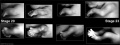
Stage 20-23 limbs
Stage 20 Optical Projection Tomography

Historic Images
| Historic Disclaimer - information about historic embryology pages |
|---|
 |
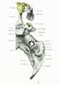
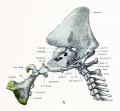
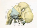
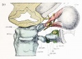
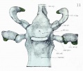
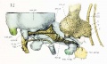
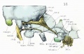
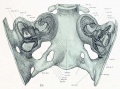
Lewis, WH. The Cartilaginous Skull Of A Human Embryo Twenty-One Millimeters In Length (1920) Contrib. to Embryol. 9:299-324.
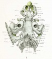
Skull - Dorsal aspect of base with the basioccipital in horizontal plane.
Skull - Right half base of cartilaginous skull.
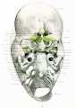
Skull - Dorsal aspect of cartilaginous and membranous skull.
Skull - Median sagittal aspect.
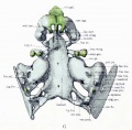
Skull - Ventral aspect of base.
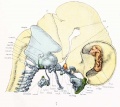
Skull - Lateral aspect and cervical vertebrae with brain and cervical cord and hypophysis.
Skull - Lateral aspect and cervical vertebra with brain, cervical cord, and nerves.
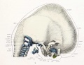
Skull - Lateral view and cervical vertebrae with overlying membranous skull and dorsal membrane.
Skull - Dorsal aspect of sphenoid cartilage.
Skull - Dorsal aspect of sphenoid cartilage.
Skull - Lateral view of right otic region.
Skull - Lateral view of right otic region showing relations of facial nerve.

Skull - Lateral view of base with deeper muscles of occipital region, mouth and pharynx.

Skull - Lateral view of part of cartilaginous and membranous skull.
Skull - Dorsal view of temporal and occipital cartilages.


Dickie JK. The Anatomy of the Head End of a 20 mm Human Embryo. J Anat Physiol. 1914 Jul;48(Pt 4):445-60. PMID 17233010
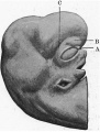
Fig 1. Profile view of the embryo
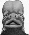
Fig 2. Front view of embryo
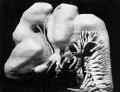
Fig 3. Lateral surface model of brain and cranial nerves
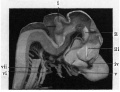
Fig 4. Shows medial aspect of model of brain
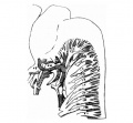
Fig 5. Sketch of nerves in occipital region
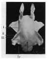
Fig 6. Model of the upper air passages
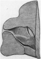
Fig 7. Lsteral wall of the nasal cavity
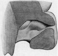
Fig 8. Medial wall of nose
Fig 9. Lateral aspect of the membranous labyrinth
Fig 10. Medial aspect of the membranous labyrinth


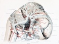
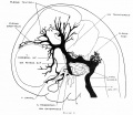
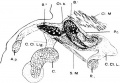
Left lateral view of larger blood-vessels of the brain.
Dural venous system.
Human embryonic shoulder girdle


- Carnegie Stages: 1 | 2 | 3 | 4 | 5 | 6 | 7 | 8 | 9 | 10 | 11 | 12 | 13 | 14 | 15 | 16 | 17 | 18 | 19 | 20 | 21 | 22 | 23 | About Stages | Timeline
Cite this page: Hill, M.A. (2024, April 25) Embryology Carnegie stage 20. Retrieved from https://embryology.med.unsw.edu.au/embryology/index.php/Carnegie_stage_20
- © Dr Mark Hill 2024, UNSW Embryology ISBN: 978 0 7334 2609 4 - UNSW CRICOS Provider Code No. 00098G