Carnegie stage 15: Difference between revisions
| Line 68: | Line 68: | ||
File:Carnegie_stage_15_OPT.jpg|Stage 15 Optical Projection Tomography | File:Carnegie_stage_15_OPT.jpg|Stage 15 Optical Projection Tomography | ||
File:External ear stages-14-23-adult.jpg|External ear Stages 14-23 and adult | File:External ear stages-14-23-adult.jpg|External ear Stages 14-23 and adult | ||
</gallery> | |||
<gallery caption = Mall (1891)> | |||
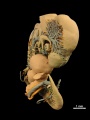
File:Mall1891 Plate01Fig01.jpg|Pl. 1 Fig. 1. External view of the embryo before it was sectioned. | |||
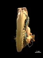
File:Mall1891 Plate01Fig02.jpg|Pl. 1 Fig. 2. Corrosion preparation of the pericardial and pleuro-peritoneal cavities. | |||
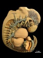
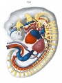
File:Mall1891 Plate02Fig01.jpg|Pl. 2 Fig. 1. Reconstruction viewed from the left side. | |||
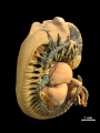
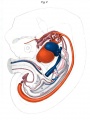
File:Mall1891 Plate02Fig02.jpg|Pl. 2 Fig. 2. he same as Fig. I. Deeper view | |||
</gallery> | </gallery> | ||
Revision as of 18:47, 29 July 2015
| Embryology - 19 Apr 2024 |
|---|
| Google Translate - select your language from the list shown below (this will open a new external page) |
|
العربية | català | 中文 | 中國傳統的 | français | Deutsche | עִברִית | हिंदी | bahasa Indonesia | italiano | 日本語 | 한국어 | မြန်မာ | Pilipino | Polskie | português | ਪੰਜਾਬੀ ਦੇ | Română | русский | Español | Swahili | Svensk | ไทย | Türkçe | اردو | ייִדיש | Tiếng Việt These external translations are automated and may not be accurate. (More? About Translations) |
Introduction

Facts
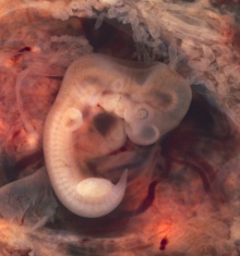
Facts: Week 5, 35 - 38 days, 7 - 9 mm
Gestational Age GA week 7
Events
- Ectoderm: sensory placodes, lens pit, otocyst, nasal pit, primary/secondary vesicles, fourth ventricle of brain,
- Mesoderm: heart prominence
- Head: 1st, 2nd and 3rd pharyngeal arch, forebrain, site of lens placode, site of otic placode, stomodeum
- Body: heart, liver, umbilical cord, mesonephric ridge
- Limb: upper and lower limb buds, hand plate
Features
- Identify: midbrain region, nasal pit, lens pit, 1st, 2nd and 3rd pharyngeal arches, 1st pharyngeal groove, maxillary and mandibular components of 1st pharyngeal arch, fourth ventricle of brain, heart prominence, cervical sinus, upper limb bud, mesonephric ridge, lower limb bud, umbilical cord Labelled Stage 15
- Links: Week 5 | Head | Lecture - Limb | Lecture - Gastrointestinal | Lecture - Head Development | Science Practical - Gastrointestinal | Science Practical - Head | Category:Carnegie Stage 15 | Stage 16
| Week: | 1 | 2 | 3 | 4 | 5 | 6 | 7 | 8 |
| Carnegie stage: | 1 2 3 4 | 5 6 | 7 8 9 | 10 11 12 13 | 14 15 | 16 17 | 18 19 | 20 21 22 23 |
- Carnegie Stages: 1 | 2 | 3 | 4 | 5 | 6 | 7 | 8 | 9 | 10 | 11 | 12 | 13 | 14 | 15 | 16 | 17 | 18 | 19 | 20 | 21 | 22 | 23 | About Stages | Timeline
Kyoto Collection

View: Lateral view. Amniotic membrane removed.
Image source: The Kyoto Collection images are reproduced with the permission of Prof. Kohei Shiota and Prof. Shigehito Yamada, Anatomy and Developmental Biology, Kyoto University Graduate School of Medicine, Kyoto, Japan for educational purposes only and cannot be reproduced electronically or in writing without permission.
Carnegie Collection


| Week: | 1 | 2 | 3 | 4 | 5 | 6 | 7 | 8 |
| Carnegie stage: | 1 2 3 4 | 5 6 | 7 8 9 | 10 11 12 13 | 14 15 | 16 17 | 18 19 | 20 21 22 23 |
| iBook - Carnegie Embryos | |
|---|---|

|
|
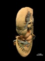
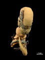
Blechschmidt Collection
| Embryo 7.5mm |
| Page | Play |
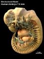
left lateral
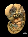
left ventrolateral
left ventral
ventral
right ventral
right lateral
right dorsolateral
right dorsal
dorsal
left dorsolateral


Photograph

|

|
Image: Dr Ed Uthman (Houston, Texas) - other pathology images
Image version links: Virtual Slide | ExtraLarge 1874 x 2000px | Large 959 x 1024px | Medium 468 x 500px
Embryo Virtual Slide
| Stage 15 - Ventral View
|
| Stage 15 | Embryo Slides |
Additional Images
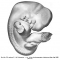
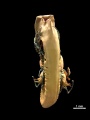
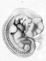
Fig. 52 of Keibel and Mall Manual of Human Embryology I (1910)
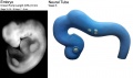
Neural tube model
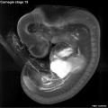
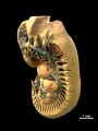
Stage 15 Optical Projection Tomography

External ear Stages 14-23 and adult


- Mall
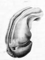
Pl. 1 Fig. 1. External view of the embryo before it was sectioned.
Pl. 1 Fig. 2. Corrosion preparation of the pericardial and pleuro-peritoneal cavities.
Pl. 2 Fig. 1. Reconstruction viewed from the left side.
Pl. 2 Fig. 2. he same as Fig. I. Deeper view


- Carnegie Stages: 1 | 2 | 3 | 4 | 5 | 6 | 7 | 8 | 9 | 10 | 11 | 12 | 13 | 14 | 15 | 16 | 17 | 18 | 19 | 20 | 21 | 22 | 23 | About Stages | Timeline
Cite this page: Hill, M.A. (2024, April 19) Embryology Carnegie stage 15. Retrieved from https://embryology.med.unsw.edu.au/embryology/index.php/Carnegie_stage_15
- © Dr Mark Hill 2024, UNSW Embryology ISBN: 978 0 7334 2609 4 - UNSW CRICOS Provider Code No. 00098G