Carnegie stage 12: Difference between revisions
mNo edit summary |
mNo edit summary |
||
| Line 85: | Line 85: | ||
==Hinrichsen Collection== | ==Hinrichsen Collection== | ||
[[File:ME50 001.jpg|400px]] | [[File:ME50 001.jpg|400px]][[File:ME50 001.jpg|400px]] | ||
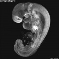
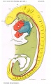
Hinrichsen collection Human Embryo ME50 ([[Carnegie stage 12|stage12]]). | Hinrichsen collection Human Embryo ME50 ([[Carnegie stage 12|stage12]]). | ||
Revision as of 15:47, 18 October 2016
| Embryology - 19 Apr 2024 |
|---|
| Google Translate - select your language from the list shown below (this will open a new external page) |
|
العربية | català | 中文 | 中國傳統的 | français | Deutsche | עִברִית | हिंदी | bahasa Indonesia | italiano | 日本語 | 한국어 | မြန်မာ | Pilipino | Polskie | português | ਪੰਜਾਬੀ ਦੇ | Română | русский | Español | Swahili | Svensk | ไทย | Türkçe | اردو | ייִדיש | Tiếng Việt These external translations are automated and may not be accurate. (More? About Translations) |
Introduction

|
FactsWeek 4, 26 - 30 days, 3 - 5 mm, Somite Number 21 - 29 Gestational Age GA week 6 Summary
Features
|
| Week: | 1 | 2 | 3 | 4 | 5 | 6 | 7 | 8 |
| Carnegie stage: | 1 2 3 4 | 5 6 | 7 8 9 | 10 11 12 13 | 14 15 | 16 17 | 18 19 | 20 21 22 23 |
- Carnegie Stages: 1 | 2 | 3 | 4 | 5 | 6 | 7 | 8 | 9 | 10 | 11 | 12 | 13 | 14 | 15 | 16 | 17 | 18 | 19 | 20 | 21 | 22 | 23 | About Stages | Timeline
Bright Field


Scanning EM


Image Source: Scanning electron micrographs of the Carnegie stages of the early human embryos are reproduced with the permission of Prof Kathy Sulik, from embryos collected by Dr. Vekemans and Tania Attié-Bitach. Images are for educational purposes only and cannot be reproduced electronically or in writing without permission.
Kyoto Collection

View: Lateral view, day 26, 27 somites, Amniotic membrane removed.
Image source: The Kyoto Collection images are reproduced with the permission of Prof. Kohei Shiota and Prof. Shigehito Yamada, Anatomy and Developmental Biology, Kyoto University Graduate School of Medicine, Kyoto, Japan for educational purposes only and cannot be reproduced electronically or in writing without permission.
Carnegie Collection


- Carnegie Stages: 1 | 2 | 3 | 4 | 5 | 6 | 7 | 8 | 9 | 10 | 11 | 12 | 13 | 14 | 15 | 16 | 17 | 18 | 19 | 20 | 21 | 22 | 23 | About Stages | Timeline
| iBook - Carnegie Embryos | |
|---|---|

|
|
Hill Collection

|

|
| right view | left view |
- Links: Hill Collection
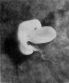
Hinrichsen Collection

Hinrichsen collection Human Embryo ME50 (stage12).
Note the developing pharyngeal arches, otic and optic placodes, and heart in this left lateral view of the embryo.
Image source: The Hinrichsen Collection images are reproduced with the permission of Prof. Beate Brand-Saberi, Head, Department of Anatomy and Molecular Embryology, Ruhr-Universität Bochum. Images are for educational purposes only and cannot be reproduced electronically or in writing without permission.
Historic Embryology
Chicago Collection H1093 and H984
H1093 and H984 are University of Chicago Collection 22 and 23 somite embryos respectively, described by Wen, I. C., The anatomy of human embryos with seventeen to twenty-three pairs of somites J. Comp. Neural, 1928, 45:301-376.
Events
- Somitogenesis - stages 12 and 13 embryo somite 1 has dispersed and is now contributing to the hypoglossal cord. Therefore “in embryos with more than 20 somites” (beginning of stage 12), the first ones visible “actually are second somites”.[1]
- hearing - otic vesicle (otocyst) is forming and connects by a narrow pore with the surface[2] Otocyst ventral wall contributes to the vestibulocochlear crest.
References
- ↑ Arey, L. B. 1938. The history of the first somite in human embryos. Carnegie Instn. Wash. Publ. 496, Contrib. Embryoi, 27, 233-269.
- ↑ Streeter GL. Developmental horizons in human embryos. Description of age group XI, 13 to 20 somites, and age group XII, 21 to 29 somites. (1942) Contrib. Embryol., Carnegie Inst. Wash. Publ. 541, 30: 211-245.
Wen, I. C., The anatomy of human embryos with seventeen to twenty-three pairs of somites J. Comp. Neural, 1928, 45:301-376.
Additional Images
Stage 12 Optical Projection Tomography

Historic Images
| Historic Disclaimer - information about historic embryology pages |
|---|
 |
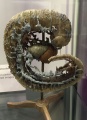
Heard model of Carnegie embryo 588
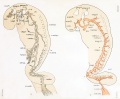
Right and left profile views of a wax-plate reconstruction of the main arteries and veins in a human embryo 4 mm.
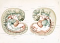
Gray's Anatomy Fig. 472
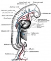
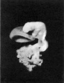
Graphic reconstruction of embryo from serial sections PMID 17232769


- Historic Papers: 22 Somites | 23 Somites | 25 Somites | 27 Somites | Brain Vascular System of the Human Embryo
Glossary Links
- Glossary: A | B | C | D | E | F | G | H | I | J | K | L | M | N | O | P | Q | R | S | T | U | V | W | X | Y | Z | Numbers | Symbols | Term Link
Cite this page: Hill, M.A. (2024, April 19) Embryology Carnegie stage 12. Retrieved from https://embryology.med.unsw.edu.au/embryology/index.php/Carnegie_stage_12
- © Dr Mark Hill 2024, UNSW Embryology ISBN: 978 0 7334 2609 4 - UNSW CRICOS Provider Code No. 00098G