Carnegie stage 11: Difference between revisions
mNo edit summary |
mNo edit summary |
||
| (60 intermediate revisions by the same user not shown) | |||
| Line 2: | Line 2: | ||
== Introduction == | == Introduction == | ||
{| | {| | ||
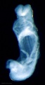
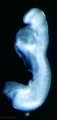
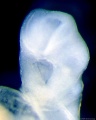
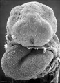
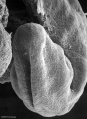
| [[File:Stage11 | | width=255px|[[File:Stage11 bf2.jpg|250px]] | ||
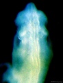
| [[File:Stage11 sem6.jpg|250px]] | | width=255px|[[File:Stage11 sem6.jpg|250px]] | ||
| | | | ||
===Facts=== | ===Facts=== | ||
| Line 9: | Line 9: | ||
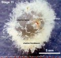
Week 4, 23 - 26 days, 2.5 - 4.5 mm, Somite Number 13 - 20 | Week 4, 23 - 26 days, 2.5 - 4.5 mm, Somite Number 13 - 20 | ||
Gestational Age - week 6 | Gestational Age {{GA}} - week 6 | ||
=== | ===Summary=== | ||
* Ectoderm: Neural tube continues to close, Rostral neuropore closes | |||
* Mesoderm: continued segmentation of paraxial mesoderm (13 - 20 somite pairs), heart tube bending | |||
This early week 4 embryonic stage shows key features of heart tube and neural plate to tube development. | |||
The embryo is still transparent enough to view the underlying somite and transverse septum development. | |||
See also [[Carnegie stage 11#Events|'''Carnegie stage 11 Events''']] | |||
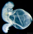
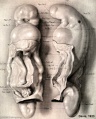
The embryo is still quite small and is comparable in size to the external yolk sac. | The embryo is still quite small and is comparable in size to the external yolk sac. | ||
There are a number of different images and resources available for this embryonic stage including: | There are a number of different images and resources available for this embryonic stage including: | ||
| Line 35: | Line 34: | ||
| | | | ||
|} | |} | ||
===Features=== | ===Features=== | ||
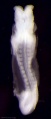
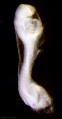
rostral neuropore closing, forebrain, neural tube in region of developing spinal cord, somites, caudal neuropore, connecting stalk, amnion | rostral neuropore closing, forebrain, neural tube in region of developing spinal cord, somites, caudal neuropore, connecting stalk, amnion | ||
Identify: heart, rostral (cranial, anterior) neuropore closing, forebrain, neural tube in region of developing spinal cord, somites, caudal neuropore, connecting stalk, amnion | Identify: heart, rostral (cranial, anterior) neuropore closing, forebrain, neural tube in region of developing spinal cord, somites, caudal neuropore, connecting stalk, amnion | ||
{{Carnegie stage 11 links}} | |||
{{Carnegie_stage_table_1}} | |||
{{Carnegie_stages}} | {{Carnegie_stages}} | ||
| Line 62: | Line 60: | ||
<gallery> | <gallery> | ||
File:Embryo-membranes stage 11.jpg|Embryo with Membranes | |||
File:Stage11 bf1.jpg|BF1 - dorsal view | File:Stage11 bf1.jpg|BF1 - dorsal view | ||
File:Stage11 bf2.jpg|BF2 - lateral view | File:Stage11 bf2.jpg|BF2 - lateral view | ||
| Line 104: | Line 103: | ||
|- | |- | ||
|} | |} | ||
{| | |||
[[File:Stage11 | ! Cranial End of Embryo | ||
|- | |||
| [[File:Stage11 sem8.jpg|400px]] | |||
| valign=top|[[File:Stage11 sem81.jpg|400px]] | |||
|} | |||
===Buccopharyngeal Membrane=== | ===Buccopharyngeal Membrane=== | ||
The degenerating buccopharyngeal membrane is shown at the floor of the stomodeum. | |||
{| | |||
| [[File:Stage11_sem3b.jpg]] | |||
| [[File:Stage11_sem3b.gif]] | |||
|} | |||
<gallery> | <gallery> | ||
File:Stage11 sem4.jpg|Low power ventral view of the Buccopharyngeal Membrane | File:Stage11 sem4.jpg|Low power ventral view of the Buccopharyngeal Membrane | ||
| Line 124: | Line 131: | ||
| [[File:Stage11_sem21.jpg|300px]] | | [[File:Stage11_sem21.jpg|300px]] | ||
| Scanning EM showing low power image of whole embryo and region shown in detail box right of [[:File:Stage11_sem21.jpg|neural crest cells]]. | | Scanning EM showing low power image of whole embryo and region shown in detail box right of [[:File:Stage11_sem21.jpg|neural crest cells]]. | ||
:'''Links:''' [[Neural Crest Development]] | :'''Links:''' [[Neural Crest Development]] | ||
| Line 130: | Line 136: | ||
|} | |} | ||
{{ | {{Necker SEM}} | ||
==Kyoto Collection== | ==Kyoto Collection== | ||
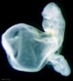
[[File:Stage11_bf7.jpg| | [[File:Stage11_bf7.jpg|400px]][[File:Stage11_bf71.jpg|400px]] | ||
View: This is a dorsolateral view of embryo. Amniotic membrane removed. | View: This is a left dorsolateral view of embryo. Amniotic membrane removed. | ||
[[File:Stage11 K17941-01.jpg|400px]] | |||
View: Transverse section (scale bar 0.5 mm) | |||
{{Kyoto collection}} | {{Kyoto collection}} | ||
| Line 150: | Line 159: | ||
{{Carnegie Embryo iBook table}} | {{Carnegie Embryo iBook table}} | ||
{{Carnegie Collection stage 11 table}} | |||
==Historic Embryology== | |||
===Harvard Collection No. 714=== | |||
* [[Harvard Collection|Harvard Collection]] '''No. 714''' described by Bremer, J. L. 1906. [[Paper - Description of a 4-mm Human Embryo|Description of a 4-mm Human Embryo]]. Amer. J. Anat., 5, 459-480. | |||
===Pfannenstiel III=== | |||
Keibel Collection {{Pfannenstiel III}} | |||
{{Ref-Keibel1908}} Shown as Embryo No. 6 (Plate 1 [[:File:Keibel1908_plate01.jpg|fig. Vr.]] and Plate 2 [[:File:Keibel1908_plate02.jpg|fig. Vv.]]). [[Book_-_Text-Book_of_Embryology_8#Fig084|Text-Book of Embryology (1921) Fig. 84]] | |||
{{Ref-Low1908}} | |||
<gallery> | |||
File:Low_plate_01.jpg|Embryo model left side | |||
File:Low_plate_02.jpg|Embryo model frontal view | |||
File:Low_plate_03.jpg|sagittal section viewed from the left | |||
</gallery> | |||
===Chicago Collection H951=== | |||
{{Ref-Wen1928}} H951 is a University of Chicago Collection 17 somite embryo. | |||
===Carnegie Collection=== | |||
{{Ref-Davis1923}} | |||
<gallery> | |||
File:Stage_11_historic-Davis1923-1.jpg | |||
File:Stage_11_historic-Davis1923-2.jpg | |||
File:Stage_11_historic-Davis1923-3.jpg | |||
File:Stage_11_historic-Davis1923-4.jpg | |||
</gallery> | |||
{{Ref-Heuser1930}} | |||
{{Ref-Atwell1930}} | |||
<gallery> | |||
File:Stage_11_historic-Heuser1930-1.jpg|1930 Dorsolateral view drawing 14 somite pairs, 24 days | |||
File:Stage_11_historic-Atwell1930-1.jpg|1930 Sagittal (midline) section drawing of 17 somite pairs | |||
File:Stage_11_historic-Atwell1930-4.jpg|1930 Ventral and lateral view drawing of 19 somite pairs, approx 25 days | |||
File:Stage 11 historic-Davis1923-1.jpg|1923 Ventral and lateral view drawing of 20 somite pairs | |||
File:Bailey084.jpg|Dorso-lateral view of human embryo with fourteen pairs of mesodermal somites (Kollmann). | |||
File:Bailey085.jpg|Lateral view of human embryo of 2.6 mm (His, from Keibel and Mall). | |||
File:Pohlman1911 plate1A.jpg|1911 Cloaca model | |||
</gallery> | |||
==Histology== | ==Histology== | ||
| Line 163: | Line 219: | ||
'''Stage 11 histology links:''' [[:File:Stage11_histology-optic pit.jpg|optic pit]] | [[:File:Stage11 histology-optic vesicle-hindbrain.jpg|optic vesicle-hindbrain]] | [[:File:Stage11 histology-neural tube roof plate 1.jpg|neural tube roof plate 1]] | [[:File:Stage11 histology-neural tube roof plate 2.jpg|neural tube roof plate 2]] | '''Stage 11 histology links:''' [[:File:Stage11_histology-optic pit.jpg|optic pit]] | [[:File:Stage11 histology-optic vesicle-hindbrain.jpg|optic vesicle-hindbrain]] | [[:File:Stage11 histology-neural tube roof plate 1.jpg|neural tube roof plate 1]] | [[:File:Stage11 histology-neural tube roof plate 2.jpg|neural tube roof plate 2]] | ||
== | ==Events== | ||
* '''{{cardiovascular}}''' - heart beating and peristaltic flow begins.<ref>{{Ref-deVries1962}}</ref> | |||
* '''{{hearing}}''' - at 16 somites the otic pit is formed and lies dorsal to the second pharyngeal groove (cleft).<ref name=Bartelmez1926>{{Ref-Bartelmez1926}}</ref> | |||
* '''{{vision}}''' - right and left optic primordia meet at the optic chiasma forming a U-shaped rim.{{#pmid:7364662|PMID7364662}} | |||
* '''{{gastrointestinal tract}}''' - cloacal membrane is located on the ventral surface of the caudal part of the body wall, in a central oval depression. | |||
* '''{{meninges}}''' - ({{spinal cord}}) - migration of cells from the somites has increased in embryos and the neural crests have undergone rapid development. Extending ventrally from the neural crests, and appearing to be continuous with them, is a single layered strand of cells. This extends along the lateral edge of the neural tube and contains cells which are more flattened and elongated than those migrating from the somite.<ref name=Sensenig1951>{{Ref-Sensenig1951}}</ref> | |||
===References=== | |||
<references/> | |||
===Additional References=== | |||
{{Ref-Mizoguti1989}} | |||
{{#pmid:3826651}} | |||
| Line 191: | Line 243: | ||
{{Footer}} | {{Footer}} | ||
[[Category:Human Embryo]] [[Category:Carnegie Stage]] [[Category:Carnegie Stage 11]] [[Category:Week 4]] | [[Category:Human Embryo]] [[Category:Carnegie Stage]] [[Category:Carnegie Stage 11]] [[Category:Week 4]] | ||
[[Category:Pfannenstiel III]] | |||
Revision as of 14:36, 12 November 2018
| Embryology - 25 Apr 2024 |
|---|
| Google Translate - select your language from the list shown below (this will open a new external page) |
|
العربية | català | 中文 | 中國傳統的 | français | Deutsche | עִברִית | हिंदी | bahasa Indonesia | italiano | 日本語 | 한국어 | မြန်မာ | Pilipino | Polskie | português | ਪੰਜਾਬੀ ਦੇ | Română | русский | Español | Swahili | Svensk | ไทย | Türkçe | اردو | ייִדיש | Tiếng Việt These external translations are automated and may not be accurate. (More? About Translations) |
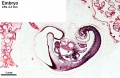
Introduction

|

|
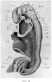
FactsWeek 4, 23 - 26 days, 2.5 - 4.5 mm, Somite Number 13 - 20 Gestational Age GA - week 6 Summary
This early week 4 embryonic stage shows key features of heart tube and neural plate to tube development. The embryo is still transparent enough to view the underlying somite and transverse septum development.
The embryo is still quite small and is comparable in size to the external yolk sac. There are a number of different images and resources available for this embryonic stage including: |
| Lateral (right) view of embryo |
Features
rostral neuropore closing, forebrain, neural tube in region of developing spinal cord, somites, caudal neuropore, connecting stalk, amnion
Identify: heart, rostral (cranial, anterior) neuropore closing, forebrain, neural tube in region of developing spinal cord, somites, caudal neuropore, connecting stalk, amnion
| Week: | 1 | 2 | 3 | 4 | 5 | 6 | 7 | 8 |
| Carnegie stage: | 1 2 3 4 | 5 6 | 7 8 9 | 10 11 12 13 | 14 15 | 16 17 | 18 19 | 20 21 22 23 |
- Carnegie Stages: 1 | 2 | 3 | 4 | 5 | 6 | 7 | 8 | 9 | 10 | 11 | 12 | 13 | 14 | 15 | 16 | 17 | 18 | 19 | 20 | 21 | 22 | 23 | About Stages | Timeline
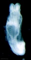
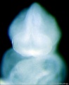
Bright Field

|

|

|
| Embryo dorsal view | Embryo ventral with 1 mm scale bar | Embryo lateral view |
Select image below to open full-size.
Embryo with Membranes
BF1 - dorsal view
BF2 - lateral view
BF3 - ventral with scale bar
BF4 - ventral view
BF5 - lateral view
BF6 - ventral view
BF8 - ventral head
BF9 - ventral head
BF10 - dorsal neural
BF11 - ventral left
BF12 - ventral right


- Stage 11 Images: BF1 - dorsal view | BF2 - lateral view | BF3 - ventral with scale bar | BF4 - ventral view | BF5 - lateral view | BF6 - ventral view | BF7 - Kyoto embryo | BF8 - ventral head | BF9 - ventral head | BF10 - dorsal neural | BF11 - ventral embryo and yolk sac | Scanning EM embryo | Carnegie stage 11
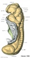
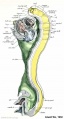
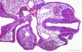
Scanning EM

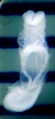
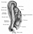
This is a scanning EM of the embryo dorsal view showing the neural tube closing with open neuropores and the paired somites visible through the thin ectoderm. Features: surface ectoderm, neural tube, cranial (anterior) neuropore, caudal (posterior) neuropore, somites, heart, cut edge of amnion, 24 days, 13 somite pairs. |

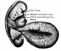
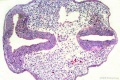
This is a scanning EM of the embryo fractured close to the midline to show the hindbrain rhombomeres. Features: surface ectoderm, neural tube, hindbrain rhombomeres, forebrain, buccopharyngeal membrane, yolk sac, 25 days, 19 somite pairs. |

This is a scanning EM of the embryo superior dorsal view showing the paired otic placodes sinking into the surface at the level of the hindbrain. Features: surface ectoderm, paired otic placodes, pharyngeal arches heart, 25 days, 19 somite pairs. |

This is a scanning EM of the embryo dorsolateral view showing the neural tube closing with open neuropores and the paired somites visible through the thin ectoderm. Features: surface ectoderm, neural tube, cranial (anterior) neuropore, caudal (posterior) neuropore, somites, heart, cut edge of amnion, 24 days, 13 somite pairs. |

This is a scanning EM of the embryo fractured to show the neural tube, notochord and somites. Features: surface ectoderm, neural tube, notochord, somites, somitocoels, dorsal aortas, gastrointestinal tract, 25 days, 19 somite pairs. |

This is a labeled version of the scanning EM of the fractured embryo. |
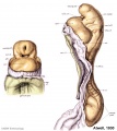
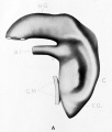
| Cranial End of Embryo | |
|---|---|

|

|
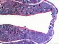
Buccopharyngeal Membrane
The degenerating buccopharyngeal membrane is shown at the floor of the stomodeum.

|

|

Low power ventral view of the Buccopharyngeal Membrane

Higher power ventrolateral view of the Buccopharyngeal Membrane

Close up view of the degenerating Buccopharyngeal Membrane


Neuropores
Anterior Neuropore
Posterior Neuropore


Neural Crest

|
Scanning EM showing low power image of whole embryo and region shown in detail box right of neural crest cells.
|
Image Source: Scanning electron micrographs of the Carnegie stages of the early human embryos are reproduced with the permission of Prof Kathy Sulik, from embryos collected by Dr. Vekemans and Tania Attié-Bitach. Images are for educational purposes only and cannot be reproduced electronically or in writing without permission.
Kyoto Collection


View: This is a left dorsolateral view of embryo. Amniotic membrane removed.

View: Transverse section (scale bar 0.5 mm)
Image source: The Kyoto Collection images are reproduced with the permission of Prof. Kohei Shiota and Prof. Shigehito Yamada, Anatomy and Developmental Biology, Kyoto University Graduate School of Medicine, Kyoto, Japan for educational purposes only and cannot be reproduced electronically or in writing without permission.
Carnegie Collection


| iBook - Carnegie Embryos | |
|---|---|

|
|
| Carnegie Collection - Stage 11 | ||||||||||
|---|---|---|---|---|---|---|---|---|---|---|
| Serial No. | Pairs of somites | Size (mm) | Grade | Fixative | Embedding Medium | Plane | Thinness (µm) | Stain | Year | Notes |
| 12 | 14 | E, 2.1 Ch, 13 | Poor | P | Transverse | 10 | Al. carm. | 1893 | ||
| 164 | 18 | E, 3.5 Ch, 14 | Good | Formalin | P | Transverse | 20 | Al. carm. | 1913 | |
| 318 | 13/14 | E, 2.5 Ch, 16 | Good | P | Transverse | 25 | Al. carm. | 1905 | ||
| 470 | 17 | E, 4.3 Ch, 16 | Good | Formalin | P | Transverse | 10 | Al. carm. . | 1910 | |
| 779 | 14 | E, 2.75 | Good | C | Transverse | 15 | Al. coch. | 1913 | Dysraphism. Noted by Dekaban (1964)[1] | |
| 1182b | E, 3 Ch, 15x12x5 | Good | Formalin | ? | Transverse | 20 | Al. carm. | 1915 | ||
| 2053 | 20 | E, 3.1 Ch, 12 | Exc. | Formalin | P | Transverse | 10 | Al. coch. | 1918 | Most advanced in group. Ag added to slide 2 Monographs by Davis (1923)[2] and Congdon (1922)[3] |
| 4315 | 17 | E, 4.7 Ch, 23x10.4X11 | Excellent | ? | C-P | Transverse | 10 | I.H. & E. | 1923 | Univ. Chicago No. 951. Wen (1928)[4] |
| 4529 | 14 | E, 2.4 Ch, 21 | Excellent | Formalin | P | Transverse | 10 | Al. coch, or. G. | 1924 | Heuser (1930)[5] |
| 4783 | 13 | E, 2.3 | Fair | ? | ? | Transverse | 5 | I.H. | 1924 | Wallin (1913)[6] |
| 4877 | 13 | E, 2 Ch, 15 | Good | Formalin | P | Transverse | 15 | Al. coch. | 1925 | |
| 5072 | 17 | E, 3 | Good | Formalin | P | Transverse | 10 | (Stain - Haematoxylin Eosin) | 1925 | Tubal Type specimen. Atwell (1930)[7] |
| 6050 | 19/21 | E.,3 Ch, 10 | Good | Formalin | C-P | Coronal | 10 | Al. coch. | 1930 | Advanced |
| 6344 | 13 | E, 2.5 Ch, 17 | Excellent | Formalin | C-P | Transverse | 6 | Al. coch. | 1931 | Least advanced in group |
| 6784 | 17 | E, 5 Ch, 16 | Excellent | Formalin | C-P | Transverse | 6 | I.H, or. G. | 1933 | |
| 7358 | 16 | E, ? Ch, 15 | Poor | Alc, formol | p | Oblique | 25 | (Stain - Haematoxylin Eosin) | 1936 | |
| 7611 | 16 | E., 2.4 Ch., 12 | Excellent | Bouin | C-P | Transverse | 8 | (Stain - Haematoxylin Eosin) | 1938 | |
| 7665 | 19 | E., 4.36 | Excellent | ? | C-P | Transverse | 6 | 1939 | Univ. Chicago No. H 1516 | |
| 7702 | 17 | E, 3.7 Ch., 14 | Good | Formalin | C-P | Transverse | 10 | Al. coch. | 1940 | Returned to B M Patten |
| 7851 | 13 | E., 4.3 Ch, 18 | Excellent | Formalin | C-P | Transverse | 8 | (Stain - Haematoxylin Eosin) | 1940 | Slightly injured |
| 8005 | 16/17 | E, 3 | Excellent | Bouin | C-P | Transverse | 8 | (Stain - Haematoxylin Eosin) | 1942 | Tubal |
| 8116 | 17 | E, 14 Ch.. 17 | Good | Formalin | p | Sagittal | 8 | Azan | 1953 | |
| 8962 | 15 | E, 1.55 | Good | ? | * | Sagittal | ? | ? | 1952 | Tubal Univ. Chicago No. H 810 |
Abbreviations
| ||||||||||
References
| ||||||||||
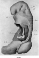
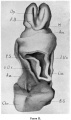
Historic Embryology
Harvard Collection No. 714
- Harvard Collection No. 714 described by Bremer, J. L. 1906. Description of a 4-mm Human Embryo. Amer. J. Anat., 5, 459-480.
Pfannenstiel III
Keibel Collection Pfannenstiel III
Keibel F. and Elze C. Normal Plates of the Development of the Human Embryo (Homo sapiens). (1908) Vol. 8 in series by Keibel F. Normal plates of the development of vertebrates (Normentafeln zur Entwicklungsgeschichte der Wirbelthiere) Fisher, Jena., Germany. Shown as Embryo No. 6 (Plate 1 fig. Vr. and Plate 2 fig. Vv.). Text-Book of Embryology (1921) Fig. 84
Low A. Description of a human embryo of 13-14 mesodermic somites. (1908) J Anat Physiol. 42(3): 237-51. PMID 17232769 | PMC1289161
Embryo model left side
Embryo model frontal view
sagittal section viewed from the left


Chicago Collection H951
Wen IC. The anatomy of human embryos with seventeen to twenty-three pairs of somites (1928) J. Comp. Neural., 45: 301-376. H951 is a University of Chicago Collection 17 somite embryo.
Carnegie Collection
Davis CL. Description of a human embryo having twenty paired somites. (1923) Carnegie Instn. Wash. Publ. 332, Contrib. Embryol., 15: 1-51.


Heuser CH. A human embryo with 14 pairs of somites. (1930) Carnegie Instn. Wash. Publ. 414, Contrib. Embryol., Carnegie Inst. Wash. 22:135-153.
Atwell WJ. A human embryo with seventeen pairs of somites. (1930) Contrib. Embryol., Carnegie Inst. Wash. Publ. 407, 21: 1-24.
1930 Dorsolateral view drawing 14 somite pairs, 24 days
1930 Sagittal (midline) section drawing of 17 somite pairs
1930 Ventral and lateral view drawing of 19 somite pairs, approx 25 days
1923 Ventral and lateral view drawing of 20 somite pairs
Dorso-lateral view of human embryo with fourteen pairs of mesodermal somites (Kollmann).
Lateral view of human embryo of 2.6 mm (His, from Keibel and Mall).
1911 Cloaca model


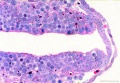
Histology
Human embryo (CRL 4.2 mm) Blechschmidt Collection
optic pit
optic vesicle-hindbrain
neural tube roof plate 1
neural tube roof plate 2


Stage 11 histology links: optic pit | optic vesicle-hindbrain | neural tube roof plate 1 | neural tube roof plate 2
Events
- cardiovascular - heart beating and peristaltic flow begins.[1]
- hearing - at 16 somites the otic pit is formed and lies dorsal to the second pharyngeal groove (cleft).[2]
- vision - right and left optic primordia meet at the optic chiasma forming a U-shaped rim.[3]
- gastrointestinal tract - cloacal membrane is located on the ventral surface of the caudal part of the body wall, in a central oval depression.
- meninges - (spinal cord) - migration of cells from the somites has increased in embryos and the neural crests have undergone rapid development. Extending ventrally from the neural crests, and appearing to be continuous with them, is a single layered strand of cells. This extends along the lateral edge of the neural tube and contains cells which are more flattened and elongated than those migrating from the somite.[4]
References
- ↑ de Vries PA. and Saunders JBdeCH. Development of the ventricles and spiral outflow tract in the human heart. (1962) Carnegie Instn. Wash. Publ. 621, Contrib. Embryol., 37: 87-114.
- ↑ Bartelmez GW. and Evans HM. Development of the human embryo during the period of somite formation, including embryos with 2 to 16 pairs of somites. (1926) Contrib. Embryol., Carnegie Inst. Wash. Publ. 362, 17: 1-67.
- ↑ Pearson AA. (1980). The development of the eyelids. Part I. External features. J. Anat. , 130, 33-42. PMID: 7364662
- ↑ Sensenig EC. The early development of the meninges of the spinal cord in human embryos. (1951) Contrib. Embryol., Carnegie Inst. Wash. Publ. 611.
Additional References
Mizoguti H. A fifteen-somite human embryo. (1989) Adv Anat Embryol Cell Biol. 116:1-102. PMID: 2610025
Müller F & O'Rahilly R. (1986). The development of the human brain and the closure of the rostral neuropore at stage 11. Anat. Embryol. , 175, 205-22. PMID: 3826651
- Carnegie Stages: 1 | 2 | 3 | 4 | 5 | 6 | 7 | 8 | 9 | 10 | 11 | 12 | 13 | 14 | 15 | 16 | 17 | 18 | 19 | 20 | 21 | 22 | 23 | About Stages | Timeline
Cite this page: Hill, M.A. (2024, April 25) Embryology Carnegie stage 11. Retrieved from https://embryology.med.unsw.edu.au/embryology/index.php/Carnegie_stage_11
- © Dr Mark Hill 2024, UNSW Embryology ISBN: 978 0 7334 2609 4 - UNSW CRICOS Provider Code No. 00098G