Carnegie stage 11: Difference between revisions
| Line 41: | Line 41: | ||
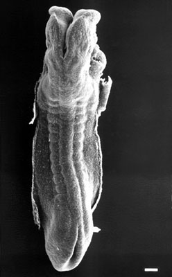
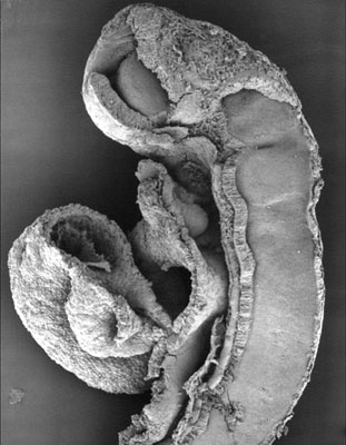
| [[File:Stage11_sem5c.jpg|frame|This is a scanning EM of the embryo dorsolateral view showing the neural tube closing with open neuropores and the paired somites visible through the thin ectoderm. Features: surface ectoderm, neural tube, cranial (anterior) neuropore, caudal (posterior) neuropore, somites, heart, cut edge of amnion, 24 days, 13 somite pairs]] | | [[File:Stage11_sem5c.jpg|frame|This is a scanning EM of the embryo dorsolateral view showing the neural tube closing with open neuropores and the paired somites visible through the thin ectoderm. Features: surface ectoderm, neural tube, cranial (anterior) neuropore, caudal (posterior) neuropore, somites, heart, cut edge of amnion, 24 days, 13 somite pairs]] | ||
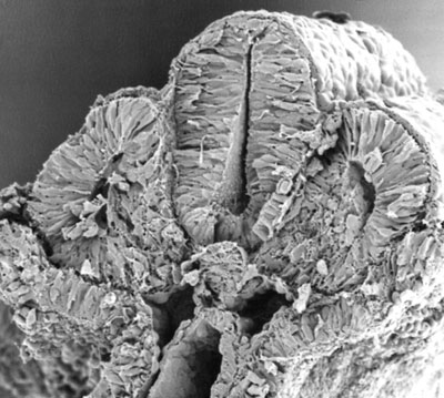
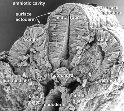
| [[File:Stage11 sem10c.jpg|frame|This is a scanning EM of the embryo fractured to show the neural tube, notochord and somites. Features: surface ectoderm, neural tube, notochord, somites, somitocoels, dorsal aortas, gastrointestinal tract, 25 days, 19 somite pairs]] | | [[File:Stage11 sem10c.jpg|frame|This is a scanning EM of the embryo fractured to show the neural tube, notochord and somites. Features: surface ectoderm, neural tube, notochord, somites, somitocoels, dorsal aortas, gastrointestinal tract, 25 days, 19 somite pairs]] | ||
| [[File:Stage11 sem100c.jpg|frame|This is a labeled version of the scanning EM of the fractured embryo]] | |||
|- | |- | ||
|} | |} | ||
Revision as of 11:57, 6 May 2010
Introduction
Facts
Week 4, 23 - 26 days, 2.5 - 4.5 mm, Somite Number 13 - 20
Events
Ectoderm: Neural tube continues to close, Rostral neuropore closes
Mesoderm: continued segmentation of paraxial mesoderm (13 - 20 somite pairs), heart tube bending
Features
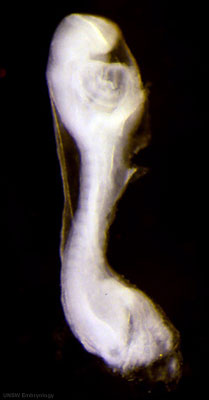
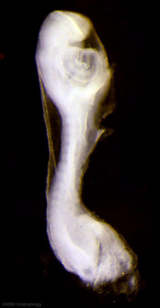
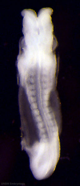
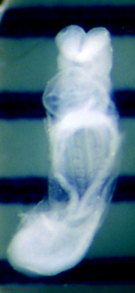
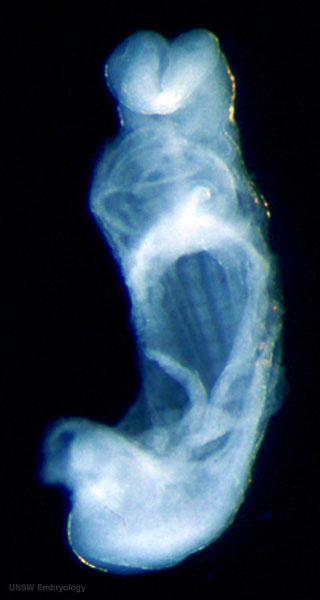
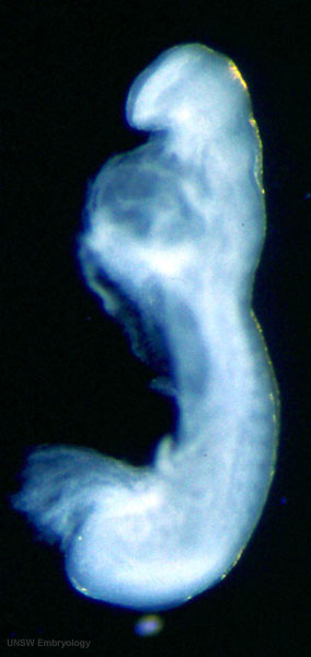
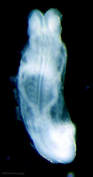
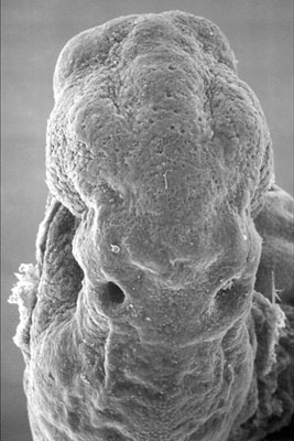
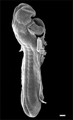
rostral neuropore closing, forebrain, neural tube in region of developing spinal cord, somites, caudal neuropore, connecting stalk, amnion
Identify: heart, rostral neuropore closing, forebrain, neural tube in region of developing spinal cord, somites, caudal neuropore, connecting stalk, amnion
- Carnegie Stages: 1 | 2 | 3 | 4 | 5 | 6 | 7 | 8 | 9 | 10 | 11 | 12 | 13 | 14 | 15 | 16 | 17 | 18 | 19 | 20 | 21 | 22 | 23 | About Stages | Timeline
Bright Field
Scanning EM
Buccopharyngeal Membrane
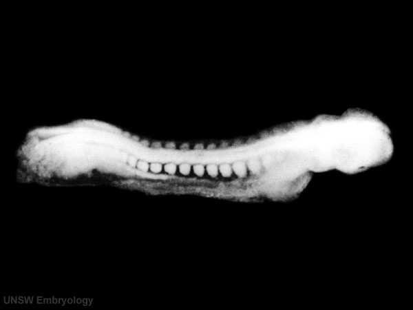
Kyoto Collection
View: This is a dorsolateral view of embryo. Amniotic membrane removed.
Image source: Embryology page Created: 19.03.1999
- Carnegie Stages: 1 | 2 | 3 | 4 | 5 | 6 | 7 | 8 | 9 | 10 | 11 | 12 | 13 | 14 | 15 | 16 | 17 | 18 | 19 | 20 | 21 | 22 | 23 | About Stages | Timeline
Image Source: Scanning electron micrographs of the Carnegie stages of the early human embryos are reproduced with the permission of Prof Kathy Sulik, from embryos collected by Dr. Vekemans and Tania Attié-Bitach. Images are for educational purposes only and cannot be reproduced electronically or in writing without permission.
Image source: The Kyoto Collection images are reproduced with the permission of Prof. Kohei Shiota and Prof. Shigehito Yamada, Anatomy and Developmental Biology, Kyoto University Graduate School of Medicine, Kyoto, Japan for educational purposes only and cannot be reproduced electronically or in writing without permission.
Cite this page: Hill, M.A. (2024, April 16) Embryology Carnegie stage 11. Retrieved from https://embryology.med.unsw.edu.au/embryology/index.php/Carnegie_stage_11
- © Dr Mark Hill 2024, UNSW Embryology ISBN: 978 0 7334 2609 4 - UNSW CRICOS Provider Code No. 00098G
