Carnegie stage 10: Difference between revisions
mNo edit summary |
mNo edit summary |
||
| Line 7: | Line 7: | ||
Week 4, 22 - 23 days, 2 - 3.5 mm, Somite number 4 - 12 | Week 4, 22 - 23 days, 2 - 3.5 mm, Somite number 4 - 12 | ||
Gestational Age - week 6 | Gestational Age {{GA}} - week 6 | ||
===Features=== | ===Features=== | ||
Somite Number 4 - 12, rostral neuropore, neural folds in region of developing brain, neural tube, somites, caudal neuropore, neural fold fuses, remnant of amniotic sac | * Somite Number 4 - 12, rostral neuropore, neural folds in region of developing brain, neural tube, somites, caudal neuropore, neural fold fuses, remnant of amniotic sac | ||
=== Events === | === Events === | ||
* Ectoderm: Neural fold deeepens, edges approach midline, neural fold fuses, neural plate folds ventrally in brain region | |||
Ectoderm: Neural fold deeepens, edges approach midline, neural fold fuses, neural plate folds ventrally in brain region | * Mesoderm: Somitogenesis, continued segmentation of paraxial mesoderm (4 - 12 somite pairs) | ||
Mesoderm: Somitogenesis, continued segmentation of paraxial mesoderm (4 - 12 somite pairs) | |||
===Identify=== | ===Identify=== | ||
| Line 24: | Line 22: | ||
:'''Links:''' [[Week 3]] | [[Gastrulation]] | [[Lecture - Week 3 Development|Lecture]] | [[ANAT2341_Lab_2|Practical]] | [[Carnegie_stage_10 gallery|Carnegie stage 10 image gallery]] | [[:Category:Carnegie_Stage_10|Category:Carnegie Stage 10]] | [[Carnegie_stage_11|Stage 11]] | :'''Links:''' [[Week 3]] | [[Gastrulation]] | [[Lecture - Week 3 Development|Lecture]] | [[ANAT2341_Lab_2|Practical]] | [[Carnegie_stage_10 gallery|Carnegie stage 10 image gallery]] | [[:Category:Carnegie_Stage_10|Category:Carnegie Stage 10]] | [[Carnegie_stage_11|Stage 11]] | ||
{{ | {{Carnegie_stage_table_1}} | ||
{{Carnegie_stages}} | |||
== Bright Field == | == Bright Field == | ||
Revision as of 14:30, 15 September 2014
| Embryology - 20 Apr 2024 |
|---|
| Google Translate - select your language from the list shown below (this will open a new external page) |
|
العربية | català | 中文 | 中國傳統的 | français | Deutsche | עִברִית | हिंदी | bahasa Indonesia | italiano | 日本語 | 한국어 | မြန်မာ | Pilipino | Polskie | português | ਪੰਜਾਬੀ ਦੇ | Română | русский | Español | Swahili | Svensk | ไทย | Türkçe | اردو | ייִדיש | Tiếng Việt These external translations are automated and may not be accurate. (More? About Translations) |
Introduction

Facts
Week 4, 22 - 23 days, 2 - 3.5 mm, Somite number 4 - 12
Gestational Age GA - week 6
Features
- Somite Number 4 - 12, rostral neuropore, neural folds in region of developing brain, neural tube, somites, caudal neuropore, neural fold fuses, remnant of amniotic sac
Events
- Ectoderm: Neural fold deeepens, edges approach midline, neural fold fuses, neural plate folds ventrally in brain region
- Mesoderm: Somitogenesis, continued segmentation of paraxial mesoderm (4 - 12 somite pairs)
Identify
The rostral neuropore, neural folds in region of developing brain, neural tube, somites (note the different number formed), caudal neuropore, neural fold fuses, cut edge of amniotic sac.
- Links: Week 3 | Gastrulation | Lecture | Practical | Carnegie stage 10 image gallery | Category:Carnegie Stage 10 | Stage 11
| Week: | 1 | 2 | 3 | 4 | 5 | 6 | 7 | 8 |
| Carnegie stage: | 1 2 3 4 | 5 6 | 7 8 9 | 10 11 12 13 | 14 15 | 16 17 | 18 19 | 20 21 22 23 |
- Carnegie Stages: 1 | 2 | 3 | 4 | 5 | 6 | 7 | 8 | 9 | 10 | 11 | 12 | 13 | 14 | 15 | 16 | 17 | 18 | 19 | 20 | 21 | 22 | 23 | About Stages | Timeline
Bright Field


Scanning EM


Image Source: Scanning electron micrographs of the Carnegie stages of the early human embryos are reproduced with the permission of Prof Kathy Sulik, from embryos collected by Dr. Vekemans and Tania Attié-Bitach. Images are for educational purposes only and cannot be reproduced electronically or in writing without permission.
Kyoto Collection
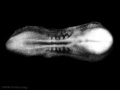
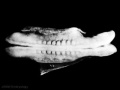
Facts: Week 4, 22 - 23 days, 2 - 3.5 mm, Somite Number 4 - 12
View: This is a dorsal view of the human embryo, the amniotic membrane has been removed. Top embryo is an early stage 10, bottom is late stage 10.


1000px

800px

600px

400px

1000px

800px

600px
400px


Image source: The Kyoto Collection images are reproduced with the permission of Prof. Kohei Shiota and Prof. Shigehito Yamada, Anatomy and Developmental Biology, Kyoto University Graduate School of Medicine, Kyoto, Japan for educational purposes only and cannot be reproduced electronically or in writing without permission.
Carnegie Collection


| iBook - Carnegie Embryos | |
|---|---|

|
|
Additional Images
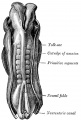
Historic drawing of human embryo, 2.11 mm. in length. (After Eternod.)
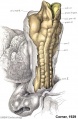
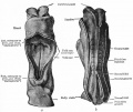
A well-preserved human embryo of 10 somites by George W. Corner
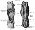
Eternod. From models by Ziegler.
Eternod. From models by Ziegler.


- Carnegie Stages: 1 | 2 | 3 | 4 | 5 | 6 | 7 | 8 | 9 | 10 | 11 | 12 | 13 | 14 | 15 | 16 | 17 | 18 | 19 | 20 | 21 | 22 | 23 | About Stages | Timeline
Cite this page: Hill, M.A. (2024, April 20) Embryology Carnegie stage 10. Retrieved from https://embryology.med.unsw.edu.au/embryology/index.php/Carnegie_stage_10
- © Dr Mark Hill 2024, UNSW Embryology ISBN: 978 0 7334 2609 4 - UNSW CRICOS Provider Code No. 00098G