Carnegie stage 10: Difference between revisions
m (→Events) |
m (→Events) |
||
| Line 123: | Line 123: | ||
* '''8 somites''' - Carnegie No. 4216. Described by Payne (1925), and very frequently cited. | * '''8 somites''' - Carnegie No. 4216. Described by Payne (1925), and very frequently cited. | ||
* '''8 somites''' - Dublin. Described by West (1930); see also Arey (1938). Photographs and models are in the Carnegie Collection, No. 4923. Bartelmez (personal communication) thinks that this distorted embryo had only 5–6 somites. | * '''8 somites''' - Dublin. Described by West (1930); see also Arey (1938). Photographs and models are in the Carnegie Collection, No. 4923. Bartelmez (personal communication) thinks that this distorted embryo had only 5–6 somites. | ||
* '''8 somites''' - [[:Category:Carnegie Embryo 391|Carnegie No. 391]]. Described by [[Paper - A Human Embryo with Seven Pairs of Somites Measuring about 2 mm in Length|Dandy]] (1910)<ref> | * '''8 somites''' - [[:Category:Carnegie Embryo 391|Carnegie No. 391]]. Described by [[Paper - A Human Embryo with Seven Pairs of Somites Measuring about 2 mm in Length|Dandy]] (1910)<ref name=Dandy1910>{{Ref-Dandy1910}}</ref> and frequently cited (cf. Bartelmez and Evans, 1926, with additional illustrations). There were neither camera drawings nor photographs of the intact specimen, and therefore the reconstructions are not entirely satisfactory. The plaster models now at the Carnegie laboratory were made by [[Embryology History - Osborne Heard|O. Heard]] under the supervision of Bartelmez for the paper by Bartelmez and Evans (1926). The apparent lack of fusion of the neural folds described by Dandy is an artifact produced by a crack. | ||
* '''8 somites''' - Carnegie No. 1201 (University of Chicago H 87). Described briefly by Evans and Bartelmez (1917); cited, with illustrations, by Bartelmez and Evans (1926). | * '''8 somites''' - Carnegie No. 1201 (University of Chicago H 87). Described briefly by Evans and Bartelmez (1917); cited, with illustrations, by Bartelmez and Evans (1926). | ||
* '''8 somites''' - Embryologisches Institut, Embryo Ct, Vienna. Fully described by Politzer (1930). Arey (1938) counts 8 paired somites in this embryo instead of 7 as stated by Politzer. | * '''8 somites''' - Embryologisches Institut, Embryo Ct, Vienna. Fully described by Politzer (1930). Arey (1938) counts 8 paired somites in this embryo instead of 7 as stated by Politzer. | ||
Revision as of 11:43, 20 August 2016
| Embryology - 19 Apr 2024 |
|---|
| Google Translate - select your language from the list shown below (this will open a new external page) |
|
العربية | català | 中文 | 中國傳統的 | français | Deutsche | עִברִית | हिंदी | bahasa Indonesia | italiano | 日本語 | 한국어 | မြန်မာ | Pilipino | Polskie | português | ਪੰਜਾਬੀ ਦੇ | Română | русский | Español | Swahili | Svensk | ไทย | Türkçe | اردو | ייִדיש | Tiếng Việt These external translations are automated and may not be accurate. (More? About Translations) |
Introduction

Facts
Week 4, 22 - 23 days, 2 - 3.5 mm, Somite number 4 - 12
Gestational Age GA - week 6
Features
- Somite Number 4 - 12, rostral neuropore, neural folds in region of developing brain, neural tube, somites, caudal neuropore, neural fold fuses, remnant of amniotic sac
Summary
- Ectoderm: Neural fold deeepens, edges approach midline, neural fold fuses, neural plate folds ventrally in brain region
- Mesoderm: Somitogenesis, continued segmentation of paraxial mesoderm (4 - 12 somite pairs)
See also Events
Identify
The rostral neuropore, neural folds in region of developing brain, neural tube, somites (note the different number formed), caudal neuropore, neural fold fuses, cut edge of amniotic sac.
- Links: Week 4 | Gastrulation | Lecture | Practical | Carnegie stage 10 image gallery | Category:Carnegie Stage 10 | Stage 11
| Week: | 1 | 2 | 3 | 4 | 5 | 6 | 7 | 8 |
| Carnegie stage: | 1 2 3 4 | 5 6 | 7 8 9 | 10 11 12 13 | 14 15 | 16 17 | 18 19 | 20 21 22 23 |
- Carnegie Stages: 1 | 2 | 3 | 4 | 5 | 6 | 7 | 8 | 9 | 10 | 11 | 12 | 13 | 14 | 15 | 16 | 17 | 18 | 19 | 20 | 21 | 22 | 23 | About Stages | Timeline
Bright Field


Scanning EM


Image Source: Scanning electron micrographs of the Carnegie stages of the early human embryos are reproduced with the permission of Prof Kathy Sulik, from embryos collected by Dr. Vekemans and Tania Attié-Bitach. Images are for educational purposes only and cannot be reproduced electronically or in writing without permission.
Kyoto Collection
Facts: Week 4, 22 - 23 days, 2 - 3.5 mm, Somite Number 4 - 12
View: This is a dorsal view of the human embryo, the amniotic membrane has been removed. Top embryo is an early stage 10, bottom is late stage 10.
| Early | Late | |
|---|---|---|

|

|

|

| Stage 10 Embryo (12202) - transverse section |
|---|
|


Image source: The Kyoto Collection images are reproduced with the permission of Prof. Kohei Shiota and Prof. Shigehito Yamada, Anatomy and Developmental Biology, Kyoto University Graduate School of Medicine, Kyoto, Japan for educational purposes only and cannot be reproduced electronically or in writing without permission.
Carnegie Collection


| Carnegie Collection - Stage 10 | ||||||||||
|---|---|---|---|---|---|---|---|---|---|---|
| Serial No. | Pairs of somites | Size (mm) | Grade | Fixative | Embedding Medium | Thinness (µm) | Stain | Year | Notes | |
| 391 | 8 | E, 2 Ch., 14 | Good | Formalin | P | 10 | Al. coch. | 1907 | Monograph by Dandy (1910)[1] | |
| 1201 | 7 | E,2 Ch.. 144 | Good | Formalin | P | 8 | H. & or. G. | 1915 | Univ. Chicago No. H 87 | |
| 2795 | 4-5 | E,2 | Poor | Alc. | P | 6 | Al coch,or.G. | 1919 | ||
| 3707 | 12 | E, 1 5 | Good | Formalin | P | 12.5 | I. H. | 1921 | Univ. Calif. No. H 197 | |
| 3709 | 4 | E. 1.4 Ch.. 14.8 | Poor | Formalin | P | 10 | Erythrosin | 1921 | Univ. Chicago No H 279 | |
| 3710 | 12 | E., 3.6 Ch., 19.0 | Good | Formalin | C-P | 10 | H. & or. G. | 1921 | Univ. Chicago No. H 392 | |
| 4216 | 8 | E, 2 Ch, 9.8 | Good | Formalin | P | 15 | ? | 1923 | Monograph by Payne (1925)[2] | |
| 5074 | 10 | E., 3.3 Ch., 10.8 | Exc. | Bouin | P | 10 | Al. coch. | 1925 | Univ. Rochester No. H 10. Monograph by Corner (1929)[3] | |
| 6330 | 7 | E, 2.83 | Good | P | 5 | Ehr. H. | 1931 | Univ. Chicago No. H 1404 | ||
| 6740 | 12 | E., 2.2 | Good | p | C-P | 8 | ? | 1933 | Litzenberg embryo. Studied by Boyden (1940) | |
| 7251 | 8 | E., 1.27 | Good | Formalin | C-P | 10 | (Stain - Haematoxylin Eosin) | 1941 | "Singapore embryo." Univ. Cambridge No. H 98. Studied by Wilson (1914)[4] | |
| 8244 | 6 | E., 1.55 Ch, 8,5 | Good | Alc. | C-P | 8 | (Stain - Haematoxylin Eosin) phlox. | 1944 | ||
| 9870 | 12 | Ch, ca. 8 | Good | Zenker | P | 5 | Various, chiefly carmine | 1952 | Univ. Chicago. No. H 637. Dicephaly | |
Abbreviations
| ||||||||||
| iBook - Carnegie Embryos | |
|---|---|

|
|
Events
For a detailed description of the stage 10 human embryos see the papers published 1910[1], 1929, [5], 1939 [6], review by Heuser in 1957[7] and in chapter by O'Rahilly and Müller in 1987.[8]
- Hearing - 10 somites first indication of otic placode invagination[5] 12 somites cells migrate from the otic disc.[9]
- Vision - optic primordia appear.[10]
| Embryo Examples |
|---|
Based on O'Rahilly and Müller (1987)[8]
|
References
- ↑ 1.0 1.1 1.2 Dandy WE. A human embryo with seven pairs of somites measuring about 2 mm in length. (1910) Amer. J Anat. 10: 85-109.
- ↑ Payne, F. 1925. General description of a 7-somite human embryo. Carnegie Instn. Wash. Publ. 361, Contrib. Embryol., 16,115-124.
- ↑ Corner GW. A well-preserved human embryo of 10 somites. (1929) Contrib. Embryol., Carnegie Inst. Wash. Publ. 394, 20:81-102.
- ↑ Wilson JT. Observations upon young human embryos. (1914) J Anat Physiol., 48(3): 315-51 PMID 17233002 PMC1288949
- ↑ 5.0 5.1 Corner GW. A well-preserved human embryo of 10 somites. (1929) Carnegie Instn. Wash. Publ. 394, Contrib. Embryol., Carnegie Inst. Wash. 20: 81-102.
- ↑ Baxter JS. and Boyd JD. Observations on the neural crest of a ten-somite human embryo. (1939) J Anat. 73: 318–326. PMID 17104759
- ↑ Heuser CH. and Corner GW. Developmental horizons in human embryos. Description of age group X, 4 to 12 somites. (1957) Carnegie Instn Wash Publ 611, Contrib. Embryol., 36: 29-39.
- ↑ 8.0 8.1 O'Rahilly R. and Müller F. Developmental Stages in Human Embryos. Contrib. Embryol., Carnegie Inst. Wash. 637 (1987).
- ↑ Bartelmez GW. and Evans HM. Development of the human embryo during the period of somite formation, including embryos with 2 to 16 pairs of somites. (1926) Contrib. Embryol., Carnegie Inst. Wash. Publ. 362, 17: 1-67.
- ↑ <pubmed>7364662</pubmed>
- ↑ Baxter JS. and Boyd JD. Observations on The Neural Crest of a Ten-Somite Human Embryo (1939) J Anat. 73:318–326. PMID 17104759
Additional Images
Historic
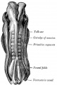
Historic drawing of human embryo, 2.11 mm. in length. (After Eternod.)
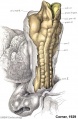
A well-preserved human embryo of 10 somites by George W. Corner
Eternod. From models by Ziegler.
Eternod. From models by Ziegler.


Dandy WE. A human embryo with seven pairs of somites measuring about 2 mm in length. (1910) Amer. J Anat. 10: 85-109.
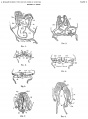
Plate 1 Sections
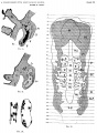
Plate 2 Sections
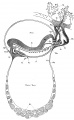
Plate 3 Sagittal view of embryo vascular system
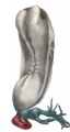
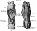
Plate 4 Dorsol-lateral view of embryo model
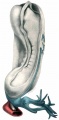
Plate 5 Dorsol-lateral view somites and mesoderm
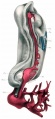
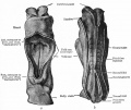
Plate 6 Dorsol-lateral view brain vesicles and aorta


Baxter JS. and Boyd JD. Observations on the neural crest of a ten-somite human embryo. (1939) J Anat. 73: 318–326. PMID 17104759
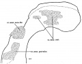
Text-fig 1 reconstruction of cephalic portion of nervous system
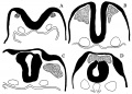
Text-fig 2 acoustico-facial neural crest primordia in certain human embryos
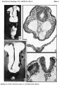
Plate 1
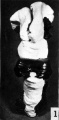
Fig 1 Reconstruction of the future brain region viewed from above and behind.
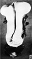
Fig 2 Reconstruction of the future brain region viewed from in front
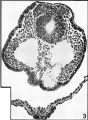
Fig 3 cranial part of the acoustico-facial primordium
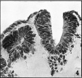
Fig 4 level of the eighth somite
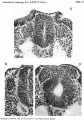
Plate 2
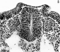
Fig 5 caudal part of the acoustico-facial primordium
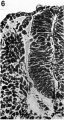
Fig 6 level of the first somite
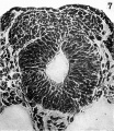
Fig 7 cranial part of the acoustico-facial neural crest primordium


Heuser CH. and Corner GW. Developmental horizons in human embryos. Description of age group X, 4 to 12 somites. (1957) Carnegie Instn Wash Publ 611, Contrib. Embryol., 36: 29-39.
- Carnegie Stages: 1 | 2 | 3 | 4 | 5 | 6 | 7 | 8 | 9 | 10 | 11 | 12 | 13 | 14 | 15 | 16 | 17 | 18 | 19 | 20 | 21 | 22 | 23 | About Stages | Timeline
Cite this page: Hill, M.A. (2024, April 19) Embryology Carnegie stage 10. Retrieved from https://embryology.med.unsw.edu.au/embryology/index.php/Carnegie_stage_10
- © Dr Mark Hill 2024, UNSW Embryology ISBN: 978 0 7334 2609 4 - UNSW CRICOS Provider Code No. 00098G