Carnegie stage 1
From Embryology

Facts: Week 1, size 0.1-0.15 mm
Features: zygote, fertilized oocyte, pronuclei, polar bodies, zona pellucida
This image shows an early human zygote where the male and female pronuclei have not yet combined to form the single zygote nucleus.
Two of the egg's polar bodies (right) are shown at the edge of the egg cytoplasm.
Additional Images
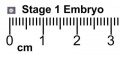
Stage1 size with ruler

- Carnegie Stages: 1 | 2 | 3 | 4 | 5 | 6 | 7 | 8 | 9 | 10 | 11 | 12 | 13 | 14 | 15 | 16 | 17 | 18 | 19 | 20 | 21 | 22 | 23 | About Stages | Timeline
Cite this page: Hill, M.A. (2024, April 18) Embryology Carnegie stage 1. Retrieved from https://embryology.med.unsw.edu.au/embryology/index.php/Carnegie_stage_1
- © Dr Mark Hill 2024, UNSW Embryology ISBN: 978 0 7334 2609 4 - UNSW CRICOS Provider Code No. 00098G