Carnegie stage 1: Difference between revisions
| Line 33: | Line 33: | ||
File:Human zygote two pronuclei 22.jpg|Human zygote two pronuclei (labelled) PMID 20579351 | File:Human zygote two pronuclei 22.jpg|Human zygote two pronuclei (labelled) PMID 20579351 | ||
File:Human-oocyte_to_blastocyst.jpg|Human oocyte to blastocyst PMID 19924284 | File:Human-oocyte_to_blastocyst.jpg|Human oocyte to blastocyst PMID 19924284 | ||
</gallery> | |||
===Electron Micrographs=== | |||
<gallery> | |||
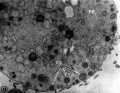
File:Human pronuclear stage EM02.jpg|Human pronuclei EM PMID 6008199 | File:Human pronuclear stage EM02.jpg|Human pronuclei EM PMID 6008199 | ||
File:Human pronuclear stage EM022.jpg|Human pronuclei EM PMID 6008199 | File:Human pronuclear stage EM022.jpg|Human pronuclei EM PMID 6008199 | ||
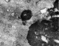
File:Human pronuclear stage EM22.jpg|Spermatozoon Components | |||
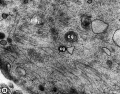
File:Human pronuclear stage EM25.jpg|Golgi complex | |||
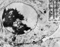
File:Human pronuclear stage EM26.jpg|First Polar Body | |||
File:Human pronuclear stage EM27.jpg|First Polar Body | |||
File:Human pronuclear stage EM28.jpg|First Polar Body | |||
File:Human pronuclear stage EM29.jpg|Second Polar Body | |||
File:Human pronuclear stage EM30.jpg|Second Polar Body | |||
</gallery> | </gallery> | ||
Revision as of 00:24, 24 February 2012
Introduction

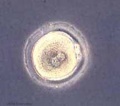
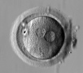
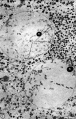
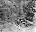
Human early zygote showing male and female pronuclei |
The early human zygote where the male and female pronuclei (centre of image) have not yet combined to form the single zygote nucleus. These pronuclei are the nuclei from the spermatozoa (sperm) and oocyte (egg) and contain all the nuclear genetic material (chromosomes, DNA, genes). Two of the egg's polar bodies (right, 3 o'clock position of image) are shown at the edge of the egg cytoplasm. These polar bodies contain the excess DNA from the meiotic divisions of the egg. The zona pellucida (edge of image) forms a specialised thick extracellular matrix layer around both the egg and the developing conceptus for the first week. Mitochondria in the cytoplasm contain additional genes and in humans these mitochondrial genes are entirely derived from the oocyte. Facts: Week 1, size 0.1-0.15 mm Features: zygote, fertilized oocyte, pronuclei, polar bodies, zona pellucida
|
- Carnegie Stages: 1 | 2 | 3 | 4 | 5 | 6 | 7 | 8 | 9 | 10 | 11 | 12 | 13 | 14 | 15 | 16 | 17 | 18 | 19 | 20 | 21 | 22 | 23 | About Stages | Timeline
Carnegie stage 1
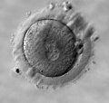
Early zygote
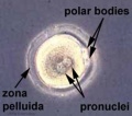
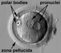
Early zygote labeled
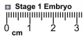
Stage 1 size with ruler
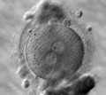
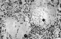
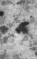
Human zygote two pronuclei PMID 20579351
Human zygote two pronuclei PMID 20579351
Human zygote two pronuclei PMID 20579351
Human zygote two pronuclei (labelled) PMID 20579351
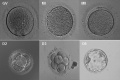
Human oocyte to blastocyst PMID 19924284


Electron Micrographs
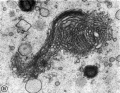
Human pronuclei EM PMID 6008199
Human pronuclei EM PMID 6008199
Spermatozoon Components
Golgi complex
First Polar Body
First Polar Body
First Polar Body
Second Polar Body
Second Polar Body


Historic Images
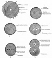
Fig. 15. Diagram of fertilization of the ovum
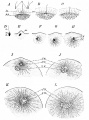
Fig. 16. Fertilization of the eggs of the star-fish and sea-urchin


References
- Carnegie Stages: 1 | 2 | 3 | 4 | 5 | 6 | 7 | 8 | 9 | 10 | 11 | 12 | 13 | 14 | 15 | 16 | 17 | 18 | 19 | 20 | 21 | 22 | 23 | About Stages | Timeline
Cite this page: Hill, M.A. (2024, April 25) Embryology Carnegie stage 1. Retrieved from https://embryology.med.unsw.edu.au/embryology/index.php/Carnegie_stage_1
- © Dr Mark Hill 2024, UNSW Embryology ISBN: 978 0 7334 2609 4 - UNSW CRICOS Provider Code No. 00098G