Cardiovascular System Development

Introduction

Development of the heart and vascular system begins very early in mesoderm both within (embryonic) and outside (extra embryonic, yolk sac and placental) the embryo. Vascular development therefore occurs in many places, the most obvious though is the early forming heart, which grows rapidly creating an externally obvious cardiac "bulge" on the early embryo. The cardiovascular system is extensively remodelled throughout development, this current page only introduces topic.
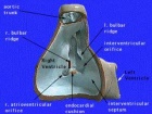
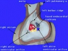
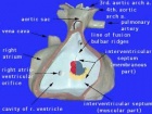
The heart forms initially in the embryonic disc as a simple paired tube inside the forming pericardial cavity, which when the disc folds, gets carried into the correct anatomical position in the chest cavity.
Throughout the mesoderm, small regions differentiate into "blood islands" which contribute both blood vessels (walls) and fetal red blood cells.
These "islands" connect together to form the first vessels which connect with the heart tube.
A detailed description of heart development is covered in the Online Heart Tutorial.
Some Recent Findings
|
Textbooks

- Human Embryology (2nd ed.) Larson Ch7 p151-188 Heart, Ch8 p189-228 Vasculature
- The Developing Human: Clinically Oriented Embryology (6th ed.) Moore and Persaud Ch14: p304-349
- Before we Are Born (5th ed.) Moore and Persaud Ch12; p241-254
- Essentials of Human Embryology Larson Ch7 p97-122 Heart, Ch8 p123-146 Vasculature
- Human Embryology Fitzgerald and Fitzgerald Ch13-17: p77-111
Heart Tutorial
| Begin Basic | Primitive Heart Tube | Embryonic Heart Divisions | Vascular Heart Connections |
| Begin Intermediate: | Primordial Heart Tube | Heart Tube Looping | Atrial Ventricular Septation | Outflow Tract | Heart Valves | Cardiac Abnormalities | Vascular Overview |
| Begin Advanced | Heart Fields | Heart Tubes | Cardiac Looping | Cardiac Septation | Outflow Tract | Valve Development | Cardiac Conduction | Cardiac Abnormalities | Molecular Development |
Timecourse

|
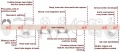
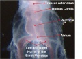
| The Human Heart from day 10 to 25 (scanning electron micrograph) |
- Forms initially in splanchnic mesoderm of prechordal plate region - cardiogenic region
- growth and folding of the embryo moves heart ventrally and downward into anatomical position
- Day 22 - 23, begins to beat in humans
- heart tube connects to blood vessels forming in splanchnic and extraembryonic mesoderm
- Week 2 - 3 pair of thin-walled tubes
- Week 3 paired heart tubes fuse, truncus arteriosus outflow, heart contracting
- Week 4 heart tube continues to elongate, curving to form S shape
- Week 5 Septation starts]], atrial and ventricular
- Septation continues, atrial septa remains open, foramen ovale
- Week 37-38 At birth, pressure difference closes foramen ovale leaving a fossa ovalis
Heart Development Movies
Animations showing aspects of heart development.
|
|
|
|
|
| Heart Cartoons | |||||||||||||||||||||||||||
|---|---|---|---|---|---|---|---|---|---|---|---|---|---|---|---|---|---|---|---|---|---|---|---|---|---|---|---|
|
|
|
|
|
|
|
|
|
Pages within the online Cardiac tutorial.
| Heart Fields | Primitive Heart Tube | Heart Tubes | Cardiac Looping | Cardiac Septation | Outflow Tract |
| Outflow Tract |
| Page | Play |
Historic animations including audio descriptions. Some of these descriptions may be currently inaccurate, the transfer is from an old class film and the audio track is of very poor quality.
| Historic Animations | |||||||||||||||
|---|---|---|---|---|---|---|---|---|---|---|---|---|---|---|---|
|
|
|
| ||||||||||||
|
|
|
|
| About Historic Animations | ||||
|---|---|---|---|---|
 The sound quality is quite poor and some of the information is now out of date, most general concepts are still correct. Please note the relatively large size (Mb) of each excerpt will effect download and viewing. March 2013
|
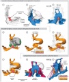
Ventricular septation rotation models.
|
|
|
Chicken Heart Development
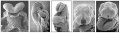
Note the images of chicken heart development[2] shown below are Hamburger Hamilton Stages of chicken development, not Carnegie stages. See also Heart 3D reconstruction.
Chicken (day 2, Stage 12)
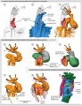
Chicken (day 3, Stage 16)
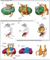
Chicken (day 4, Stage 21)
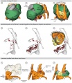
Chicken (day 5, Stage 25)


Pharyngeal Arch Arteries

In the head region of the embryo, each pharyngeal arch initially has paired arch arteries. These are extensively remodelled through development and give rise to a range of different arterial structures, as shown in the list below.
- Arch 1 - mainly lost, form part of maxillary artery.
- Arch 2 - stapedial arteries.
- Arch 3 - common carotid arteries, internal carotid arteries.
- Arch 4 - left forms part of aortic arch, right forms part right subclavian artery.
- Arch 6 - left forms part of left pulmonary artery , right forms part of right pulmonary artery.
- Links: Head Development
Renal Venous Development
The renal arterial and venous systems are also reorganised extensively throughout development with changing kidney position.

|

|
| Embryo renal venous | Adult renal venous |
- Links: Renal Development
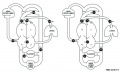
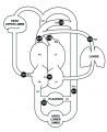
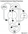
Fetal Blood Flow

Mean Late Fetal Blood Flows[3]
(8 subjects) in the major vessels of the human fetal circulation by phase contrast MRI. (median gestational age 37 weeks, age range of 30–39 weeks)
| (left) Mean flows in ml/kg/min | (right) Proportions of the combined ventricular output in the major vessels of the human fetal circulation by phase contrast MRI. |
|
|
- Cardiovascular Links: Fetal Blood Flow values | Mean Fetal Blood Flow | Proportions Ventricular Output | Ventricular Output (colour) | heart | blood | cardiovascular
References
- ↑ <pubmed>18682987</pubmed>
- ↑ <pubmed>21779373</pubmed>| PLoS One
- ↑ <pubmed>23181717</pubmed>| J Cardiovasc Magn Reson.
Reviews
<pubmed></pubmed> <pubmed></pubmed> <pubmed></pubmed> <pubmed>22449840</pubmed> <pubmed>21593862</pubmed> <pubmed>18607112</pubmed> <pubmed>16565980</pubmed> <pubmed>16236564</pubmed> <pubmed>15614842</pubmed>
Articles
<pubmed>21808168</pubmed> <pubmed>21732277</pubmed> <pubmed>21541028</pubmed> <pubmed>21540552</pubmed> <pubmed>21364285</pubmed> <pubmed>18057862</pubmed>
Search Pubmed
Search May 2010
- Cardiovascular System Development All (63457) Review (10735) Free Full Text (15717)
Search Pubmed: Cardiovascular System Development
Additional Images
See also Category:Heart ILP and Category:Heart
Historic image
Heart Development Timeline
Human heart SEM
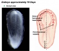
Early Heart Tube (Dorsal)
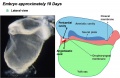
Early Heart Tube (Lateral)
Heart Tube Segments
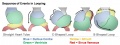
Heart Looping Sequence
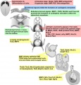
Molecular & Genetic Cardiac Development Factors
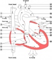
Adult heart blood flow cartoon
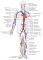
Adult human cardiovascular system cartoon
Fetal Blood Flow
Fetal Blood Flow
Fetal Blood Flow


.jpg)
.jpg)


External Links
External Links Notice - The dynamic nature of the internet may mean that some of these listed links may no longer function. If the link no longer works search the web with the link text or name. Links to any external commercial sites are provided for information purposes only and should never be considered an endorsement. UNSW Embryology is provided as an educational resource with no clinical information or commercial affiliation.
- Australia Heart Foundation
- USA National Heart, Lung, and Blood Institute - Congenital Heart Defects | Heart and Vascular Information
| System Links: Introduction | Cardiovascular | Coelomic Cavity | Endocrine | Gastrointestinal Tract | Genital | Head | Immune | Integumentary | Musculoskeletal | Neural | Neural Crest | Placenta | Renal | Respiratory | Sensory | Birth |
Glossary Links
- Glossary: A | B | C | D | E | F | G | H | I | J | K | L | M | N | O | P | Q | R | S | T | U | V | W | X | Y | Z | Numbers | Symbols | Term Link
Cite this page: Hill, M.A. (2024, April 16) Embryology Cardiovascular System Development. Retrieved from https://embryology.med.unsw.edu.au/embryology/index.php/Cardiovascular_System_Development
- © Dr Mark Hill 2024, UNSW Embryology ISBN: 978 0 7334 2609 4 - UNSW CRICOS Provider Code No. 00098G