Cardiovascular System Development: Difference between revisions
mNo edit summary |
mNo edit summary |
||
| (21 intermediate revisions by the same user not shown) | |||
| Line 1: | Line 1: | ||
[[File:Heart Tube Fusion.jpg|right|500px]] | {{Header}}[[File:Heart Tube Fusion.jpg|right|500px]] | ||
==Introduction== | ==Introduction== | ||
[[File:Heart Tube Segments.jpg|thumb|The embryo stage 10 heart tube]] | [[File:Heart Tube Segments.jpg|thumb|The embryo stage 10 heart tube]] | ||
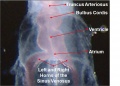
Development of the heart and vascular system begins very early in mesoderm both within (embryonic) and outside (extra embryonic, yolk sac and placental) the embryo. Vascular development therefore occurs in many places, the most obvious though is the early forming heart, which grows rapidly creating an externally obvious cardiac "bulge" on the early embryo. The cardiovascular system is extensively remodelled throughout development, this current page only introduces topic. | Development of the {{heart}} and vascular system is often described together as the {{cardiovascular}} system, with the heart being the first functional organ that forms in the embryo. Development begins very early in {{mesoderm}} both within (embryonic) and outside (extra embryonic, yolk sac and placental) the embryo. Vascular development therefore occurs in many places, the most obvious though is the early forming heart, which grows rapidly creating an externally obvious cardiac "bulge" on the early embryo. The {{cardiovascular}} system is extensively remodelled throughout development, this current page only introduces topic. | ||
The heart forms initially in the embryonic disc as a simple paired tube inside the forming pericardial cavity, which when the disc folds, gets carried into the correct anatomical position in the chest cavity. | The heart forms initially in the embryonic disc as a simple paired tube inside the forming pericardial cavity, which when the disc folds, gets carried into the correct anatomical position in the chest cavity. | ||
Throughout the mesoderm, small regions differentiate into "blood islands" which contribute both blood vessels (walls) and fetal red blood cells. | Throughout the mesoderm, small regions differentiate into "blood islands" which contribute both blood vessels (walls) and fetal red blood cells. | ||
These "islands" connect together to form the first vessels which connect with the heart tube. | These "islands" connect together to form the first vessels which connect with the heart tube. | ||
| Line 20: | Line 21: | ||
|-bgcolor="F5FAFF" | |-bgcolor="F5FAFF" | ||
| | | | ||
* ''' | * '''Development and Morphology of the Ventricular Outflow Tracts'''{{#pmid:27587491|PMID27587491}} "It is customary, at the current time, to consider many, if not most, of the lesions involving the ventricular outflow tract in terms of conotruncal malformations. This reflects the introduction, in the early 1940s, of the terms conus and truncus to describe the components of the developing outflow tract. The definitive outflow tracts in the postnatal heart, however, possess three, rather than two, components. These are the intrapericardial arterial trunks, the arterial roots, and the subvalvar ventricular outflow tracts." | ||
* '''A detailed comparison of mouse and human cardiac development'''{{#pmid:25167202|PMID25167202}} "Episcopic fluorescence image capture (EFIC) was performed on 66 wild-type mouse embryos from embryonic day (E) 9.5 to birth; 2-dimensional and 3-dimensional datasets were compared with EFIC and magnetic resonance images from a study of 52 human fetuses (Carnegie stage 13-23). Time course of atrial, ventricular, and outflow septation were outlined and followed a similar sequence in both species. Bilateral venae cavae and prominent atrial appendages were seen in the mouse fetus; in human fetuses, atrial appendages were small, and a single right superior vena cava was present. In contrast to humans with separate pulmonary vein orifices, a pulmonary venous confluence with one orifice enters the left atrium in mice. The cardiac developmental sequences observed in mouse and human fetuses are comparable, with minor differences in atrial and venous morphology. These comparisons of mouse and human cardiac development strongly support that mouse morphogenesis is a good model for human development." [[Mouse Development]] | [[Kyoto Collection]] | |||
* '''Assembly of the Cardiac Intercalated Disk during Pre- and Postnatal Development of the Human Heart'''{{#pmid:24733085|PMID24733085}} "In cardiac muscle, the intercalated disk (ID) at the longitudinal cell-edges of cardiomyocytes provides as a macromolecular infrastructure that integrates mechanical and electrical coupling within the heart. ...Our data on developmental maturation of the ID in human heart indicate that generation of the mechanical junctions at the ID precedes that of the electrical junctions with a significant difference in time. In addition arrival of the electrical junctions (Nav1.5 and Cx43) is not uniform since sodium channels localize much earlier than gap junction channels." | |||
|} | |} | ||
{| class="wikitable mw-collapsible mw-collapsed" | |||
! More recent papers | |||
|- | |||
| [[File:Mark_Hill.jpg|90px|left]] {{Most_Recent_Refs}} | |||
Search term: [http://www.ncbi.nlm.nih.gov/pubmed/?term=Cardiovascular+Embryology ''Cardiovascular Embryology''] | [http://www.ncbi.nlm.nih.gov/pubmed/?term=Cardiovascular+Development ''Cardiovascular Development''] | |||
|} | |||
{| class="wikitable mw-collapsible mw-collapsed" | |||
! Older papers | |||
|- | |||
| {{Older papers}} | |||
* '''Endothelial cell lineages of the heart.''' {{#pmid:18682987|PMID18682987}} "During early gastrulation, vertebrate embryos begin to produce endothelial cells (ECs) from the mesoderm. ECs first form primitive vascular plexus de novo and later differentiate into arterial, venous, capillary, and lymphatic ECs. In the heart, the five distinct EC types (endocardial, coronary arterial, venous, capillary, and lymphatic) have distinct phenotypes. For example, coronary ECs establish a typical vessel network throughout the myocardium, whereas endocardial ECs form a large epithelial sheet with no angiogenic sprouting into the myocardium. Neither coronary arteries, veins, and capillaries, nor lymphatic vessels fuse with the endocardium or open to the heart chamber. The developmental stage during which the specific phenotype of each cardiac EC type is determined remains unclear. The mechanisms involved in EC commitment and diversity can however be more precisely defined by tracking the migratory patterns and lineage decisions of the precursors of cardiac ECs." | |||
|} | |||
==Textbooks== | ==Textbooks== | ||
[[File:Cardiac_muscle_histology.jpg|thumb|Cardiac muscle histology]] | [[File:Cardiac_muscle_histology.jpg|thumb|Cardiac muscle histology]] | ||
| Line 54: | Line 72: | ||
** Septation continues, atrial septa remains open, foramen ovale | ** Septation continues, atrial septa remains open, foramen ovale | ||
* '''Week 37-38''' At birth, pressure difference closes foramen ovale leaving a fossa ovalis | * '''Week 37-38''' At birth, pressure difference closes foramen ovale leaving a fossa ovalis | ||
<br> | |||
{{Madrid Heart timeline table}} | |||
==Heart Development Movies== | ==Heart Development Movies== | ||
===Animations=== | |||
Animations showing aspects of heart development. | Animations showing aspects of heart development. | ||
{{CVS cartoons}} | |||
===Tutorials=== | |||
Pages within the [[Cardiac_Embryology|online Cardiac tutorial]]. | |||
{{heart cartoons}} | |||
{| border='0px' | {| border='0px' | ||
| valign="bottom"|{{Cardiovascular stage 13 movie}} | |||
| valign="bottom"|{{Cardiovascular stage 22 movie}} | |||
| | |||
| | |||
|} | |} | ||
===Historic=== | |||
Historic animations including audio descriptions. Some of these descriptions may be currently inaccurate, the transfer is from an old class film and the audio track is of very poor quality. | Historic animations including audio descriptions. Some of these descriptions may be currently inaccurate, the transfer is from an old class film and the audio track is of very poor quality. | ||
{{Historic Heart}} | {{Historic Heart}} | ||
===Septation Models=== | |||
Ventricular septation rotation models. | |||
{| | |||
| valign="bottom"|{{Heart ventricular septum model 1}} | |||
| valign="bottom"|{{Heart ventricular septum model 2}} | |||
| valign="bottom"|{{Heart ventricular septum model 3}} | |||
|} | |||
==Animal Models== | |||
Amphibians and reptiles have a three-chambered heart with a single ventricle. Blood leaves the heart ventricle through either the pulmonary artery to the lungs or the aorta to supply the body. The pulmonary artery in amphibians also supplies the skin. | |||
Mammals and birds have a four-chambered heart with a two ventricles. The right ventricle supplies the pulmonary artery to the lungs, the left ventricle supplies the aorta to the body. | |||
==Chicken Heart Development== | ===Chicken Heart Development=== | ||
Note the images of chicken heart development | Note the images of chicken heart development{{#pmid:21779373|PMID21779373}} shown below are [[Hamburger Hamilton Stages]] of chicken development, not Carnegie stages. See also [[:File:Chicken_heart_3D_reconstruction_from_sections.jpg|Heart 3D reconstruction]]. | ||
<gallery> | <gallery> | ||
| Line 168: | Line 152: | ||
'''Mean Late Fetal Blood Flows''' | '''Mean Late Fetal Blood Flows'''{{#pmid:23181717|PMID23181717}} | ||
(8 subjects) in the major vessels of the human fetal circulation by phase contrast MRI. (median gestational age 37 weeks, age range of 30–39 weeks) | (8 subjects) in the major vessels of the human fetal circulation by phase contrast MRI. (median gestational age 37 weeks, age range of 30–39 weeks) | ||
| Line 201: | Line 185: | ||
===Reviews=== | ===Reviews=== | ||
{{#pmid:28003417}} | |||
{{#pmid:22449840}} | |||
{{#pmid:21593862}} | |||
{{#pmid:18607112}} | |||
{{#pmid:16565980}} | |||
{{#pmid:16236564}} | |||
{{#pmid:15614842}} | |||
===Articles=== | ===Articles=== | ||
{{#pmid:21808168}} | |||
{{#pmid:21732277}} | |||
{{#pmid:21541028}} | |||
{{#pmid:21540552}} | |||
{{#pmid:21364285}} | |||
{{#pmid:18057862}} | |||
===Search Pubmed=== | |||
'''Search Pubmed:''' [http://www.ncbi.nlm.nih.gov/sites/entrez?db=pubmed&cmd=search&term=Cardiovascular%20System%20Development Cardiovascular System Development] | '''Search Pubmed:''' [http://www.ncbi.nlm.nih.gov/sites/entrez?db=pubmed&cmd=search&term=Cardiovascular%20System%20Development Cardiovascular System Development] | ||
{{References footer}} | |||
==Additional Images== | ==Additional Images== | ||
See also [[:Category:Heart ILP]] and [[:Category:Heart]] | See also [[:Category:Heart ILP]] and [[:Category:Heart]] | ||
[[Embryology_History_-_Ziegler_Models|Ziegler Models]] | |||
<gallery> | |||
Ziegler_model_18.jpg| | |||
Ziegler model 12.jpg| | |||
</gallery> | |||
<gallery> | <gallery> | ||
| Line 247: | Line 245: | ||
File:Fetal_blood_flow_02.jpg|Fetal Blood Flow | File:Fetal_blood_flow_02.jpg|Fetal Blood Flow | ||
File:Fetal_blood_flow_03.jpg|Fetal Blood Flow | File:Fetal_blood_flow_03.jpg|Fetal Blood Flow | ||
File:Windle1940 fig16.jpg|Heart at birth | |||
</gallery> | </gallery> | ||
| Line 255: | Line 254: | ||
* '''USA National Heart, Lung, and Blood Institute''' - [http://www.nhlbi.nih.gov/health/dci/Diseases/chd/chd_what.html Congenital Heart Defects] | [http://www.nhlbi.nih.gov/health/public/heart/index.htm Heart and Vascular Information] | * '''USA National Heart, Lung, and Blood Institute''' - [http://www.nhlbi.nih.gov/health/dci/Diseases/chd/chd_what.html Congenital Heart Defects] | [http://www.nhlbi.nih.gov/health/public/heart/index.htm Heart and Vascular Information] | ||
{{Systems}} | |||
{{Glossary}} | |||
{{Footer}} | {{Footer}} | ||
[[Category:System Development]] [[Category:Cardiovascular]] [[Category:Heart]] | [[Category:System Development]] [[Category:Cardiovascular]] [[Category:Heart]] | ||
Revision as of 12:09, 18 December 2018
| Embryology - 19 Apr 2024 |
|---|
| Google Translate - select your language from the list shown below (this will open a new external page) |
|
العربية | català | 中文 | 中國傳統的 | français | Deutsche | עִברִית | हिंदी | bahasa Indonesia | italiano | 日本語 | 한국어 | မြန်မာ | Pilipino | Polskie | português | ਪੰਜਾਬੀ ਦੇ | Română | русский | Español | Swahili | Svensk | ไทย | Türkçe | اردو | ייִדיש | Tiếng Việt These external translations are automated and may not be accurate. (More? About Translations) |

Introduction

Development of the heart and vascular system is often described together as the cardiovascular system, with the heart being the first functional organ that forms in the embryo. Development begins very early in mesoderm both within (embryonic) and outside (extra embryonic, yolk sac and placental) the embryo. Vascular development therefore occurs in many places, the most obvious though is the early forming heart, which grows rapidly creating an externally obvious cardiac "bulge" on the early embryo. The cardiovascular system is extensively remodelled throughout development, this current page only introduces topic.
The heart forms initially in the embryonic disc as a simple paired tube inside the forming pericardial cavity, which when the disc folds, gets carried into the correct anatomical position in the chest cavity.
Throughout the mesoderm, small regions differentiate into "blood islands" which contribute both blood vessels (walls) and fetal red blood cells.
These "islands" connect together to form the first vessels which connect with the heart tube.
A detailed description of heart development is covered in the Online Heart Tutorial.
Some Recent Findings
|
| More recent papers |
|---|
 This table allows an automated computer search of the external PubMed database using the listed "Search term" text link.
More? References | Discussion Page | Journal Searches | 2019 References | 2020 References Search term: Cardiovascular Embryology | Cardiovascular Development |
| Older papers |
|---|
| These papers originally appeared in the Some Recent Findings table, but as that list grew in length have now been shuffled down to this collapsible table.
See also the Discussion Page for other references listed by year and References on this current page.
|
Textbooks

- Human Embryology (2nd ed.) Larson Ch7 p151-188 Heart, Ch8 p189-228 Vasculature
- The Developing Human: Clinically Oriented Embryology (6th ed.) Moore and Persaud Ch14: p304-349
- Before we Are Born (5th ed.) Moore and Persaud Ch12; p241-254
- Essentials of Human Embryology Larson Ch7 p97-122 Heart, Ch8 p123-146 Vasculature
- Human Embryology Fitzgerald and Fitzgerald Ch13-17: p77-111
Heart Tutorial
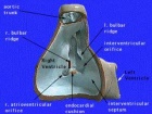
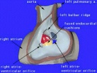
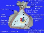
| Begin Basic | Primitive Heart Tube | Embryonic Heart Divisions | Vascular Heart Connections |
| Begin Intermediate: | Primordial Heart Tube | Heart Tube Looping | Atrial Ventricular Septation | Outflow Tract | Heart Valves | Cardiac Abnormalities | Vascular Overview |
| Begin Advanced | Heart Fields | Heart Tubes | Cardiac Looping | Cardiac Septation | Outflow Tract | Valve Development | Cardiac Conduction | Cardiac Abnormalities | Molecular Development |
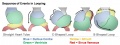
Timecourse

|
| The Human Heart from day 10 to 25 (scanning electron micrograph) |
- Forms initially in splanchnic mesoderm of prechordal plate region - cardiogenic region
- growth and folding of the embryo moves heart ventrally and downward into anatomical position
- Day 22 - 23, begins to beat in humans
- heart tube connects to blood vessels forming in splanchnic and extraembryonic mesoderm
- Week 2 - 3 pair of thin-walled tubes
- Week 3 paired heart tubes fuse, truncus arteriosus outflow, heart contracting
- Week 4 heart tube continues to elongate, curving to form S shape
- Week 5 Septation starts]], atrial and ventricular
- Septation continues, atrial septa remains open, foramen ovale
- Week 37-38 At birth, pressure difference closes foramen ovale leaving a fossa ovalis
| Characteristic | Carnegie stage: | 13 | 14 | 15 | 16 | 17 | 18 | 19 | 20 | 21 | 22 | 23 |
|---|---|---|---|---|---|---|---|---|---|---|---|---|
| Septum primum | ||||||||||||
| Foramen primum | ||||||||||||
| Atrioventricular bundle | ||||||||||||
| Atrioventricular cushions | ||||||||||||
| Conotruncal ridges | ||||||||||||
| Foramen secundum | ||||||||||||
| Semilunar cusps | ||||||||||||
| Conotruncal septum; atria | ||||||||||||
| Closure primum foramen | ||||||||||||
| Fusion atrioventricular cushions | ||||||||||||
| Septum secundum and foramen ovale | ||||||||||||
| Closure secondary interventricular foramen | ||||||||||||
| Chordae tendineae | ||||||||||||
| Colour Coding: | beginning to appear | present | Table data[5] Links: heart | Madrid Collection | |||||||||
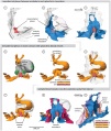
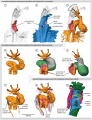
Heart Development Movies
Animations
Animations showing aspects of heart development.
|
|
|
|
|
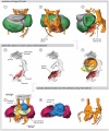
Tutorials
Pages within the online Cardiac tutorial.
| Heart Cartoons | |||||||||||||||||||||||||||
|---|---|---|---|---|---|---|---|---|---|---|---|---|---|---|---|---|---|---|---|---|---|---|---|---|---|---|---|
|
|
|
|
|
|
|
|
|
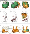
Historic
Historic animations including audio descriptions. Some of these descriptions may be currently inaccurate, the transfer is from an old class film and the audio track is of very poor quality.
| Historic Animations | |||||||||||||||
|---|---|---|---|---|---|---|---|---|---|---|---|---|---|---|---|
|
|
|
| ||||||||||||
|
|
|
|
| About Historic Animations | ||||
|---|---|---|---|---|
The sound quality is quite poor and some of the information is now out of date, most general concepts are still correct. Please note the relatively large size (Mb) of each excerpt will effect download and viewing. March 2013
|
Septation Models
Ventricular septation rotation models.
|
|
|
Animal Models
Amphibians and reptiles have a three-chambered heart with a single ventricle. Blood leaves the heart ventricle through either the pulmonary artery to the lungs or the aorta to supply the body. The pulmonary artery in amphibians also supplies the skin.
Mammals and birds have a four-chambered heart with a two ventricles. The right ventricle supplies the pulmonary artery to the lungs, the left ventricle supplies the aorta to the body.
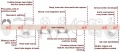
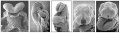
Chicken Heart Development
Note the images of chicken heart development[6] shown below are Hamburger Hamilton Stages of chicken development, not Carnegie stages. See also Heart 3D reconstruction.
Chicken (day 2, Stage 12)
Chicken (day 3, Stage 16)
Chicken (day 4, Stage 21)
Chicken (day 5, Stage 25)


Pharyngeal Arch Arteries

In the head region of the embryo, each pharyngeal arch initially has paired arch arteries. These are extensively remodelled through development and give rise to a range of different arterial structures, as shown in the list below.
- Arch 1 - mainly lost, form part of maxillary artery.
- Arch 2 - stapedial arteries.
- Arch 3 - common carotid arteries, internal carotid arteries.
- Arch 4 - left forms part of aortic arch, right forms part right subclavian artery.
- Arch 6 - left forms part of left pulmonary artery , right forms part of right pulmonary artery.
- Links: Head Development
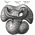
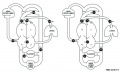
Renal Venous Development
The renal arterial and venous systems are also reorganised extensively throughout development with changing kidney position.

|

|
| Embryo renal venous | Adult renal venous |
- Links: Renal Development
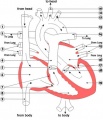
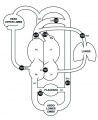
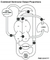
Fetal Blood Flow

Mean Late Fetal Blood Flows[7]
(8 subjects) in the major vessels of the human fetal circulation by phase contrast MRI. (median gestational age 37 weeks, age range of 30–39 weeks)
| (left) Mean flows in ml/kg/min | (right) Proportions of the combined ventricular output in the major vessels of the human fetal circulation by phase contrast MRI. |
|
|
- Cardiovascular Links: Fetal Blood Flow values | Mean Fetal Blood Flow | Proportions Ventricular Output | Ventricular Output (colour) | heart | blood | cardiovascular
References
- ↑ Anderson RH, Mori S, Spicer DE, Brown NA & Mohun TJ. (2016). Development and Morphology of the Ventricular Outflow Tracts. World J Pediatr Congenit Heart Surg , 7, 561-77. PMID: 27587491 DOI.
- ↑ Krishnan A, Samtani R, Dhanantwari P, Lee E, Yamada S, Shiota K, Donofrio MT, Leatherbury L & Lo CW. (2014). A detailed comparison of mouse and human cardiac development. Pediatr. Res. , 76, 500-7. PMID: 25167202 DOI.
- ↑ Vreeker A, van Stuijvenberg L, Hund TJ, Mohler PJ, Nikkels PG & van Veen TA. (2014). Assembly of the cardiac intercalated disk during pre- and postnatal development of the human heart. PLoS ONE , 9, e94722. PMID: 24733085 DOI.
- ↑ Ishii Y, Langberg J, Rosborough K & Mikawa T. (2009). Endothelial cell lineages of the heart. Cell Tissue Res. , 335, 67-73. PMID: 18682987 DOI.
- ↑ Arráez-Aybar LA, Turrero-Nogués A & Marantos-Gamarra DG. (2008). Embryonic cardiac morphometry in Carnegie stages 15-23, from the Complutense University of Madrid Institute of Embryology Human Embryo Collection. Cells Tissues Organs (Print) , 187, 211-20. PMID: 18057862 DOI.
- ↑ van den Berg G & Moorman AF. (2011). Development of the pulmonary vein and the systemic venous sinus: an interactive 3D overview. PLoS ONE , 6, e22055. PMID: 21779373 DOI.
- ↑ Seed M, van Amerom JF, Yoo SJ, Al Nafisi B, Grosse-Wortmann L, Jaeggi E, Jansz MS & Macgowan CK. (2012). Feasibility of quantification of the distribution of blood flow in the normal human fetal circulation using CMR: a cross-sectional study. J Cardiovasc Magn Reson , 14, 79. PMID: 23181717 DOI.
Reviews
Jensen B, Spicer DE, Sheppard MN & Anderson RH. (2017). Development of the atrial septum in relation to postnatal anatomy and interatrial communications. Heart , 103, 456-462. PMID: 28003417 DOI.
Kelly RG. (2012). The second heart field. Curr. Top. Dev. Biol. , 100, 33-65. PMID: 22449840 DOI.
Carmeliet P & Jain RK. (2011). Molecular mechanisms and clinical applications of angiogenesis. Nature , 473, 298-307. PMID: 21593862 DOI.
Degani S. (2008). Fetal cerebrovascular circulation: a review of prenatal ultrasound assessment. Gynecol. Obstet. Invest. , 66, 184-96. PMID: 18607112 DOI.
Tchirikov M, Schröder HJ & Hecher K. (2006). Ductus venosus shunting in the fetal venous circulation: regulatory mechanisms, diagnostic methods and medical importance. Ultrasound Obstet Gynecol , 27, 452-61. PMID: 16565980 DOI.
Kiserud T. (2005). Physiology of the fetal circulation. Semin Fetal Neonatal Med , 10, 493-503. PMID: 16236564 DOI.
Kiserud T & Acharya G. (2004). The fetal circulation. Prenat. Diagn. , 24, 1049-59. PMID: 15614842 DOI.
Articles
Jiji RS & Kramer CM. (2011). Cardiovascular magnetic resonance: applications in daily practice. Cardiol Rev , 19, 246-54. PMID: 21808168 DOI.
Ribatti D & Djonov V. (2011). Angiogenesis in development and cancer today. Int. J. Dev. Biol. , 55, 343-4. PMID: 21732277 DOI.
Cammarato A, Ahrens CH, Alayari NN, Qeli E, Rucker J, Reedy MC, Zmasek CM, Gucek M, Cole RN, Van Eyk JE, Bodmer R, O'Rourke B, Bernstein SI & Foster DB. (2011). A mighty small heart: the cardiac proteome of adult Drosophila melanogaster. PLoS ONE , 6, e18497. PMID: 21541028 DOI.
Min JK, Park H, Choi HJ, Kim Y, Pyun BJ, Agrawal V, Song BW, Jeon J, Maeng YS, Rho SS, Shim S, Chai JH, Koo BK, Hong HJ, Yun CO, Choi C, Kim YM, Hwang KC & Kwon YG. (2011). The WNT antagonist Dickkopf2 promotes angiogenesis in rodent and human endothelial cells. J. Clin. Invest. , 121, 1882-93. PMID: 21540552 DOI.
Guo C, Sun Y, Zhou B, Adam RM, Li X, Pu WT, Morrow BE, Moon A & Li X. (2011). A Tbx1-Six1/Eya1-Fgf8 genetic pathway controls mammalian cardiovascular and craniofacial morphogenesis. J. Clin. Invest. , 121, 1585-95. PMID: 21364285 DOI.
Arráez-Aybar LA, Turrero-Nogués A & Marantos-Gamarra DG. (2008). Embryonic cardiac morphometry in Carnegie stages 15-23, from the Complutense University of Madrid Institute of Embryology Human Embryo Collection. Cells Tissues Organs (Print) , 187, 211-20. PMID: 18057862 DOI.
Search Pubmed
Search Pubmed: Cardiovascular System Development
NCBI - Policies and Guidelines | PubMed | Help:Reference Tutorial
Additional Images
See also Category:Heart ILP and Category:Heart


Historic image
Heart Development Timeline
Human heart SEM
Early Heart Tube (Dorsal)
Early Heart Tube (Lateral)
Heart Tube Segments
Heart Looping Sequence
Molecular & Genetic Cardiac Development Factors
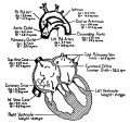
Adult heart blood flow cartoon
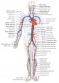
Adult human cardiovascular system cartoon
Fetal Blood Flow
Fetal Blood Flow
Fetal Blood Flow
Heart at birth


.jpg)
.jpg)


External Links
External Links Notice - The dynamic nature of the internet may mean that some of these listed links may no longer function. If the link no longer works search the web with the link text or name. Links to any external commercial sites are provided for information purposes only and should never be considered an endorsement. UNSW Embryology is provided as an educational resource with no clinical information or commercial affiliation.
- Australia Heart Foundation
- USA National Heart, Lung, and Blood Institute - Congenital Heart Defects | Heart and Vascular Information
| System Links: Introduction | Cardiovascular | Coelomic Cavity | Endocrine | Gastrointestinal Tract | Genital | Head | Immune | Integumentary | Musculoskeletal | Neural | Neural Crest | Placenta | Renal | Respiratory | Sensory | Birth |
Glossary Links
- Glossary: A | B | C | D | E | F | G | H | I | J | K | L | M | N | O | P | Q | R | S | T | U | V | W | X | Y | Z | Numbers | Symbols | Term Link
Cite this page: Hill, M.A. (2024, April 19) Embryology Cardiovascular System Development. Retrieved from https://embryology.med.unsw.edu.au/embryology/index.php/Cardiovascular_System_Development
- © Dr Mark Hill 2024, UNSW Embryology ISBN: 978 0 7334 2609 4 - UNSW CRICOS Provider Code No. 00098G