Cardiovascular System - Heart Valve Development: Difference between revisions
mNo edit summary |
mNo edit summary |
||
| Line 6: | Line 6: | ||
The heart valves form between the atria and ventricles (mitral valve, tricuspid valve) and between the atria and blood vessels (aortic valve, pulmonary valve). The cardiac cushions in the atrioventricular (AV) canal contain cells that are the primordia of the cardiac valves. The atrioventricular valves are attached to papillary muscles by chordae tendineae. | The heart valves form between the atria and ventricles (mitral valve, tricuspid valve) and between the atria and blood vessels (aortic valve, pulmonary valve). The cardiac cushions in the atrioventricular (AV) canal contain cells that are the primordia of the cardiac valves. The atrioventricular valves are attached to papillary muscles by chordae tendineae. | ||
Scleraxis (SCX) is a transcription factor involved in tendon and ligament development and has been identified as also expressed in early heart valve development. | Scleraxis (SCX) is a transcription factor involved in tendon and ligament development and has been identified as also expressed in early heart valve development.{{#pmid:24983472|PMID24983472}} | ||
Mitral valve also called the "bicuspid valve". | Mitral valve also called the "bicuspid valve". | ||
| Line 19: | Line 19: | ||
|-bgcolor="F5FAFF" | |-bgcolor="F5FAFF" | ||
| | | | ||
* '''Twist1 directly regulates genes that promote cell proliferation and migration in developing heart valves''' | * '''Review - Calcific Aortic Valve Disease: a Developmental Biology Perspective'''{{#pmid:29520694|PMID29520694}} "Calcification of the aortic valve is an active process characterized by calcific nodule formation on the aortic surface leading to a less supple and more stiffened cusp, thereby limiting movement and causing clinical stenosis. The mechanisms underlying these pathogenic changes are largely unknown, but emerging studies have suggested that signaling pathways common to valvulogenesis and bone development play significant roles and include Transforming Growth Factor-β (TGF-β), bone morphogenetic protein (BMP), Wnt, Notch, and Sox9." | ||
* '''Hemodynamic patterning of the avian atrioventricular valve''' | |||
* '''Twist1 directly regulates genes that promote cell proliferation and migration in developing heart valves'''{{#pmid:22242143|PMID22242143}} "Twist1, a basic helix-loop-helix transcription factor, is expressed in mesenchymal precursor populations during embryogenesis and in metastatic cancer cells. In the developing heart, Twist1 is highly expressed in endocardial cushion (ECC) valve mesenchymal cells and is down regulated during valve differentiation and remodeling. Previous studies demonstrated that Twist1 promotes cell proliferation, migration, and expression of primitive extracellular matrix (ECM) molecules in ECC mesenchymal cells. Furthermore, Twist1 expression is induced in human pediatric and adult diseased heart valves." | |||
* '''Hemodynamic patterning of the avian atrioventricular valve'''{{#pmid:21181939|PMID21181939}} "In this study, we develop an innovative approach to rigorously quantify the evolving hemodynamic environment of the atrioventricular (AV) canal of avian embryos. Ultrasound generated velocity profiles were imported into Micro-Computed Tomography generated anatomically precise cardiac geometries between Hamburger-Hamilton (HH) stages 17 and 30. Computational fluid dynamic simulations were then conducted and iterated until results mimicked in vivo observations. Blood flow in tubular hearts (HH17) was laminar with parallel streamlines, but strong vortices developed simultaneous with expansion of the cushions and septal walls. For all investigated stages, highest wall shear stresses (WSS) are localized to AV canal valve-forming regions. Peak WSS increased from 19.34 dynes/cm(2) at HH17 to 287.18 dynes/cm(2) at HH30, but spatiotemporally averaged WSS became 3.62 dynes/cm(2) for HH17 to 9.11 dynes/cm(2) for HH30. Hemodynamic changes often preceded and correlated with morphological changes. These results establish a quantitative baseline supporting future hemodynamic analyses and interpretations." | |||
|} | |} | ||
{| class="wikitable mw-collapsible mw-collapsed" | {| class="wikitable mw-collapsible mw-collapsed" | ||
| Line 61: | Line 64: | ||
==Molecular== | ==Molecular== | ||
'''Scleraxis''' (Scx) - basic helix–loop–helix transcription factor expressed in the progenitors and cells of all tendon tissues (mouse). | '''Scleraxis''' (Scx) - basic helix–loop–helix transcription factor expressed in the progenitors and cells of all tendon tissues (mouse).{{#pmid:18802027|PMID18802027}} | ||
'''Periostin''' - regulates lineage commitment of valve precursor cells (chicken). | '''Periostin''' - regulates lineage commitment of valve precursor cells (chicken).{{#pmid:19334280|PMID19334280}} | ||
Gata4 and Gata6 | Gata4 and Gata6 | ||
| Line 96: | Line 99: | ||
===Noonan syndrome=== | ===Noonan syndrome=== | ||
An autosomal dominant single-gene cause of congenital heart disease. Patients also have proportionate short stature, facial abnormalities, and an increased risk of myeloproliferative disease. About half the patients have mutations in PTPN11, encoding the protein tyrosine phosphatase SHP2. A recent study in mice has identified PTPN11 acting in endocardium to enhance endocardial-mesenchymal transformation. | An autosomal dominant single-gene cause of congenital heart disease. Patients also have proportionate short stature, facial abnormalities, and an increased risk of myeloproliferative disease. About half the patients have mutations in PTPN11, encoding the protein tyrosine phosphatase SHP2. A recent study in mice has identified PTPN11 acting in endocardium to enhance endocardial-mesenchymal transformation.{{#pmid:19251646|PMID19251646}} | ||
===Calcific Aortic Valve Disease=== | |||
Calcification of the aortic valve shows calcific nodule formation on the aortic surface, leading to a less supple and more stiffened cusp, limiting movement and causing clinical stenosis.{{#pmid:29520694|PMID29520694}} | |||
Pathogenic mechanism is unknown but may involve signaling pathways common to valvulogenesis and bone development: | |||
* Transforming Growth Factor-β (TGF-β) | |||
* bone morphogenetic protein (BMP) | |||
* Wnt | |||
* Notch | |||
* Sox9 | |||
{{Factors}} | |||
==References== | ==References== | ||
| Line 102: | Line 117: | ||
===Reviews=== | ===Reviews=== | ||
{{#pmid:20809794}} | |||
{{#pmid:20201901}} | |||
{{#pmid:14567955}} | |||
{{#pmid:12768658}} | |||
===Articles=== | ===Articles=== | ||
{{#pmid:17549728}} | |||
{{#pmid:16914500}} | |||
{{#pmid:17104787}} | |||
===Search PubMed=== | ===Search PubMed=== | ||
Revision as of 09:28, 13 March 2018
| Embryology - 20 Apr 2024 |
|---|
| Google Translate - select your language from the list shown below (this will open a new external page) |
|
العربية | català | 中文 | 中國傳統的 | français | Deutsche | עִברִית | हिंदी | bahasa Indonesia | italiano | 日本語 | 한국어 | မြန်မာ | Pilipino | Polskie | português | ਪੰਜਾਬੀ ਦੇ | Română | русский | Español | Swahili | Svensk | ไทย | Türkçe | اردو | ייִדיש | Tiếng Việt These external translations are automated and may not be accurate. (More? About Translations) |
Introduction


The heart valves form between the atria and ventricles (mitral valve, tricuspid valve) and between the atria and blood vessels (aortic valve, pulmonary valve). The cardiac cushions in the atrioventricular (AV) canal contain cells that are the primordia of the cardiac valves. The atrioventricular valves are attached to papillary muscles by chordae tendineae.
Scleraxis (SCX) is a transcription factor involved in tendon and ligament development and has been identified as also expressed in early heart valve development.[1]
Mitral valve also called the "bicuspid valve".
Some Recent Findings
|
| More recent papers |
|---|
 This table allows an automated computer search of the external PubMed database using the listed "Search term" text link.
More? References | Discussion Page | Journal Searches | 2019 References | 2020 References Search term: Heart Valve Embryology <pubmed limit=5>Heart Valve Embryology</pubmed> |
Textbooks
- Human Embryology (2nd ed.) Larson Ch7 p151-188 Heart
- The Developing Human: Clinically Oriented Embryology (6th ed.) Moore and Persaud Ch14: p304-349
- Before we Are Born (5th ed.) Moore and Persaud Ch12; p241-254
- Essentials of Human Embryology Larson Ch7 p97-122 Heart
- Human Embryology Fitzgerald and Fitzgerald Ch13-17: p77-111
Tutorial Images
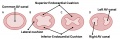
Canal Division (Superior View)
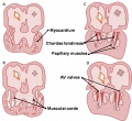
Development of the mitral and tricuspid valves
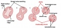
Development of the semilunar valves
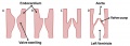
Development of the semilunar cusps


Fetal Heart Valve Sounds
<mp3player>File:Week17 fetal heart rate.mp3</mp3player>
Audio recording of the Second Trimester fetal heart (GA week 17).
The characteristic "lub-dup" sounds are associated with closing of heart valves.
- First sound (lub) occurs as atrioventricular valves close and signifies beginning of systole (contraction)
- Second sound (dup) occurs when semilunar valves close at the beginning of ventricular diastole (relaxation)
- Links: Fetal Heart Sounds Audio
Molecular
Scleraxis (Scx) - basic helix–loop–helix transcription factor expressed in the progenitors and cells of all tendon tissues (mouse).[5]
Periostin - regulates lineage commitment of valve precursor cells (chicken).[6]
Gata4 and Gata6
Tbx5
Images
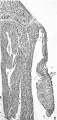
Historic
| Historic Disclaimer - information about historic embryology pages |
|---|
|
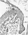
Odgers PNB. The development of the atrio-ventricular valves in man. (1939) J Anat. 73: 643-57. PMID 17104787
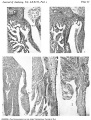
Plate 1
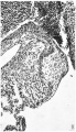
1 right A.-V. orifice in an 11.2 mm. embryo
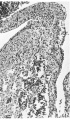
2 right lateral cusp in a 17.5 mm. embryo
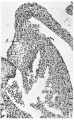
3 left lateral cusp in a 17.5 mm. embryo
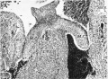
4 central cusps in a 23 mm. embryo
5 right lateral cusp in a 28.5 mm. embryo
Plate 2
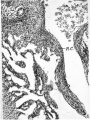
6 right lateral cusp in a 46 mm. embryo
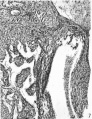
7 right lateral cusp in a 61 mm. embryo
8 right lateral cusp in an 85 mm. foetus
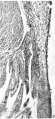
9 right lateral cusp in a 210 mm. foetus
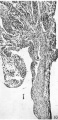
right lateral cusp of a full time foetus


Abnormalities
Noonan syndrome
An autosomal dominant single-gene cause of congenital heart disease. Patients also have proportionate short stature, facial abnormalities, and an increased risk of myeloproliferative disease. About half the patients have mutations in PTPN11, encoding the protein tyrosine phosphatase SHP2. A recent study in mice has identified PTPN11 acting in endocardium to enhance endocardial-mesenchymal transformation.[7]
Calcific Aortic Valve Disease
Calcification of the aortic valve shows calcific nodule formation on the aortic surface, leading to a less supple and more stiffened cusp, limiting movement and causing clinical stenosis.[2]
Pathogenic mechanism is unknown but may involve signaling pathways common to valvulogenesis and bone development:
- Transforming Growth Factor-β (TGF-β)
- bone morphogenetic protein (BMP)
- Wnt
- Notch
- Sox9
References
- ↑ Barnette DN, VandeKopple M, Wu Y, Willoughby DA & Lincoln J. (2014). RNA-seq analysis to identify novel roles of scleraxis during embryonic mouse heart valve remodeling. PLoS ONE , 9, e101425. PMID: 24983472 DOI.
- ↑ 2.0 2.1 Dutta P & Lincoln J. (2018). Calcific Aortic Valve Disease: a Developmental Biology Perspective. Curr Cardiol Rep , 20, 21. PMID: 29520694 DOI.
- ↑ Lee MP & Yutzey KE. (2011). Twist1 directly regulates genes that promote cell proliferation and migration in developing heart valves. PLoS ONE , 6, e29758. PMID: 22242143 DOI.
- ↑ Yalcin HC, Shekhar A, McQuinn TC & Butcher JT. (2011). Hemodynamic patterning of the avian atrioventricular valve. Dev. Dyn. , 240, 23-35. PMID: 21181939 DOI.
- ↑ Levay AK, Peacock JD, Lu Y, Koch M, Hinton RB, Kadler KE & Lincoln J. (2008). Scleraxis is required for cell lineage differentiation and extracellular matrix remodeling during murine heart valve formation in vivo. Circ. Res. , 103, 948-56. PMID: 18802027 DOI.
- ↑ Norris RA, Potts JD, Yost MJ, Junor L, Brooks T, Tan H, Hoffman S, Hart MM, Kern MJ, Damon B, Markwald RR & Goodwin RL. (2009). Periostin promotes a fibroblastic lineage pathway in atrioventricular valve progenitor cells. Dev. Dyn. , 238, 1052-63. PMID: 19334280 DOI.
- ↑ Araki T, Chan G, Newbigging S, Morikawa L, Bronson RT & Neel BG. (2009). Noonan syndrome cardiac defects are caused by PTPN11 acting in endocardium to enhance endocardial-mesenchymal transformation. Proc. Natl. Acad. Sci. U.S.A. , 106, 4736-41. PMID: 19251646 DOI.
Reviews
Hinton RB & Yutzey KE. (2011). Heart valve structure and function in development and disease. Annu. Rev. Physiol. , 73, 29-46. PMID: 20809794 DOI.
Markwald RR, Norris RA, Moreno-Rodriguez R & Levine RA. (2010). Developmental basis of adult cardiovascular diseases: valvular heart diseases. Ann. N. Y. Acad. Sci. , 1188, 177-83. PMID: 20201901 DOI.
Délot EC. (2003). Control of endocardial cushion and cardiac valve maturation by BMP signaling pathways. Mol. Genet. Metab. , 80, 27-35. PMID: 14567955
Barnett JV & Desgrosellier JS. (2003). Early events in valvulogenesis: a signaling perspective. Birth Defects Res. C Embryo Today , 69, 58-72. PMID: 12768658 DOI.
Articles
van den Berg G, Somi S, Buffing AA, Moorman AF & van den Hoff MJ. (2007). Patterns of expression of the Follistatin and Follistatin-like1 genes during chicken heart development: a potential role in valvulogenesis and late heart muscle cell formation. Anat Rec (Hoboken) , 290, 783-7. PMID: 17549728 DOI.
Rivera-Feliciano J, Lee KH, Kong SW, Rajagopal S, Ma Q, Springer Z, Izumo S, Tabin CJ & Pu WT. (2006). Development of heart valves requires Gata4 expression in endothelial-derived cells. Development , 133, 3607-18. PMID: 16914500 DOI.
Odgers PN. (1939). The development of the atrio-ventricular valves in man. J. Anat. , 73, 643-57. PMID: 17104787
Search PubMed
Search Pubmed: heart valve development | heart valve morphogenesis | Valvulogenesis
External Links
External Links Notice - The dynamic nature of the internet may mean that some of these listed links may no longer function. If the link no longer works search the web with the link text or name. Links to any external commercial sites are provided for information purposes only and should never be considered an endorsement. UNSW Embryology is provided as an educational resource with no clinical information or commercial affiliation.
Glossary Links
- Glossary: A | B | C | D | E | F | G | H | I | J | K | L | M | N | O | P | Q | R | S | T | U | V | W | X | Y | Z | Numbers | Symbols | Term Link
Cite this page: Hill, M.A. (2024, April 20) Embryology Cardiovascular System - Heart Valve Development. Retrieved from https://embryology.med.unsw.edu.au/embryology/index.php/Cardiovascular_System_-_Heart_Valve_Development
- © Dr Mark Hill 2024, UNSW Embryology ISBN: 978 0 7334 2609 4 - UNSW CRICOS Provider Code No. 00098G