Cardiovascular System - Heart Development: Difference between revisions
mNo edit summary |
mNo edit summary |
||
| Line 1: | Line 1: | ||
{{Header}} | |||
==Introduction== | ==Introduction== | ||
[[File:Human-heart-E3L.jpg|thumb|300px|Human embryo heart (day 54 - 56)]]In human embryos the heart begins to beat at about 22-23 days, with blood flow beginning in the 4th week. The heart is therefore one of the earliest differentiating and functioning organs. | [[File:Human-heart-E3L.jpg|thumb|300px|Human embryo heart (day 54 - 56)]]In human embryos the heart begins to beat at about 22-23 days, with blood flow beginning in the 4th week. The heart is therefore one of the earliest differentiating and functioning organs. | ||
The best place for students to start is the [[Cardiac_Embryology|Heart Tutorial]]. | |||
The heart begins very early in mesoderm within the trilaminar embryonic disc. The heart forms initially in the embryonic disc as a simple paired tube inside the forming pericardial cavity, which when the disc folds, gets carried into the correct anatomical position in the chest cavity. | The heart begins very early in mesoderm within the trilaminar embryonic disc. The heart forms initially in the embryonic disc as a simple paired tube inside the forming pericardial cavity, which when the disc folds, gets carried into the correct anatomical position in the chest cavity. | ||
| Line 136: | Line 139: | ||
<nowiki>*</nowiki> Table data is "embryonic age" while original reference used "gestational age" (from LMP). The stroke volume (SV) can be calculated from ultrasound measurement of end diastole volume (EDV) minus end systole volume (ESV); (SV = EDV - ESV). (Data: [http://www.ncbi.nlm.nih.gov:80/entrez/query.fcgi?cmd=Retrieve&db=pubmed&dopt=Abstract&list_uids=18634132 Molina FS, Faro C, Sotiriadis A, Dagklis T, Nicolaides KH.] Heart stroke volume and cardiac output by four-dimensional ultrasound in normal fetuses. Ultrasound Obstet Gynecol. 2008 Aug;32(2):181-7. | <nowiki>*</nowiki> Table data is "embryonic age" while original reference used "gestational age" (from LMP). The stroke volume (SV) can be calculated from ultrasound measurement of end diastole volume (EDV) minus end systole volume (ESV); (SV = EDV - ESV). (Data: [http://www.ncbi.nlm.nih.gov:80/entrez/query.fcgi?cmd=Retrieve&db=pubmed&dopt=Abstract&list_uids=18634132 Molina FS, Faro C, Sotiriadis A, Dagklis T, Nicolaides KH.] Heart stroke volume and cardiac output by four-dimensional ultrasound in normal fetuses. Ultrasound Obstet Gynecol. 2008 Aug;32(2):181-7. | ||
==Historic Images== | ==Additional Images== | ||
===Historic Images=== | |||
<gallery> | <gallery> | ||
File:Kollmann528.jpg|Fig. 528. Heart of a Rabbit Embryo seen from behind at 3.4 mm head length | File:Kollmann528.jpg|Fig. 528. Heart of a Rabbit Embryo seen from behind at 3.4 mm head length | ||
| Line 154: | Line 159: | ||
<references/> | <references/> | ||
<pubmed>15797462</pubmed> | |||
<pubmed>15977172</pubmed> | |||
<pubmed>12807866</pubmed> | |||
<pubmed>12860885</pubmed> | |||
<pubmed>12781678</pubmed> | |||
<pubmed>11891982</pubmed> | |||
<pubmed>11920381</pubmed> | |||
<pubmed>10948449</pubmed> | |||
| Line 174: | Line 179: | ||
{{ | {{Glossary}} | ||
{{Footer}} | |||
[[Category:Cardiovascular]] [[Category:Heart]] | [[Category:Cardiovascular]] [[Category:Heart]] | ||
Revision as of 12:31, 13 October 2014
| Embryology - 19 Apr 2024 |
|---|
| Google Translate - select your language from the list shown below (this will open a new external page) |
|
العربية | català | 中文 | 中國傳統的 | français | Deutsche | עִברִית | हिंदी | bahasa Indonesia | italiano | 日本語 | 한국어 | မြန်မာ | Pilipino | Polskie | português | ਪੰਜਾਬੀ ਦੇ | Română | русский | Español | Swahili | Svensk | ไทย | Türkçe | اردو | ייִדיש | Tiếng Việt These external translations are automated and may not be accurate. (More? About Translations) |
Introduction

In human embryos the heart begins to beat at about 22-23 days, with blood flow beginning in the 4th week. The heart is therefore one of the earliest differentiating and functioning organs.
The best place for students to start is the Heart Tutorial.
The heart begins very early in mesoderm within the trilaminar embryonic disc. The heart forms initially in the embryonic disc as a simple paired tube inside the forming pericardial cavity, which when the disc folds, gets carried into the correct anatomical position in the chest cavity.
A key aspect of heart development is the septation of the heart into separate chambers. As the embryonic/fetal circulation is different to the neonatal circulation (lung/pulmonary activation), several defects of heart septation may only become apparent on this transition. One septal "defect" occurs in us all, the foramen ovale (between the 2 atria) which in general closes in the neonate over time.
Embryonic Heart Rate (EHR), early in development the heart starts to spontaneously beat and a recent study by Wisser and Dirschedl in dated human embryos showed an increase up to 63 postmenstrual days or 22 mm greatest length. Thereafter a steady decrease of EHR was noted. Maximal EHR is reached when morphological development of the embryonic heart is completed.
The best place for students to start is the Heart Tutorial.
Basic Heart
Advanced Heart
Textbooks

|
Citation: UNSW Embryology (13th ed.) Hill, M.A., 2013. Sydney: UNSW.
|

|
Citation: The Developing Human: Clinically Oriented Embryology 9th ed. Keith L. Moore, T.V.N. Persaud, Mark G. Torchia. Philadelphia, PA: Saunders, 2011. (links available to UNSW students) |

|
Citation: Larsen's Human Embryology 4th ed. Schoenwolf, Gary C; Larsen, William J, (William James). Philadelphia, PA : Elsevier/Churchill Livingstone, c2009. (links available to UNSW students) |
- Human Embryology (2nd ed.) Larson Ch7 p151-188 Heart
- The Developing Human: Clinically Oriented Embryology (6th ed.) Moore and Persaud Ch14: p304-349
- Before we Are Born (5th ed.) Moore and Persaud Ch12; p241-254
- Essentials of Human Embryology Larson Ch7 p97-122 Heart
- Human Embryology Fitzgerald and Fitzgerald Ch13-17: p77-111
- Links: Embryology Textbooks
Movies
Cardiovascular System Development | Heart Tutorial
|
|
|
|
|
| Heart Cartoons | |||||||||||||||||||||||||||
|---|---|---|---|---|---|---|---|---|---|---|---|---|---|---|---|---|---|---|---|---|---|---|---|---|---|---|---|
|
|
|
|
|
|
|
|
|
|
| Historic Animations | |||||||||||||||
|---|---|---|---|---|---|---|---|---|---|---|---|---|---|---|---|
|
|
|
| ||||||||||||
|
|
|
|
| About Historic Animations | ||||
|---|---|---|---|---|
 The sound quality is quite poor and some of the information is now out of date, most general concepts are still correct. Please note the relatively large size (Mb) of each excerpt will effect download and viewing. March 2013
|
Human Development Timecourse

.jpg)
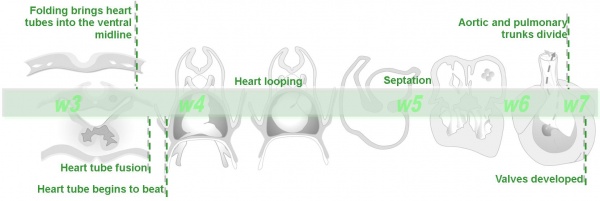
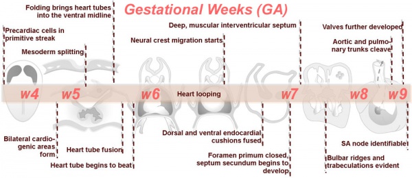
- Cardiogenic region - in splanchnic mesenchyme of prechordal plate region
- Week 2 pair of thin-walled tubes
- Week 3 tubes fused, truncus arteriosus outflow, heart contracting
- Week 4 heart tube continues to elongate, curving to form S shape
- Week 5 Septation starts, atrial and ventricular
- Septation continues, atrial septa remains open, foramen ovale
- Week 40 At birth pressure difference closes foramen ovale leaving a fossa ovalis
Link: Timeline human development
Heart Layers
- pericardium - covers the heart, formed by 3 layers consisting of a fibrous pericardium and a double layered serous pericardium (parietal layer and visceral epicardium layer).
- myocardium - muscular wall of the heart, thickest layer formed by spirally arranged cardiac muscle cells.
- endocardium - lines the heart, epithelial tissue lining the inner surface of heart chambers and valves.
Heart Volume
| Week* | Heart Volume (ml) | Lung Volume (ml) |
| 10 | 0.6 | 1.6 |
| 18 | 4.3 | 10.9 |
| 30 | 26.6 | 49.3 |
* Table data is "embryonic age" while original reference used "gestational age" (from LMP)(Data: Peralta CF, Cavoretto P, Csapo B, Falcon O, Nicolaides KH. Lung and heart volumes by three-dimensional ultrasound in normal fetuses at 12-32 weeks' gestation. Ultrasound Obstet Gynecol. 2006 Feb;27(2):128-33.)
Stroke Volume
| Week* | Left Stroke Volume (ml) | Right Stroke Volume (ml) | Cardiac Output L/R (ml/min) |
| 10 | 0.02 | 0.01 | 2.39 / 1.8 |
| 18 | 0.32 | 0.30 | 43.46 / 46.72 |
| 32 | 2.07 | 2.67 | 284.71 / 365.99 |
* Table data is "embryonic age" while original reference used "gestational age" (from LMP). The stroke volume (SV) can be calculated from ultrasound measurement of end diastole volume (EDV) minus end systole volume (ESV); (SV = EDV - ESV). (Data: Molina FS, Faro C, Sotiriadis A, Dagklis T, Nicolaides KH. Heart stroke volume and cardiac output by four-dimensional ultrasound in normal fetuses. Ultrasound Obstet Gynecol. 2008 Aug;32(2):181-7.
Additional Images
Historic Images
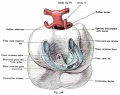
Fig. 528. Heart of a Rabbit Embryo seen from behind at 3.4 mm head length
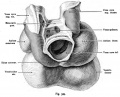
Fig. 529. The heart of a 24 mm Embryo
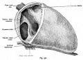
Fig. 530. Fetal heart (6 months) in normal situation
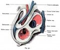
Fig. 531. Heart included in the pericardium of a human embryo of 7.5 mm body length
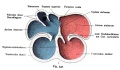
Fig. 532. Development of the heart chambers and septa
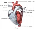
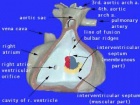
Fig. 533. Heart of a newborn viewed from the front and placed in the vertical direction
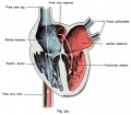
Fig. 534. Fetal heart, dorsal half with the afferent paths, open and colored according to the physiological condition of the blood
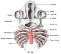
Fig. 535. The aortic arch in the shark embryo (Pristiurus)
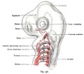
Fig. 536. The arteries of the gill arch region of a shark embryo (Pristiurus)
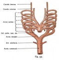
Fig. 537. Aortic arch of mammals and man
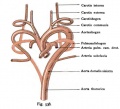
Fig. 538. Arteries in mammals and humans from the Aortic Arch


References
- ↑ <pubmed>20571530</pubmed>| Mol Syst Biol.
<pubmed>15797462</pubmed> <pubmed>15977172</pubmed> <pubmed>12807866</pubmed> <pubmed>12860885</pubmed> <pubmed>12781678</pubmed> <pubmed>11891982</pubmed> <pubmed>11920381</pubmed> <pubmed>10948449</pubmed>
Terms
- fibrous trigone - (trigonum fibrosum) term describing the dense connective tissue between the aortic ring and the atrioventricular ring and has a left and right component. The right fibrous trigone (trigonum fibrosum dextrum) lies between the aortic ring and the right atrioventricular ring. The left fibrous trigone (trigonum fibrosum sinistrum) lies between the aortic ring and the left atrioventricular ring.
External Links
External Links Notice - The dynamic nature of the internet may mean that some of these listed links may no longer function. If the link no longer works search the web with the link text or name. Links to any external commercial sites are provided for information purposes only and should never be considered an endorsement. UNSW Embryology is provided as an educational resource with no clinical information or commercial affiliation.
- Embryo Images - Embryo Images Online Early Cell Populations (cardiogenic section) | Cardiovascular Development | Week 3 Development | Week 4 Development | Heart Chambers and Outflow Tract | Atrioventricular Septation | Outflow Tract Septation | Ventricular Septation | Atrial Septation | Atrial Walls Aortic Arch Vessels | Changes at Birth
Glossary Links
- Glossary: A | B | C | D | E | F | G | H | I | J | K | L | M | N | O | P | Q | R | S | T | U | V | W | X | Y | Z | Numbers | Symbols | Term Link
Cite this page: Hill, M.A. (2024, April 19) Embryology Cardiovascular System - Heart Development. Retrieved from https://embryology.med.unsw.edu.au/embryology/index.php/Cardiovascular_System_-_Heart_Development
- © Dr Mark Hill 2024, UNSW Embryology ISBN: 978 0 7334 2609 4 - UNSW CRICOS Provider Code No. 00098G