Cardiovascular System - Double Outlet Right Ventricle: Difference between revisions

From Embryology
mNo edit summary
Line 7: Line 7:
* Arrangement of the atrioventricular valves and the ventriculoarterial connections are variable.
* Clinical manifestations variable.


{{Heart Abnormal}}
{{Heart Abnormal}}

Revision as of 16:23, 18 February 2017

Embryology - 19 Apr 2024    Facebook link Pinterest link Twitter link  Expand to Translate  
Google Translate - select your language from the list shown below (this will open a new external page)

العربية | català | 中文 | 中國傳統的 | français | Deutsche | עִברִית | हिंदी | bahasa Indonesia | italiano | 日本語 | 한국어 | မြန်မာ | Pilipino | Polskie | português | ਪੰਜਾਬੀ ਦੇ | Română | русский | Español | Swahili | Svensk | ไทย | Türkçe | اردو | ייִדיש | Tiếng Việt    These external translations are automated and may not be accurate. (More? About Translations)

Introduction

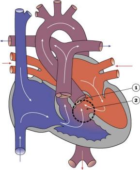
Double Outlet Right Ventricle cartoon
Double Outlet Right Ventricle
  • 1-1.5% of Congenital Heart Disease
  • Both large arteries arise wholly or mainly from the right ventricle.
  • Arrangement of the atrioventricular valves and the ventriculoarterial connections are variable.
  • Clinical manifestations variable.


Heart Abnormal: Tutorial Abnormalities | atrial septal defects | double outlet right ventricle | hypoplastic left heart | patent ductus arteriosus‎ | transposition of the great vessels | Tetralogy of Fallot | ventricular septal defects | coarctation of the aorta | Category ASD | Category PDA | Category ToF | Category VSD | ICD10 - Cardiovascular | ICD11


Cardiovascular Links: cardiovascular | Heart Tutorial | Lecture - Early Vascular | Lecture - Heart | Movies | 2016 Cardiac Review | heart | coronary circulation | heart valve | heart rate | Circulation | blood | blood vessel | blood vessel histology | heart histology | Lymphatic | ductus venosus | spleen | Stage 22 | cardiovascular abnormalities | OMIM | 2012 ECHO Meeting | Category:Cardiovascular
Historic Embryology - Cardiovascular 
1902 Vena cava inferior | 1905 Brain Blood Vessels | 1909 Cervical Veins | 1909 Dorsal aorta and umbilical veins | 1912 Heart | 1912 Human Heart | 1914 Earliest Blood-Vessels | 1915 Congenital Cardiac Disease | 1915 Dura Venous Sinuses | 1916 Blood cell origin | 1916 Pars Membranacea Septi | 1919 Lower Limb Arteries | 1921 Human Brain Vascular | 1921 Spleen | 1922 Aortic-Arch System | 1922 Pig Forelimb Arteries | 1922 Chicken Pulmonary | 1923 Head Subcutaneous Plexus | 1923 Ductus Venosus | 1925 Venous Development | 1927 Stage 11 Heart | 1928 Heart Blood Flow | 1935 Aorta | 1935 Venous valves | 1938 Pars Membranacea Septi | 1938 Foramen Ovale | 1939 Atrio-Ventricular Valves | 1940 Vena cava inferior | 1940 Early Hematopoiesis | 1941 Blood Formation | 1942 Truncus and Conus Partitioning | Ziegler Heart Models | 1951 Heart Movie | 1954 Week 9 Heart | 1957 Cranial venous system | 1959 Brain Arterial Anastomoses | Historic Embryology Papers | 2012 ECHO Meeting | 2016 Cardiac Review | Historic Disclaimer

Some Recent Findings

  • Are There Head Volume Alterations at 11 to 14 Weeks in Fetuses with Congenital Heart Defects? A First Trimester Case Series[1] "This study aims to assess head volume (HV) alterations at 11 to 14 weeks in fetuses with congenital heart defects (CHD). Methods A retrospective case-control study on 100 normal and 26 CHD fetuses was conducted. ...Despite the small sample size, our case series suggests that alterations in HV may potentially be apparent as early as 11 to 14 weeks in CHD fetuses, particularly those with HLH. Larger prospective studies are needed to validate our findings."
More recent papers  
Mark Hill.jpg
PubMed logo.gif

This table allows an automated computer search of the external PubMed database using the listed "Search term" text link.

  • This search now requires a manual link as the original PubMed extension has been disabled.
  • The displayed list of references do not reflect any editorial selection of material based on content or relevance.
  • References also appear on this list based upon the date of the actual page viewing.


References listed on the rest of the content page and the associated discussion page (listed under the publication year sub-headings) do include some editorial selection based upon both relevance and availability.

More? References | Discussion Page | Journal Searches | 2019 References | 2020 References

Search term: Double Outlet Right Ventricle

<pubmed limit=5>Double Outlet Right Ventricle</pubmed>

Anatomy

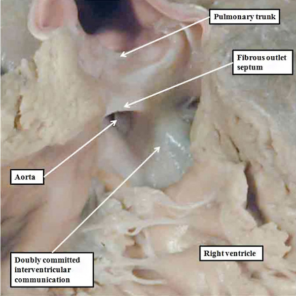
Anderson2016-fig47.jpg

fig 47 Double Outlet from Right Ventricle


References

  1. <pubmed>27308099</pubmed>

Reviews

<pubmed></pubmed> <pubmed></pubmed> <pubmed>25663264</pubmed> <pubmed>25836709</pubmed> <pubmed>25917992</pubmed> <pubmed>8489880</pubmed> <pubmed>2659180</pubmed>

Articles

<pubmed></pubmed> <pubmed></pubmed> <pubmed></pubmed> <pubmed></pubmed>

Search Pubmed

Search Pubmed: Search PubMed

External Links

External Links Notice - The dynamic nature of the internet may mean that some of these listed links may no longer function. If the link no longer works search the web with the link text or name. Links to any external commercial sites are provided for information purposes only and should never be considered an endorsement. UNSW Embryology is provided as an educational resource with no clinical information or commercial affiliation.