Cardiovascular System - Developmental Shunts: Difference between revisions
mNo edit summary |
|||
| (12 intermediate revisions by one other user not shown) | |||
| Line 1: | Line 1: | ||
{{Header}} | |||
<div style="background:#F5FFFA; border: 1px solid #CEF2E0; padding: 1em; margin: auto; width: 98%; float:left;"><div style="margin:0;background-color:#cef2e0;font-family:sans-serif;font-size:120%;font-weight:bold;border:1px solid #a3bfb1;text-align:left;color:#000;padding-left:0.4em;padding-top:0.2em;padding-bottom:0.2em;">Notice - Mark Hill</div>Currently this page is only a template and will be updated (this notice removed when completed).</div> | <div style="background:#F5FFFA; border: 1px solid #CEF2E0; padding: 1em; margin: auto; width: 98%; float:left;"><div style="margin:0;background-color:#cef2e0;font-family:sans-serif;font-size:120%;font-weight:bold;border:1px solid #a3bfb1;text-align:left;color:#000;padding-left:0.4em;padding-top:0.2em;padding-bottom:0.2em;">Notice - Mark Hill</div>Currently this page is only a template and will be updated (this notice removed when completed).</div> | ||
==Introduction== | ==Introduction== | ||
Before birth there are three identified "shunts" in the cardiovascular system: | |||
[[File:Stage_22_image_083.jpg|thumb|300px| Week 8 Human embryo (stage 22) Ductus Venosus]] | |||
Before birth there are three identified "shunts" in the mammalian cardiovascular system: | |||
# the foramen ovale, within the heart between the atria | # the foramen ovale, within the heart between the atria | ||
# the ductus arteriosus, within the aortic arch | # the ductus arteriosus, within the aortic arch | ||
| Line 9: | Line 14: | ||
{{Heart Links}} | |||
==Some Recent Findings== | ==Some Recent Findings== | ||
| Line 17: | Line 22: | ||
* '''Prenatal cardiovascular shunts in amniotic vertebrates'''<ref><pubmed>21513818</pubmed></ref> "During amniotic vertebrate development, the embryo and fetus employ a number of cardiovascular shunts. These shunts provide a right-to-left shunt of blood and are essential components of embryonic life ensuring proper blood circulation to developing organs and fetal gas exchanger, as well as bypassing the pulmonary circuit and the unventilated, fluid filled lungs. In this review we examine and compare the embryonic shunts available for fetal mammals and embryonic reptiles, including lizards, crocodilians, and birds. These groups have either a single ductus arteriosus (mammals) or paired ductus arteriosi that provide a right-to-left shunt of right ventricular output away from the unventilated lungs. The mammalian foramen ovale and the avian atrial foramina function as a right-to-left shunt of blood between the atria. The presence of atrial shunts in non-avian reptiles is unknown. Mammals have a venous shunt, the ductus venosus that diverts umbilical venous return away from the liver and towards the inferior vena cava and foramen ovale. Reptiles do not have a ductus venosus during the latter two thirds of development. While the fetal shunts are well characterized in numerous mammalian species, much less is known about the developmental physiology of the reptilian embryonic shunts. In the last years, the reactivity and the process of closure of the ductus arteriosus have been characterized in the chicken and the emu. In contrast, much less is known about embryonic shunts in the non-avian reptiles. It is possible that the single ventricle found in lizards, snakes, and turtles and the origin of the left aorta in the crocodilians play a significant role in the right-to-left embryonic shunt in these species." | * '''Prenatal cardiovascular shunts in amniotic vertebrates'''<ref><pubmed>21513818</pubmed></ref> "During amniotic vertebrate development, the embryo and fetus employ a number of cardiovascular shunts. These shunts provide a right-to-left shunt of blood and are essential components of embryonic life ensuring proper blood circulation to developing organs and fetal gas exchanger, as well as bypassing the pulmonary circuit and the unventilated, fluid filled lungs. In this review we examine and compare the embryonic shunts available for fetal mammals and embryonic reptiles, including lizards, crocodilians, and birds. These groups have either a single ductus arteriosus (mammals) or paired ductus arteriosi that provide a right-to-left shunt of right ventricular output away from the unventilated lungs. The mammalian foramen ovale and the avian atrial foramina function as a right-to-left shunt of blood between the atria. The presence of atrial shunts in non-avian reptiles is unknown. Mammals have a venous shunt, the ductus venosus that diverts umbilical venous return away from the liver and towards the inferior vena cava and foramen ovale. Reptiles do not have a ductus venosus during the latter two thirds of development. While the fetal shunts are well characterized in numerous mammalian species, much less is known about the developmental physiology of the reptilian embryonic shunts. In the last years, the reactivity and the process of closure of the ductus arteriosus have been characterized in the chicken and the emu. In contrast, much less is known about embryonic shunts in the non-avian reptiles. It is possible that the single ventricle found in lizards, snakes, and turtles and the origin of the left aorta in the crocodilians play a significant role in the right-to-left embryonic shunt in these species." | ||
* '''Preferential streaming of the ductus venosus and inferior caval vein towards the right heart is associated with left heart underdevelopment in human fetuses with left-sided diaphragmatic hernia'''<ref><pubmed>20702536</pubmed></ref> "Left heart underdevelopment is commonly observed in fetuses with left diaphragmatic hernia. This finding has been attributed to compression of the left atrium by herniated abdominal organs, redistribution of fetal cardiac output and/or low pulmonary venous return. As preferential right or left heart underdevelopment is usually not a feature of right diaphragmatic hernia, we searched for an alternative mechanism. Since in normal fetuses the major fraction of left heart filling is provided by the ductus venosus via the inferior caval vein and oval foramen, our study focused in particular on the streaming direction of these structures." | * '''Preferential streaming of the ductus venosus and inferior caval vein towards the right heart is associated with left heart underdevelopment in human fetuses with left-sided diaphragmatic hernia'''<ref><pubmed>20702536</pubmed></ref> "Left heart underdevelopment is commonly observed in fetuses with left diaphragmatic hernia. This finding has been attributed to compression of the left atrium by herniated abdominal organs, redistribution of fetal cardiac output and/or low pulmonary venous return. As preferential right or left heart underdevelopment is usually not a feature of right diaphragmatic hernia, we searched for an alternative mechanism. Since in normal fetuses the major fraction of left heart filling is provided by the ductus venosus via the inferior caval vein and oval foramen, our study focused in particular on the streaming direction of these structures." | ||
|} | |||
{| class="wikitable mw-collapsible mw-collapsed" | |||
! More recent papers | |||
|- | |||
| [[File:Mark_Hill.jpg|90px|left]] {{Most_Recent_Refs}} | |||
Search term: [http://www.ncbi.nlm.nih.gov/pubmed/?term=Cardiovascular+Developmental+Shunts ''Cardiovascular Developmental Shunts''] | |||
<pubmed limit=5>Cardiovascular Developmental Shunts</pubmed> | |||
Search term: [http://www.ncbi.nlm.nih.gov/pubmed/?term=ductus+venosus ''ductus venosus''] | |||
<pubmed limit=5>ductus venosus</pubmed> | |||
Search term: [http://www.ncbi.nlm.nih.gov/pubmed/?term=ductus+arteriosus ''ductus arteriosus''] | |||
<pubmed limit=5>ductus arteriosus</pubmed> | |||
Search term: [http://www.ncbi.nlm.nih.gov/pubmed/?term=foramen+ovale ''foramen ovale''] | |||
<pubmed limit=5>foramen ovale</pubmed> | |||
|} | |} | ||
==Foramen Ovale== | ==Foramen Ovale== | ||
{| | |||
[[File:Stage 13 image 068.jpg|300px]] | | [[File:Stage 13 image 068.jpg|300px]] | ||
| [[File:Stage 22 image 079.jpg|300px]] | |||
Ostium Primum (week 4, stage 13) | |- | ||
| Ostium Primum (week 4, stage 13) | |||
| Atrial septum (week 8, stage 22) | |||
|} | |||
==Ductus Arteriosus== | ==Ductus Arteriosus== | ||
| Line 32: | Line 61: | ||
==Ductus Venosus== | ==Ductus Venosus== | ||
The [[V#vitelline|vitelline]] blood vessel lying within the liver that connects (shunts) the portal and [[U#umbilical vein|umbilical veins]] to the inferior vena cava and also acts to protect the [[F#fetus|fetus]] from placental overcirculation. | |||
Absence can cause [[H#hydrops fetalis|hydrops fetalis]] and the umbilical vein then drains directly into the inferior vena cava or right atrium. | |||
Postnatally this shunt functionally closes then structurally closes and degenerates to form it the [[L#ligamentum venosus|ligamentum venosum]]. | |||
{| | {| | ||
| [[File:Stage 13 image 075.jpg|300px]] | | [[File:Stage 13 image 075.jpg|300px]] | ||
| Line 57: | Line 92: | ||
==Abnormalities== | ==Abnormalities== | ||
===Atrial Septal Defect=== | ===Atrial Septal Defect=== | ||
Foramen ovale | [[File:Atrial Septal Defect.jpg|thumb|Atrial Septal Defect]] | ||
* Foramen ovale defects are generally classes as atrial septal defects. | |||
* Atrial Septal Defects (ASD) are a group of common (1% of cardiac) congenital anomolies defects occuring in a number of different forms and more often in females. | |||
:'''Links:''' [[Cardiovascular_System_-_Abnormalities#Atrial_Septal_Defects|Atrial Septal Defects]] | |||
===Patent ductus arteriosus=== | ===Patent ductus arteriosus=== | ||
Ductus arteriosus | * Ductus arteriosus | ||
[[File:Patent Ductus Arteriosus.jpg|thumb|Patent ductus arteriosus]] | |||
[[File:Patent_ductus_arteriosus_classification.jpg|600px]] | |||
Patent ductus arteriosus classification | |||
:'''Links:''' [[Cardiovascular_System_-_Abnormalities#Patent_Ductus_Arteriosus|thumb|Patent Ductus Arteriosu]] | |||
==References== | ==References== | ||
| Line 79: | Line 127: | ||
'''Search Pubmed:''' [http://www.ncbi.nlm.nih.gov/sites/entrez?db=pubmed&cmd=search&term=heart%20shunt heart | '''Search Pubmed:''' [http://www.ncbi.nlm.nih.gov/sites/entrez?db=pubmed&cmd=search&term=foramen%20ovale foramen ovale] | [http://www.ncbi.nlm.nih.gov/sites/entrez?db=pubmed&cmd=search&term=ductus%20arteriosus ductus arteriosus] | [http://www.ncbi.nlm.nih.gov/sites/entrez?db=pubmed&cmd=search&term=ductus%20venosus ductus venosus] | [http://www.ncbi.nlm.nih.gov/sites/entrez?db=pubmed&cmd=search&term=heart%20shunt heart shunt] | [http://www.ncbi.nlm.nih.gov/sites/entrez?db=pubmed&cmd=search&term=cardiovascular%20shunts cardiovascular shunts] | ||
Latest revision as of 13:22, 17 September 2015
| Embryology - 16 Apr 2024 |
|---|
| Google Translate - select your language from the list shown below (this will open a new external page) |
|
العربية | català | 中文 | 中國傳統的 | français | Deutsche | עִברִית | हिंदी | bahasa Indonesia | italiano | 日本語 | 한국어 | မြန်မာ | Pilipino | Polskie | português | ਪੰਜਾਬੀ ਦੇ | Română | русский | Español | Swahili | Svensk | ไทย | Türkçe | اردو | ייִדיש | Tiếng Việt These external translations are automated and may not be accurate. (More? About Translations) |
Introduction
Before birth there are three identified "shunts" in the mammalian cardiovascular system:
- the foramen ovale, within the heart between the atria
- the ductus arteriosus, within the aortic arch
- the ductus venosus, within the liver
Some Recent Findings
|
| More recent papers |
|---|
|
This table allows an automated computer search of the external PubMed database using the listed "Search term" text link.
More? References | Discussion Page | Journal Searches | 2019 References | 2020 References Search term: Cardiovascular Developmental Shunts <pubmed limit=5>Cardiovascular Developmental Shunts</pubmed> Search term: ductus venosus <pubmed limit=5>ductus venosus</pubmed> Search term: ductus arteriosus <pubmed limit=5>ductus arteriosus</pubmed> Search term: foramen ovale <pubmed limit=5>foramen ovale</pubmed> |
Foramen Ovale
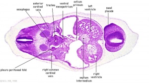
|
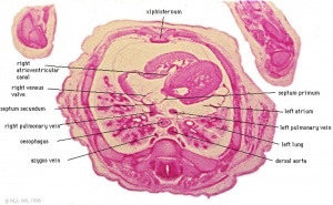
|
| Ostium Primum (week 4, stage 13) | Atrial septum (week 8, stage 22) |
Ductus Arteriosus
Week 8 (stage 22) Aortic Arch
Ductus Venosus
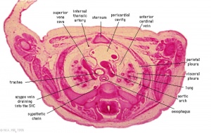
The vitelline blood vessel lying within the liver that connects (shunts) the portal and umbilical veins to the inferior vena cava and also acts to protect the fetus from placental overcirculation.
Absence can cause hydrops fetalis and the umbilical vein then drains directly into the inferior vena cava or right atrium.
Postnatally this shunt functionally closes then structurally closes and degenerates to form it the ligamentum venosum.
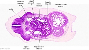
|
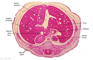
|
| Week 4 embryo (stage 13) Ductus Venosus | Week 8 Human embryo (stage 22) Ductus Venosus |
11254155
8841232
Textbooks
- Human Embryology (2nd ed.) Larson Ch7 p151-188 Heart
- The Developing Human: Clinically Oriented Embryology (6th ed.) Moore and Persaud Ch14: p304-349
- Before we Are Born (5th ed.) Moore and Persaud Ch12; p241-254
- Essentials of Human Embryology Larson Ch7 p97-122 Heart
- Human Embryology Fitzgerald and Fitzgerald Ch13-17: p77-111
Molecular
Abnormalities
Atrial Septal Defect
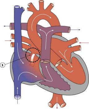
- Foramen ovale defects are generally classes as atrial septal defects.
- Atrial Septal Defects (ASD) are a group of common (1% of cardiac) congenital anomolies defects occuring in a number of different forms and more often in females.
- Links: Atrial Septal Defects
Patent ductus arteriosus
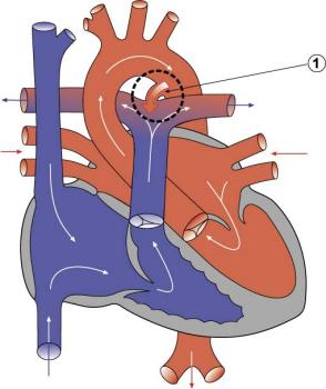
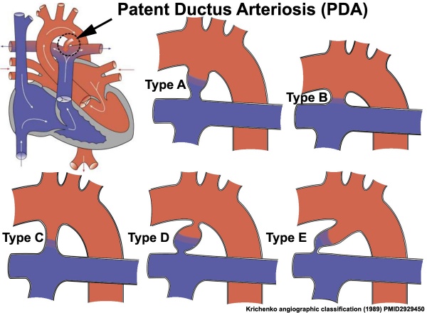
- Ductus arteriosus
Patent ductus arteriosus classification
References
Reviews
<pubmed>21513818</pubmed>
Articles
<pubmed>16565980</pubmed> <pubmed>12589721</pubmed> <pubmed>6832717</pubmed>
17984953
Search PubMed
Search Pubmed: foramen ovale | ductus arteriosus | ductus venosus | heart shunt | cardiovascular shunts
Glossary Links
- Glossary: A | B | C | D | E | F | G | H | I | J | K | L | M | N | O | P | Q | R | S | T | U | V | W | X | Y | Z | Numbers | Symbols | Term Link
Cite this page: Hill, M.A. (2024, April 16) Embryology Cardiovascular System - Developmental Shunts. Retrieved from https://embryology.med.unsw.edu.au/embryology/index.php/Cardiovascular_System_-_Developmental_Shunts
- © Dr Mark Hill 2024, UNSW Embryology ISBN: 978 0 7334 2609 4 - UNSW CRICOS Provider Code No. 00098G

