|
|
| Line 11: |
Line 11: |
| :{{Template:Heart Links}} | [http://embryology.med.unsw.edu.au/Notes/heart.htm original page] | | :{{Template:Heart Links}} | [http://embryology.med.unsw.edu.au/Notes/heart.htm original page] |
|
| |
|
|
| |
| :{{Template:Systems}}
| |
|
| |
|
| ==Some Recent Findings== | | ==Some Recent Findings== |
| Line 21: |
Line 19: |
| |} | | |} |
|
| |
|
| ==Textbooks==
| |
| [[File:Cardiac_muscle_histology.jpg|thumb|Cardiac_muscle_histology]]
| |
|
| |
| * '''Human Embryology''' (2nd ed.) Larson Ch7 p151-188 Heart, Ch8 p189-228 Vasculature
| |
| * '''The Developing Human: Clinically Oriented Embryology''' (6th ed.) Moore and Persaud Ch14: p304-349
| |
| * '''Before we Are Born''' (5th ed.) Moore and Persaud Ch12; p241-254
| |
| * '''Essentials of Human Embryology''' Larson Ch7 p97-122 Heart, Ch8 p123-146 Vasculature
| |
| * '''Human Embryology''' Fitzgerald and Fitzgerald Ch13-17: p77-111
| |
|
| |
| ==Timecourse==
| |
| {|
| |
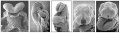
| | [[File:Human heart SEM1.jpg|The Human Heart from day 10 to 25 (scanning electron micrograph)]]
| |
| |-
| |
| | The Human Heart from day 10 to 25 (scanning electron micrograph)
| |
| |}
| |
|
| |
| * forms initially in splanchnic mesoderm of prechordal plate region - cardiogenic region
| |
| ** growth and folding of the embryo moves heart ventrally and downward into anatomical position
| |
| * day 22-23, begins to beat in Humans
| |
| * heart tube connects to blood vessels forming in splanchnic and extraembryonic mesoderm
| |
|
| |
| * Week 2-3 pair of thin-walled tubes
| |
| * Week 3 paired heart tubes fuse, truncus arteriosus outflow, heart contracting
| |
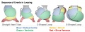
| * Week 4 heart tube continues to elongate, curving to form S shape
| |
| * Week 5 Septation starts, atrial and ventricular
| |
|
| |
|
| * Septation continues, atrial septa remains open, foramen ovale
| |
|
| |
| * Week 37-38 At birth, pressure difference closes foramen ovale leaving a fossa ovalis
| |
|
| |
|
| |
| ==Heart Development Movies==
| |
| Animations showing aspects of heart development.
| |
| {| border='0px'
| |
|
| |
| |-
| |
| | [[File:Heart1_looping icon.jpg|90px|link=Development_Animation_-_Heart Looping]]
| |
| | [[File:Heart1_realign icon.jpg|90px|link=Development_Animation_-_Heart Realign]]
| |
| | [[File:Heart1_atrium icon.jpg|90px|link=Development_Animation_-_Heart Atrial Septation]]
| |
| | [[File:heart1 ventricle icon.jpg|90px|link=Development_Animation_-_Heart Outflow Septation]]
| |
| |-
| |
| | [[Development_Animation_-_Heart Looping|Heart Looping]]
| |
| | [[Development_Animation_-_Heart Realign|Heart Realign]]
| |
| | [[Development_Animation_-_Heart Atrial Septation|Heart Atrial Septation]]
| |
| | [[Development_Animation_-_Heart Outflow Septation|Heart Outflow Septation]]
| |
| |-
| |
| |}
| |
|
| |
| {| border='0px'
| |
|
| |
| |-
| |
| | [[File:Stage13-CVS-icon.jpg|90px|link=Movie_-_Cardiovascular_3D_stage_13]]
| |
| | [[File:Stage22-CVS-icon.jpg|90px|link=Movie_-_Cardiovascular_3D_stage_22]]
| |
| |-
| |
| | [[Movie_-_Cardiovascular_3D_stage_13|Stage 13]]
| |
| | [[Movie_-_Cardiovascular_3D_stage_22|Stage 22]]
| |
| |-
| |
| |}
| |
|
| |
| Pages within the [[Cardiac_Embryology|online Cardiac tutorial]].
| |
|
| |
| {| border='0px'
| |
|
| |
| |-
| |
| | [[File:Heart_fields_001_icon.jpg|90px|link=Advanced_-_Heart_Fields]]
| |
| | [[File:Heart folding 002 icon.jpg|90px|link=Basic - Primitive Heart Tube]]
| |
| | [[File:Heart_folding_001_icon.jpg|90px|link=Advanced_-_Heart_Tubes]]
| |
| | [[File:Heart looping 006 icon.jpg|90px|link=Advanced - Cardiac Looping]]
| |
| | [[File:Heart septation 001 icon.jpg|90px|link=Advanced - Cardiac Septation]]
| |
| | [[File:Outflow tract 001 icon.jpg|90px|link=Advanced - Outflow Tract]]
| |
| |-
| |
| | [[Advanced_-_Heart_Fields|Heart Fields]]
| |
| | [[Basic - Primitive Heart Tube|Primitive Heart Tube]]
| |
| | [[Advanced_-_Heart_Tubes|Heart Tubes]]
| |
| | [[Advanced - Cardiac Looping|Cardiac Looping]]
| |
| | [[Advanced - Cardiac Septation|Cardiac Septation]]
| |
| | [[Advanced - Outflow Tract|Outflow Tract]]
| |
| |-
| |
| |}
| |
|
| |
|
| |
|
| |
| Historic animations including audio descriptions. Some of these descriptions may be currently inaccurate, the transfer is from an old class film and the audio track is of very poor quality.
| |
|
| |
| {| border='0px'
| |
|
| |
| |-
| |
| | [[File:Heart_historic_001 icon.jpg|90px|link=Historic_Animation_-_Heart_01]]
| |
| | [[File:Heart_historic_002 icon.jpg|90px|link=Historic_Animation_-_Heart_02]]
| |
| | [[File:Heart_historic_003 icon.jpg|90px|link=Historic_Animation_-_Heart_03]]
| |
| | [[File:Heart_historic_004 icon.jpg|90px|link=Historic_Animation_-_Heart_04]]
| |
| |-
| |
| | [[Historic_Animation_-_Heart_01| Part 1]]
| |
| | [[Historic_Animation_-_Heart_02| Part 2]]
| |
| | [[Historic_Animation_-_Heart_03| Part 3]]
| |
| | [[Historic_Animation_-_Heart_04| Part 4]]
| |
| |-
| |
| | [[File:Heart_historic_005 icon.jpg|90px|link=Historic_Animation_-_Heart_05]]
| |
| | [[File:Heart_historic_006 icon.jpg|90px|link=Historic_Animation_-_Heart_06]]
| |
| | [[File:Heart_historic_007 icon.jpg|90px|link=Historic_Animation_-_Heart_07]]
| |
| | [[File:Heart_historic_008 icon.jpg|90px|link=Historic_Animation_-_Heart_08]]
| |
| |-
| |
| | [[Historic_Animation_-_Heart_05| Part 5]]
| |
| | [[Historic_Animation_-_Heart_06| Part 6]]
| |
| | [[Historic_Animation_-_Heart_07| Part 7]]
| |
| | [[Historic_Animation_-_Heart_08| Part 8]]
| |
| |-
| |
| |}
| |
|
| |
| Ventricular septation rotation models.
| |
|
| |
| {| border='0px'
| |
|
| |
| |-
| |
| | [[File:Heart-ventricular-septum-01.jpg|90px|link=Movie_-_Ventricular_Septum_01]]
| |
| | [[File:Heart-ventricular-septum-03.jpg|90px|link=Movie_-_Ventricular_Septum_02]]
| |
| | [[File:Heart-ventricular-septum-03.jpg|90px|link=Movie_-_Ventricular_Septum_03]]
| |
| |-
| |
| | [[Movie_-_Ventricular_Septum_01|Part 1]]
| |
| | [[Movie_-_Ventricular_Septum_02|Part 2]]
| |
| | [[Movie_-_Ventricular_Septum_03|Part 3]]
| |
| |-
| |
| |}
| |
|
| |
|
| ==References== | | ==References== |
| Line 159: |
Line 35: |
|
| |
|
|
| |
|
| '''Search Pubmed:''' [http://www.ncbi.nlm.nih.gov/sites/entrez?db=pubmed&cmd=search&term=Cardiovascular%20System%20Development Cardiovascular System Development] | | '''Search Pubmed:''' [http://www.ncbi.nlm.nih.gov/sites/entrez?db=pubmed&cmd=search&term=Coronary%20Circulation%20Development Coronary Circulation Development] |
|
| |
|
| ==Additional Images== | | ==Additional Images== |
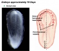
Introduction
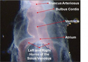
The embryo stage 10 heart tube
Development of the heart and vascular system begins very early in mesoderm both within (embryonic) and outside (extra embryonic) the embryo. Vascular development therefore occurs in many places, the most obvious though is the early forming heart, which grows rapidly creating an externally obvious cardiac "bulge" on the early embryo.
The heart forms initially in the embryonic disc as a simple paired tube inside the forming pericardial cavity, which when the disc folds, gets carried into the correct anatomical position in the chest cavity.
Throughout the mesoderm, small regions differentiate into "blood islands" which contribute both blood vessels (walls) and fetal red blood cells.
These "islands" connect together to form the first vessels which connect with the heart tube.
| original page
Some Recent Findings
- Endothelial cell lineages of the heart. [1] "During early gastrulation, vertebrate embryos begin to produce endothelial cells (ECs) from the mesoderm. ECs first form primitive vascular plexus de novo and later differentiate into arterial, venous, capillary, and lymphatic ECs. In the heart, the five distinct EC types (endocardial, coronary arterial, venous, capillary, and lymphatic) have distinct phenotypes. For example, coronary ECs establish a typical vessel network throughout the myocardium, whereas endocardial ECs form a large epithelial sheet with no angiogenic sprouting into the myocardium. Neither coronary arteries, veins, and capillaries, nor lymphatic vessels fuse with the endocardium or open to the heart chamber. The developmental stage during which the specific phenotype of each cardiac EC type is determined remains unclear. The mechanisms involved in EC commitment and diversity can however be more precisely defined by tracking the migratory patterns and lineage decisions of the precursors of cardiac ECs."
|
References
- ↑ <pubmed>18682987</pubmed>
Reviews
Articles
Search Pubmed
Search May 2010
- Cardiovascular System Development All (63457) Review (10735) Free Full Text (15717)
Search Pubmed: Coronary Circulation Development
Additional Images
See also Category:Heart ILP and Category:Heart
Heart Development Timeline
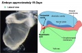
Early Heart Tube (Dorsal)
Early Heart Tube (Lateral)
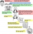
Molecular & Genetic Cardiac Development Factors
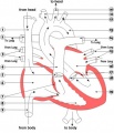
Adult heart blood flow cartoon
External Links
External Links Notice - The dynamic nature of the internet may mean that some of these listed links may no longer function. If the link no longer works search the web with the link text or name. Links to any external commercial sites are provided for information purposes only and should never be considered an endorsement. UNSW Embryology is provided as an educational resource with no clinical information or commercial affiliation.
Glossary Links
- Glossary: A | B | C | D | E | F | G | H | I | J | K | L | M | N | O | P | Q | R | S | T | U | V | W | X | Y | Z | Numbers | Symbols | Term Link
Cite this page: Hill, M.A. (2024, April 25) Embryology Cardiovascular System - Coronary Circulation Development. Retrieved from https://embryology.med.unsw.edu.au/embryology/index.php/Cardiovascular_System_-_Coronary_Circulation_Development
- What Links Here?
- © Dr Mark Hill 2024, UNSW Embryology ISBN: 978 0 7334 2609 4 - UNSW CRICOS Provider Code No. 00098G


.jpg)
.jpg)

